histology digestive system
 Call Kai
 Learn
 Practice Test
 Spaced Repetition
 Match
 Flashcards
 Knowt Play
1/103

There's no tags or description
Looks like no tags are added yet.
104 Terms

what part of the digestive system is this?
Pharynx

what part of the digestive system is this, and what is each number labeling?
esophagus
squamous mucosa
mucosa
muscularis mucosa

what part of the digestive system is this, and what is each number labeling?
esophagus
squamous mucosa
submucosa
muscularis externa
adventitia/serosa
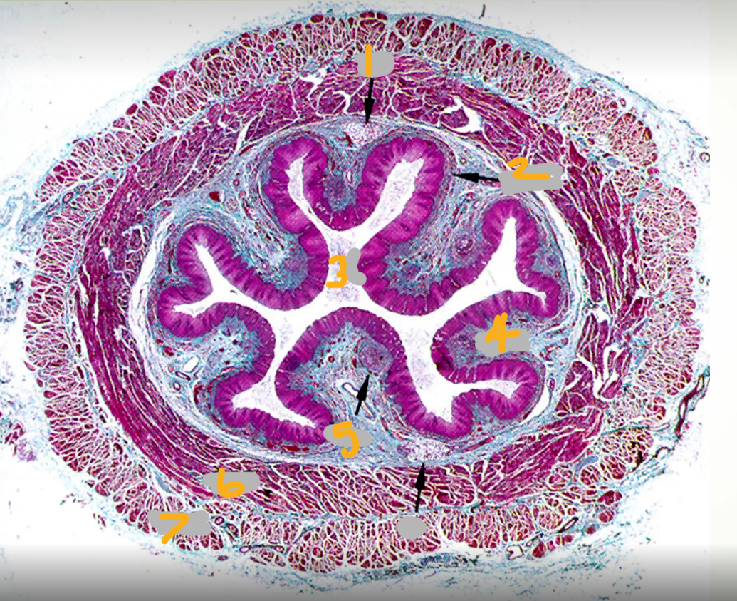
what part of the digestive system is this, and what is each number labeling?
esophagus
glands
extensive muscularis mucosae
epithelium (nonkeratinized stratified squamous)
sm
?
inner circular muscle
outer longitudinal muscle

what part of the digestive system is this, and what is each number labeling?
esophagus
epithelium
blood vessels
lamina propria
muscularis mucosae

what part of the digestive system is this, and what is each number labeling?
esophagus
mucosa
muscularis mucosae
submucosa
duct
muscularis externa

what part of the digestive system is this, and what is each number labeling?
muscularis externa
inner circular muscle
myenteric plexus (Auerbach’s plexus)
outer longitudinal muscle

what is this slide showing?


what part of the digestive system is this, and what is each number labeling?
Gastroesophageal Junction
stomach
esophagus
fundic/gastric glands
cardiac glands
muscularis externa

what part of the digestive system is this, and what is each number labeling?
stomach
rugae
pits

what is this slide showing?
fundic/gastric glands

what part of the digestive system is this, and what is each number labeling?
cardiac gland
pit
gastric pits
cardiac glands
esophageal epithelium

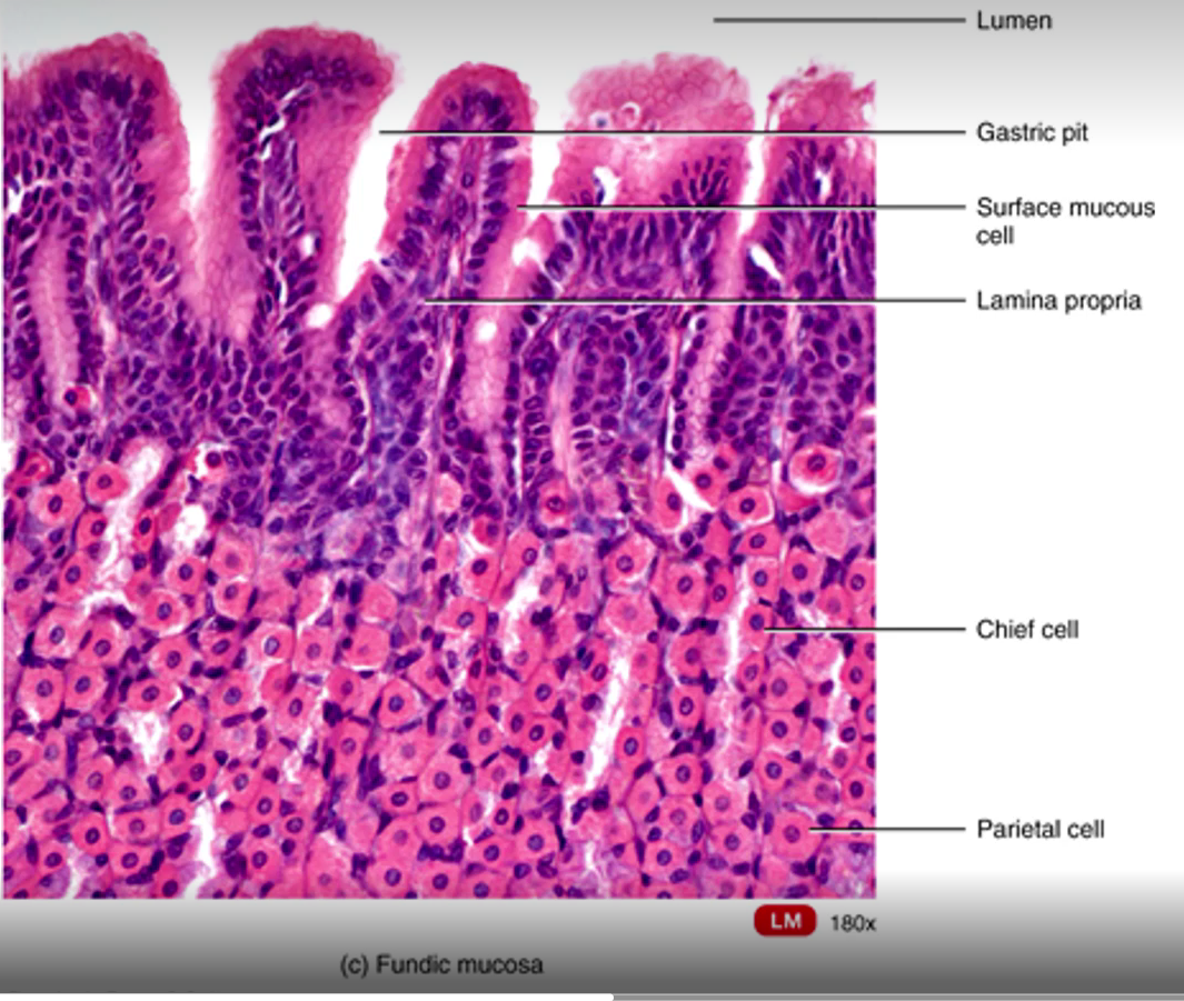
what part of the digestive system is this, and what is each line labeling?
fundic mucosa/gland

what part of the digestive system is this, and what is each number labeling?
Pylorus - Stomach
gastric pits
pyloric glands
muscularis mucosae
submucosa

what part of the digestive system is this, and what is each number labeling?
*mm = muscularis mucosae
sm = submucosa


what part of the digestive system is this showing?
transition from pylorus to duodenum

what part of the digestive system is this, and what is each number labeling?


what part of the digestive system is this, and what is each number labeling?
small intestine


what part of the digestive system is this, and what is each number labeling?
duodenum


what part of the digestive system is this, and what are the green circles labeling?
Paneth cells of small intestine

what part of the digestive system is this, and what are the green circles labeling?
Paneth cells of the small intestine

what part of the digestive system is this, and what is each number labeling?
Small intestine: Paneth cells


what part of the digestive system is this, and what is each number labeling?

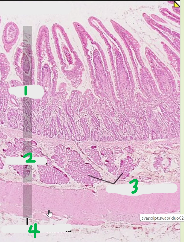
what part of the digestive system is this, and what is each number labeling?


what part of the digestive system is this, and what are the green lines labeling?
jejunum
goblet cells

what part of the digestive system is this, and what is each number labeling?


what part of the digestive system is this, and what is each number labeling?


what part of the digestive system is this, and what is each number labeling?
Ileum


what part of the digestive system is this slide showing?
Ileum

what part of the digestive system is this, and what is each number labeling?
Ileum


what part of the digestive system is this, and what is each arrow/line labeling?
colon (large intestine)


what part of the digestive system is this slide showing?
Colon (large intestine)

what part of the digestive system is this, and what is each number labeling?
Colon


what part of the digestive system is this, and what is each number labeling?
Colon


what part of the digestive system is this, and what is each number labeling?
Colon (large intestine)


what part of the digestive system is this slide showing?
Colon

what part of the digestive system is this, and what is the black arrow labeling?
appendix
lymphatic nodules

what part of the digestive system is this, and what is each number labeling?
Appendix


what part of the digestive system is this, and what is each number labeling?
Lip
glands
skeletal muscle
skin
vermillion border
oral mucosa
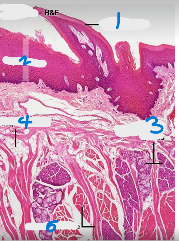
what part of the digestive system is this, and what is each number labeling?
tongue

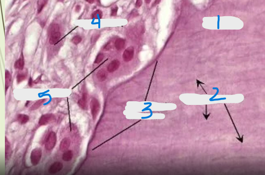
what part of the digestive system is this, and what is each number labeling?
teeth

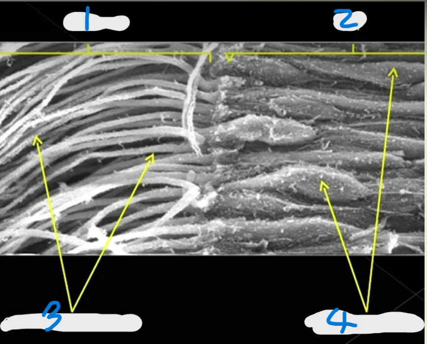
what part of the digestive system is this, and what is each number labeling?
tooth

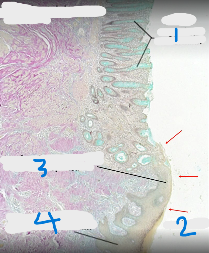
what part of the digestive system is this, and what is each number labeling?
Anorectal junction

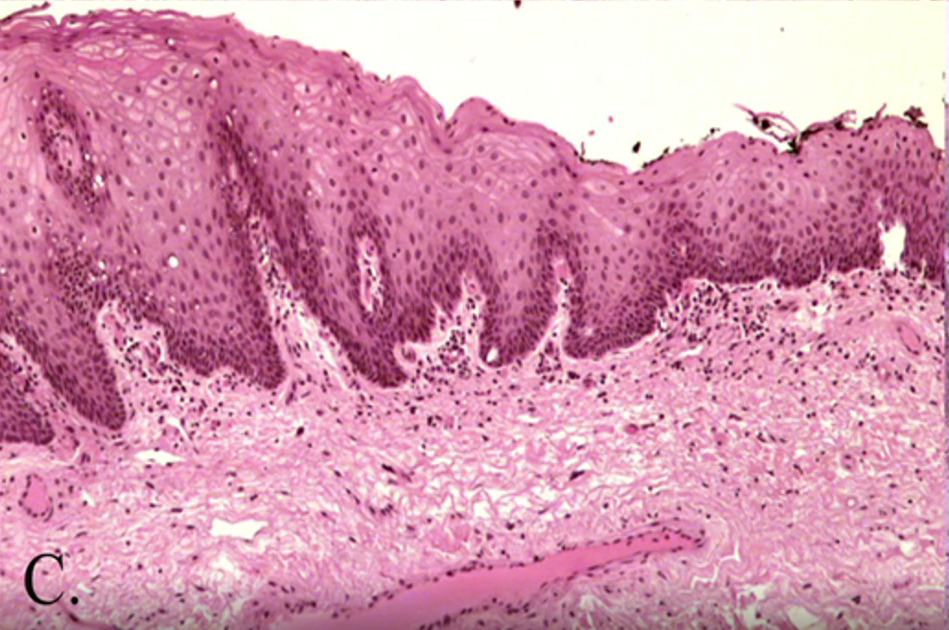
what part of the digestive system is this?
anal canal

what part of the digestive system is this, and what is the green arrow labeling?
Pancreas
Main Duct

what part of the digestive system is this, and what are the orange arrows labeling?
Pancreas
Arrows: Pancreatic Acini

what part of the digestive system is this, and what is each number labeling?
Liver
capillary
portal vein
hepatocyte
bile duct
hepatic artery

what part of the digestive system is this, and what is each number labeling?
Gallbladder


what part of the digestive system is this, and what is each box labeling?
Gallbladder


what part of the digestive system is this, and what is each box labeling?
Gallbladder
Pharynx structure
epithelium: nonkeratinized stratified squamous
CT: areolar CT and elastic fibers in lamina propria
NO submucosa, muscularis mucosa, or adventitia serosa
Muscle: skeletal muscle wall
esophageal muscularis
first and second section of esophagus are skeletal muscle, by the third section this changes to smooth muscle
between w 2 layers of smooth muscle in third section are nervous cells/myenteric plexus
1st and 2nd sections have adventitia, 3rd section has serosa
esophagus overall structure
3 sections, all nonkeratinized stratified squamous, all sections have same mucosa and have extensive muscularis mucosa
tubular acinar glands line tract (mostly in submucosa), but this changes when get to gastroesophageal junction
2 sphincters (upper esophageal at top and stays closed when breathing, inferior esophageal before entering stomach to keep stomach contents in stomach)
esophageal visual ID
thick muscle and glands (different from urinary ureters)
tubuloacinar glands in esophagus to secrete mucous vs cardiac glands which are closer to junction/stomach
3rd section of esophagus has special glands
Myenteric/Auerbach’s Plexus
fxn: one of only places where nervous tissue is found in digestive system
structure: myelinated, loose areolar CT, visible neurons
location: between muscle layers
Gastroesophageal Junction
fxn: separate stomach and esophagus, important in trying to prevent esophageal cancer
structure:
simple columnar epithelium running up and down pits (abrupt change from nonkeratinized strat squam in esophagus to simple columnar in stomach)
start seeing cardiac glands
adipose in submucosa and dense IR CT for multidirectional tension
location: between esophagus and stomach
Cardiac Glands
fxn: counter acidity of stomach by releasing more neutralizing/basic, important in trying to prevent esophageal cancer
structure: simple columnar and nonkeratinized epithelium, deep pits, homogenous
location: gastroesophageal junction and cardia portion of stomach
4 areas of stomach and their glands
cardia - cardiac glands
fundus - gastric glands
corpus - gastric glands
pylorus - pyloric glands
rugae
fxn: increase SA
structure: folds, gastric pits running in these
location: mostly in fundus and corpus of stomach
Gastric glands (fundic glands)
fxn: lots of cells doing different jobs
structure: shallow pits, heterogenous, rugae
location: fundus and corpus of stomach
pyloric glands
simple columnar epithelium, deep pits that are homogenous and they go all the way down to the muscle layer
(difference bw these and cardiac glands are that pyloric glands are simple columnar to simple columnar, while cardiac glands are simple columnar and nonkeratinized strat squam)
Pylorus of Stomach
end of stomach, pyloric sphincter, no abrupt junction of different epithelium, pyloric glands, mostly mucous cells, fairly prominent submucosa
*visual ID: look for smooth muscle change
Gastroduodenal Junction
going from simple columnar epi in stomach to simple columnar with microvilli
submucosa layer getting larger and pits getting shallower
pylorus has deeper pits while duodenum has shallow pit
submucosa has lots of glands and vessels
going from no glands in stomach to lots of glands in the intestine
Plicae Circulares
fxn: big projections in small intestine that give more SA
structure: villi (with crypts between) coming off of these projections, simple columnar epithelium lining surface villi (enterocyte, each enterocyte has tons of microvilli)
Small Intestine
fxn: absorption
structure: areolar CT, crypts of Lieberkuhn, cardio and lymph vasculature, lymph nodules and goblet cells
How to tell if vasculature in small intestine is lymph or cardio
look if there’s a RBC in capillary -
if yes = regular cardio vasculature
if no = lacteal
slits = lymph
what are the specialized cells in the small intestines?
enteroendocrine cells - Paneth cells and AUPD cells
Paneth cells
fxn: release bacterial resistant, precursor to lysosome, immune protective
structure: red-looking cells at very bottom of crypts, may have some visible granules depending on stain used
location: crypts of small intestine
AUPD cells
fxn: endocrine function, release hormones to bloodstream, associate with gastrin
location: crypts in small intestine
what are the three sections of the small intestine?
duodenum, jujenum, and ileum
duodenum
fxn: neutralizing acid from stomach
structure: very long villi, Paneth cells, thick submucosa, BRUNNER’S GLANDS, 2 layers smooth muscle (1 circular and 1 longitudinal), serosa, lots of goblet cells
location: 1st section of small intestine
Jujenum
fxn: absorption
structure: NO brunner’s glands, tongue shaped villi (much shorter and fatter/pyramid shaped), small crypts, thin layer of submucosa
location: middle section of small intestine
Ileum
fxn: protection and stopping things that need to be stopped
structure: dome shaped villi, have Peyer’s Patches (bunch of lymphatic nodules) in lamina propria, have specialized M cells
location: bottom third of small intestine nearest the large intestine
M cell in ileum
specialized cells on the surface that present antigens to lymphocyte and macrophages in order to upregulate immune system; big bubble appearance
Large Intestine
fxn: last area for absorption
structure:
epithelium: lots of simple columnar epithelium with lots of goblet cells
stem cells and lymph nodules (b/c lots of bacteria)
thick and continuous muscle called taenia coli, thick cord/drawstring-type longitudinal muscle layer
pulls in one direction
front of large intestine has serosa while back area has adventitia
deep crypts, muscularis mucosa, thick submucosa
dense IR CT and vessels
still have Auerbach’s plexus if zoomed in enough
Appendix
fxn: special area of colon that acts as first line of defense for the colon/immune receptor
structure: crypts, absorptive cells, goblet cells, M cells carry over from small intestine (good differentiating structure), lots of lymph nodules, serosa
*can see clear muscle layers and can see whole cross section on one slide → great differentiating factor between appendix and colon
location: entrance into large intestine from small intestine
Rectum
simple columnar, small lumen, lamina propria, thick circular muscle layer, adventitia, start seeing valves, LOTS of goblet cells to help with movement
Rectoanal Junction and Anus
going from simple columnar (rectum) to nonkeratinized stratified squamous (anal canal), then transition to keratinized stratified squamous when merged with integument on outside of body
2 sphincters with smooth and skeletal muscle next to each other
internal sphincter = extension of circular smooth muscle layer
external sphincter = what we control, skeletal muscle
Pancreas
fxn: both endocrine and exocrine functions
endocrine: insulin, glucagon, etc. → islets releasing hormones
exocrine: secreting enzymes
structure: lots of simple cuboidal epithelium, NO muscle, tightly packed cells with loose areolar CT, NO clear/mucous cells, have a big main duct
*easy to mix up with salivary glands
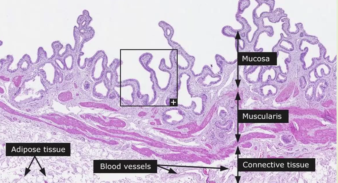
Gallbladder
fxn: store and concentrate bile, regulate release of concentrated bile, lots of osmosis
structure: small crypts, 3 layers
mucosae: simple columnar epithelium, rugae (as gallbladder stretches the rugae go away), microvilli, lamina propria, no submucosa
muscle: interwoven layer of smooth muscle (not distinct, interwoven)
adventitia/serosa
Gall Stones
water follows sodium, so pumping sodium out means water will follow and leave = concentrating the bile → when bile from gallbladder gets too concentrated and calcified it forms gall stones
Liver
fxn: largest gland in body, filters stuff, main function is nutrient interconversion
structure: capsule surrounding organ, lobules
Portal triad of liver
structure: composed of portal vein, bile duct, and hepatic artery
location: at the corners of hexagonal lobules of liver
Bile duct
part of portal triad in liver, simple cuboidal epithelial tissue
hepatic artery
part of portal triad in liver, delivers oxygen and has smooth muscle
Lobules of liver
composed of central vein, portal triad, hepatic sinusoids, Kupffer cells, and hepatocytes
Hepatic sinusoids
sinusoids large, between cells
Kupffer Cells (Stellate Reticuloendothelial Cells)
fxn: specialized macrophages for liver
structure: flat/elongated
location: in walls/linings of sinusoids
Hepatocytes
fxn: make bile
structure: lines and rows of hepatocytes, bile canaliculi connect hepatocytes and collect/transport bile towards bile duct
layering of the GI tract (innermost to outermost)
mucosa, submucosa, muscularis externa, adventitia or serosa
adventitia vs serosa
adventitia - covering of GI tract found outside the peritoneal (abdominal) cavity, lacks a mesothelium and the connective tissue blends with surrounding connective tissues
serosa - covering of GI tract within the peritoneal cavity, continuous with mesentery and peritoneal lining of abdominal cavity, contains a mesothelial lining (simple squamous epithelial lining)
Lips
structure: internal surface is nonkeratinized stratified squamous and external surface is covered in thin skin, skeletal muscle
thin skin of lips
thin skin may contain hair follicles and sebaceous glands (but lacks sweat glands)
vermillion border (red vermillion zone)
area of lip that’s the junction of skin and mucosa
Papillae of tongue
mucosal structures lining dorsal surface of tongue and base of tongue, where tastebuds are located (except filiform papillae, these are the only ones without tastebuds)
dorsal surface papillae: filiform papillae, fungiform papillae, foliate papillae
base papillae: vallate papillae
what cells make up tastebuds
gustatory cells, supportive cells, basal stem cells, and immature gustatory cells
regions of the tooth
crown - exposed tooth above gumline
neck - narrowing at gumline
root - extends below gumline into alveolar bone socket
alveolus - tooth socket in bone
alveolus of tooth
periodontal ligament: attach bone to bone, has Sharpeys fibers (collagen), anchors tooth firmly into alveolus, and allows slight movement of tooth under pressure of chewing
Gingiva/Gum: covers alveolar bone
Dentin
calcified tissue that makes up most of tooth; has odontoblasts and odontoblast processes
layers of dentin
odontoblasts: elongated cells that produce matrix of dentin; derived from mesenchymal cells of pulp cavity
odontoblast processes: extensions of odontoblast which are used in secretion of pre-dentin (this is then mineralized into dentin)