Bio 403 1
 Call Kai
 Learn
 Practice Test
 Spaced Repetition
 Match
 Flashcards
 Knowt Play
1/126

There's no tags or description
Looks like no tags are added yet.
127 Terms
Transverse plane
Cross section across anterior (towards head) and posterior (towards feet)

Horizontal/coronal plane
divided from dorsal and ventral side of the body

Midsagittal plane
divided laterally (left and right) of the plane

Cleavage
Rapid cell divisions divide the zygote cytoplasm into many
progressively smaller cells, with no increase in total volume. The size and volune of the embryo does not significantly increase until organogenesis has started, so as the amount of cells increase the cells become smaller to stay the same volume.
In early cell division: Rapid synchronous cell divisions
during cleavage are followed by asynchronous and
relatively slow cell divisions during gastrulation.
Basic difference between Cleavage, Gasturlation and Oragongensis
cleavage (cell sizes get smaller, synchronous division), gastrulation (cells move to enter embryo) , and organogenesis when identifiable structures (like the nervous system) begin to appear.
what is the vegetal pole and animal pole in egg
VEGETAL POLE (i.e. the towards the yolk The vegetal pole divides more slowly because yolk can physically interfere with cell division) ANIMAL POLE (other side from the yolk. The animal pole divides faster because it contains less yolk.)
BLASTOMERE def
a cell of the cleavage stage embryo
Morula
cleavage stage embryo when there is no cavity in the embryo.
Blastula
cleavage stage embryo when there IS a cavity in the embryo. (Called the blastocyst stage in mammals)
Gastrula
name for an embryo undergoing gastrulation. Start showing the dorsal blastopore lip through which the mesodermal and some endoderm cells migrate.
Neurula
name for an embryo as the neural tube is forming (rarely used for mammals).
2 types of cleavage
holoblastic (complete cleavage) and meroblastic (incomplete) cleavage
2 types of holoblastic cleavage
The cleavage furrow extends all the way through the egg. complete cleavage where the entire embryo is divided (all the cells are apart of the embryo). theres isolecithal and mesolecithal
isolecithal cleavage
complete cleavage where the entire embryo is divided and the cells are the same size when divided. the yolk is evenly divided.
Mesolecithal cleavage
complete cleavage. the yolk isnt divided evenly and sets up symmetry.
2 types of meroblastic cleavage
incomplete cleavage where the cleavage plane extends only to the accumulated yolk: Telolecithal (called Discoidal in fish) or Centrolecithal
Telolecithal cleavage
incomplete cleavage where the yold is so thick it cant fully divide
Centrolecithal cleavage
incomplete cleavage where the yolk is concentrated in the center of the egg. instead of the cell dividing the nuclei repricate and migrate to the edge of the cell bc the yolk is in the middle. this is common in insects.
what specialized cells do the distinct embryonic germ layers form
The germ cells (precursors of the sperm and egg) are set aside early in development and do not arise from any particular germ layer.

notochord
(outcome of gastrulation) mesodermal strucutre necessary for nervous system formation. The notochord instructs the ectoderm above it to become the nervous system (the neural tube at this stage of development). To either side of the notochord and the neural tube are the mesodermal masses called somites, which will form vertebrae, ribs, and skeletal muscles.
what are the 2 basic cell types in the embryo
epithelial cells which are attached in sheets and mesenchymal cells which are individualized cells that can move. these two cell types can go through transition to form one another.
Epithelial to mesenchymal transformation (or transition) (EMT)
Mesenchymal to epithelial transformation (or transition) (MET
Types of cell movement during gastrulation
1)invagination
2)involution
3)ingression
4)delamination
5)epiboly
6)convergent extension
what is invagination (gastrulation)
infolding of a epithelium sheet of cells into a hollow indention
what is involution(gastrulation)
inward movement of an expanding outer layer so that it spreads over the internal surface of the remaining external cells. the cells are dividing rapidly.
what is ingression (gastrulation)
migration of individual cells from the surface into the embryos interior. those cells become mensechymal and migrate seperately
what is delamination (gastrulation)
splitting of one cellular sheet into two more or less parallel sheets. while on a cellular basis it resembles ingression, the result is the formation of a new additional epithelial sheet of cells. (no mensechymal identity)
what is epiboly (gastrulation)

what is convergent extension (gastrulation)

define commitment
Describes a state in which a cell’s developmental fate has become restricted even though it is not yet displaying overt changes in cellular biochemistry and function
The stages of cell commitment during development
1) specification
2) determination
3) differentiation
What is specification
When a cell is committed to a given fate, but this commitment can still be reversed. In this stage a cell will differentiate along a certain path unless signals or its environment changes. When a cell can differentiate by itself (autonomously) into a specific cell type in a neutral environment (like in the embryo), but commitment is flexible, labile.
what is Determination
When a cell can differentiate autonomously and its fate cannot be reversed by signals from neighbouring cells. It can often be difficult to know if a cell is determined or “just” specified
Autonomous specification
autonomous specification blastomeres acquire determination factors from the egg cytoplasm.
in invertabrates
Autonomous specification is a mechanism in which embryonic cells (blastomeres) develop according to intrinsic, inherited instructions rather than external signals. The cell-intrinsic properties arise from a cleavage of a cell with asymmetrically expressed maternal cytoplasmic determinants (proteins, small regulatory RNAs and mRNA). Thus, the fate of the cell depends on factors secreted into its cytoplasm during cleavage.
Xenopus Nanos mRNA marks the germplasm, blastomeres that
acquire the germplasm become the germ cells
Conditional specification
In conditional specification embryonic cells are specified by signals from their neighboring cells
conditional specification is a cell-extrinsic process that relies on cues and interactions between cells

What is a fate map
fate refers to a cells future path of differentiation in an undisturbed embryo. This is always a statistical likelihood. And it depends on the species if a fate map can be created.
mosaic development
this refers to Autonomous specification of blastomeres
Autonomous specification in tunicates (invertabrate subphylum of filter feeders) Isolated blastomeres produce only part of the organism, like the tiles of a mosaic.
regulative development
this refers to Conditional specification of blastomeres. The embryo can respond to removal of parts.
Conditional specification of blastomeres. In vertebrates the early blastomeres are totipotent. Isolated blastomeres are said to REGULATE their development to produce a complete organism. If blastomeres in mice (or humans!) are split into two units each generates a complete embryo, i.e. twins
Hans Driesch--Early demonstration of conditional specification (1892) each blastomere regulated its development .When one removes the 4-cell embryo from its fertilization envelope and isolates each of the four cells, each cell can form a smaller, but normal, pluteus larva. (All larvae are drawn to the same scale.) Note that the four larvae are not identical, despite each one’s ability to generate all the necessary cell types. Such variation is also seen in adult sea urchins derived in this way
totipotent
An isolated cell that is able to produce a fertile adult individual. An isolated cell is transferred to a uterus (after inserting into an empty zona pellucida or after developing to the blastocyst stage in culture) and it gives rise to a fertile adult. Zygotes. Some early cleavage-stage blastomeres.
Syncytial specification
type of cell fate specification that occurs when multiple nuclei share the same cytoplasm, before individual cell membranes form around them.
syncytium
A syncytium is a structure containing many nuclei within one shared cytoplasm, without cell membranes separating the nuclei.
How does syncytial specification work?
morphogens (signalling molecules) form concentration gradients across the embryo.
Different nuclei experience different concentrations of these morphogens depending on their position.
For example, a nucleus near the anterior end experiences a different morphogen concentration than a nucleus near the posterior end.
These concentration differences activate different sets of genes, causing different regions of the embryo to acquire different developmental fates.
For example, nuclei exposed to high Bicoid concentrations activate genes involved in anterior development, caudal are posterior
morphogens
a long range signalling molecule that forms a gradient in the embryo and specification /differentiation of a cell depends n the concentration of the molecule
difference between morphogens and dietermiments
(Bicoid is a transcription factor—special situation where a transcription factor can act as a signaling molecule because it isn’t hindered by cell membranes. In all other cases besides a syncytium, a transcription factor CANNOT act as a morphogen. This is the exception.)
Determinant: Influences the fate of the cell in which it is located or produced.
Morphogen: Influences the developmental fates of other cells by spreading and creating a concentration gradient
Contrast how protein localization differs from its mRNA
In Drosophila (fruit fly) embryos:
Bicoid mRNA is tethered at the anterior (head) end of the egg.
The mRNA remains localized there rather than spreading throughout the embryo.
Ribosomes translate the mRNA into Bicoid protein.
The Bicoid protein can spread through the shared cytoplasm, forming a concentration gradient.
This gradient helps determine which parts of the embryo develop into anterior structures.
Transcription Factors
proteins that regulate gene expression. Genes must be turned on at the right time and place for development to proceed
Secreted signaling molecules (usually proteins):
involved in cell-cell communication via signal transduction cascades
Cell surface receptor proteins
responsible for receiving signals and propagating them intracellularly.
histones role is nucleosomes
The histone “tails” that extend from the core are the sites of acetylation and methylation, which may disrupt or stabilize, respectively, the formation of nucleosome assemblages. (B) Histone H1 can draw nucleosomes together into compact forms. About 147 base pairs of DNA encircle each histone octamer, and about 60–80 base pairs of DNA link the nucleosomes together. (C) Model for the arrangement of nucleosomes in the highly compacted solenoidal chromatin structure. Histone tails protruding from the nucleosome subunits allow for the attachment of chemical groups.
Epigenetic regulation by histone modification.
Methyl groups condense nucleosomes more tightly, preventing access to promoter sites and thus preventing gene transcription. Acetylation loosens nucleosome packing, exposing the DNA to RNA polymerase II and transcription factors that will activate the genes. acetylation neutralizes the positive charge of lysine, loosening it’s attraction to the negatively charged DNA making them more accessible to transcription factor binding.
Histone methylations on histone H3
The tail of histone H3 (its amino-terminal sequence, at the beginning of the protein) sticks out from the nucleosome and is capable of being methylated or acetylated. Here, lysines can be methylated and recognized by particular proteins. Methylated lysine residues at positions 4, 38, and 79 are associated with gene activation, whereas methylated lysines at positions 9 and 27 are associated with repression. The proteins binding these sites (not shown to scale) are represented above the methyl group.

acetylation of lysine
acetylation neutralizes the positive charge of lysine, loosening it’s attraction to the negatively charged DNA making them more accessible to transcription factor binding.
Acetylation and methylation at the same lysine are mutually exclusive. Both acetylation and methylation modify the same nitrogen on lysine. Once that nitrogen is methylated, it’s no longer the substrate the acetyltransferase (KAT) expects.
Acetylation by HATs
enzymes called Histone Acetyltransferases (HATs) add an acetyl group (–COCH₃) to histone proteins. This usually causes chromatin to become less tightly packed, making DNA more accessible for transcription (gene expression).
Histones contain positively charged lysine residues.
DNA has a negatively charged phosphate backbone.
The positive histones and negative DNA attract each other.
HATs add an acetyl group to lysine, neutralizing its positive charge.
This weakens histone–DNA interactions and generally makes chromatin more accessible.
Deacetylation by HDs
enzymes called Histone Deacetylases Remove acetyl groups, Restore lysine's positive charge, Generally promote tighter chromatin, Usually repress gene expression
The central dogma of biology
1. Transcription: The gene (DNA) is transcribed into pre-mRNA containing exons, introns, untranslated regions, a 5′ cap and a poly-A tail.
2. RNA processing: Introns are removed, while exons are joined together to produce mature mRNA.
3. Translation: Ribosomes read the mature mRNA to produce the β-globin protein.
4. Post-translational modification and assembly: β-globin becomes part of functional hemoglobin after modification and association with α-globin and heme.
The bridge between enhancer and promoter can be made by transcription factors
Transcription factors can form a bridge between enhancer and promoter. Certain transcription factors, called basal transcription factors, bind to DNA on the promoter (where RNA polymerase II will initiate transcription). Other transcription factors bind to the enhancer (which regulates when and where transcription can occur). Some transcriptional regulators do not bind to the DNA; rather, they link the transcription factors that have bound to the enhancer and promoter sequences. In this way, the chromatin loops to bring the enhancer to the promoter. The example shown here is the mouse β-globin gene. (A) Transcription factors assemble on the enhancer, but the promoter is not used until the Gata1 transcription factor binds to the promoter. (B) Gata1 can recruit several other factors, including Ldb1, which forms a link uniting the enhancer-bound factors to the promoter-bound factors. (After W. Deng et al. 2012. Cell 149: 1233–1244.)

an enhancer
Enhancers are often tissue-specific, i.e. they activate gene transcription in one tissue. Therefore, mutations in an enhancer can cause a very tissue-specific phenotype. An enhancer is a noncoding DNA sequence that binds transcription factors and activates transcription of a specific gene in cis, from distances as great as one million base pairs from the gene’s promoter.
what do “in cis” and “in trans” mean
"in cis" refers to genetic elements, such as enhancers, that regulate the expression of genes located on the same molecule of DNA. So, when it's stated that an enhancer "activates transcription of a specific gene in cis," it means that the enhancer influences the transcription of genes that are nearby on the same DNA molecule. This is in contrast to "in trans" effects, where regulatory elements affect genes on different DNA molecules, such as on another chromosome.

Enhancer modularity
in genetics refers to the idea that different enhancer elements (specific DNA sequences) can independently regulate the expression of a single gene in distinct spatiotemporal patterns, depending on the needs of the organism. Each enhancer acts as a separate, modular regulatory unit that responds to specific signals, thereby controlling gene expression in different tissues, at different times, or in response to different environmental cues.
The main concept of this slide is that one gene can have multiple enhancers, and each enhancer can control the expression of that same gene in a different tissue.
Brain-specific transcription factors are present.
These transcription factors bind to the brain enhancer.
The enhancer-bound proteins interact with Mediator, helping connect the enhancer to the transcription machinery.
Mediator helps stabilize RNA polymerase II at the promoter, while nucleosomes near the promoter are modified.
Gene A is transcribed in the brain
Enhancer Modularity — Control of Pax6 Expression
one gene can have multiple enhancers that control its expression in different tissues.
the Pax6 gene is involved in the development of several different tissues, and separate enhancers control where Pax6 is expressed.
How does the pancreas enhancer work?
two transcription factors: Pbx1 and Meis. The pancreas-specific enhancer contains DNA binding sites for both. For Pax6 to be activated in the pancreas, both transcription factors must be present and bind the enhancer. When the lacZ reporter gene (which codes for β-galactosidase) is fused to the Pax6 enhancers for expression in the pancreas and lens/cornea, β-galactosidase enzyme activity (blue) is seen in those tissues.
combinatorial regulation by Pbx1 and Meis and using lacZ as a reporter to identify enhancer activity.
Families of Transcription Factors (TFs)
transcription factors are grouped into families based on their structural similarities, especially their DNA-binding domains.
Transcription factors (TFs) are proteins that regulate gene expression by binding to specific DNA sequences.
Different transcription factors have different structures. Scientists group TFs into families based on shared structural features and other properties.
For example, transcription factors belonging to the same family often have similar DNA-binding domains.
A DNA-binding domain is the part of the transcription factor that recognizes and binds a specific DNA sequence.
A homeodomain is a specific type of DNA-binding domain found in a family of transcription factors called homeodomain transcription factors.
Its function is to allow a transcription factor to recognize and bind specific DNA sequences so that it can regulate gene expression.
three major domains of transcription factors
1.DNA binding domain (The DNA-binding domains are located toward the amino termini (N-termini) at the bottom of the figure. They recognize an 11-base-pair DNA sequence containing the core sequence. Transcription factors don't bind randomly to DNA. Their DNA-binding domains recognize particular sequences.)
2. trans-activating domain (This domain is located immediately above the DNA-binding region.
It allows the two identical MITF proteins to interact and form a homodimer.
Remember:
Homodimer: Two identical protein molecules.
Heterodimer: Two different protein molecules.
The helix-loop-helix structure contributes to the interaction between the MITF proteins.)
3. Protein-protein interaction domain.
(Its function is to interact with transcriptional co-regulators.
DNA Methylation as a Mechanism to Silence Transcription
adding methyl groups to DNA, particularly at gene promoters, is generally associated with decreased gene transcription. Importantly, DNA methylation does not change the DNA base sequence. It is an epigenetic modification that can affect whether a gene is expressed.The promoter is the region of DNA where the transcription machinery assembles to initiate transcription.When a promoter becomes methylated, it can interfere with transcription factor binding or recruit proteins that promote a repressive chromatin structure. As a result, transcription is reduced.
In the example on your slide, the methyl group is added to cytosine, producing 5-methylcytosine.

Modifying nucleosomes through methylated DNA
Modifying nucleosomes through methylated DNA. MeCP2 (Methyl CpG Binding Protein 2.) recognizes the methylated cytosines of DNA. It binds to the DNA and is thereby able to recruit (A) histone deacetylases (which take acetyl groups off the histones) or (B) histone methyltransferases (which add methyl groups to the histones). Both modifications promote the stability of the nucleosome and the tight packing of DNA, thereby repressing gene expression in these regions of DNA methylation.
Mutations of MeCP2 cause Rett syndrome, a severe debilitating neurological disorder affecting girls.
What are the two DNA methyltransferases, how do they function, and what are their roles in DNA methylation?
The two important DNA methyltransferases are Dnmt3 and Dnmt1, which are enzymes that add methyl groups to cytosine bases in DNA. Dnmt3 is a de novo methyltransferase that adds methyl groups to previously unmethylated cytosines, establishing new DNA methylation patterns. Dnmt1 is a perpetuating methyltransferase that recognizes methylated cytosines on one DNA strand and methylates the corresponding cytosines at CG sites on the opposite strand, maintaining existing DNA methylation patterns. DNA methylation at gene promoters generally restricts transcription, allowing Dnmt3 to establish and Dnmt1 to maintain methylation patterns associated with gene silencing.
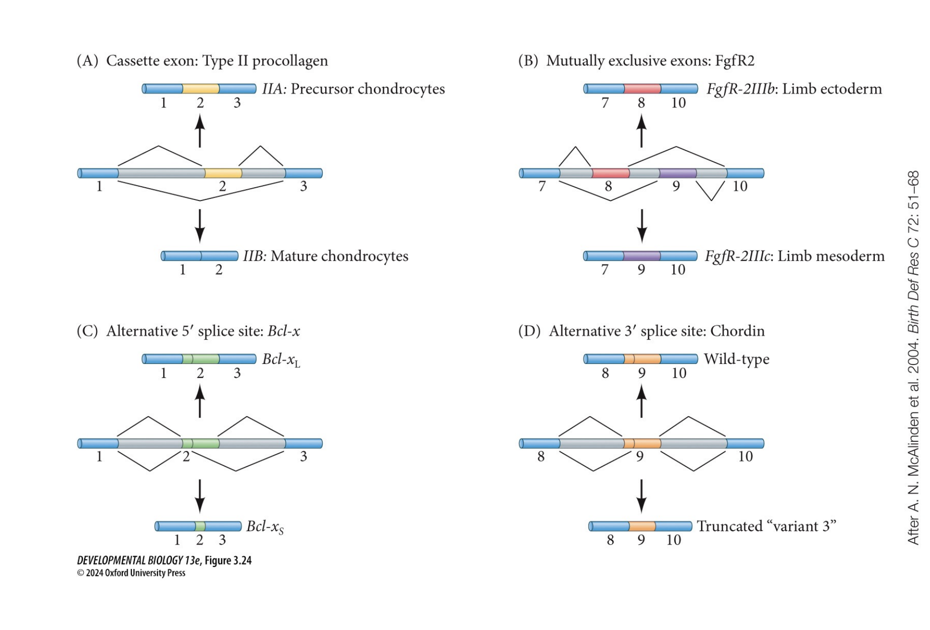
What is alternative RNA splicing, and what are the four types?
Alternative RNA splicing is a process in which the same pre-mRNA can be spliced in different ways to produce different mature mRNAs and protein isoforms from a single gene. The four types are (1) Cassette exon splicing, where an exon can either be included or skipped, as seen in type II collagen in chondrocyte precursors and mature chondrocytes; (2) Mutually exclusive exon splicing, where only one of two alternative exons is included, as seen in fibroblast growth factor receptors in the limb ectoderm and mesoderm; (3) Alternative 5′ splice site selection, where different 5′ splice sites are used to produce different protein isoforms, such as the large and small forms of Bcl-X; and (4) Alternative 3′ splice site selection, where different 3′ splice sites are used, producing different protein forms, such as normal and truncated Chordin.
What is the role of cadherins during embryonic development, and how does SLUG promote epithelial-to-mesenchymal transition (EMT)?
Cadherins are transmembrane cell adhesion proteins that hold epithelial cells together and allow cells to sort and segregate based on the types of cadherins they express, contributing to morphogenesis. During epithelial-to-mesenchymal transition (EMT), the transcription factor SLUG, a member of the SNAIL zinc-finger family of transcriptional repressors, represses E-cadherin gene expression. This reduces cell adhesion, allowing epithelial cells to detach, become migratory mesenchymal cells, and undergo ingression during gastrulation to form the mesoderm.
what is a SLUG transcription factor
SLUG is a transcription factor that represses E-cadherin gene expression, allowing epithelial cells to undergo epithelial-to-mesenchymal transition (EMT). Normally, epithelial cells express E-cadherin, a cell adhesion protein that helps neighbouring cells stick together. SLUG represses E-cadherin gene expression, reducing the amount of E-cadherin produced. With less E-cadherin, cells lose some of their cell-to-cell adhesion. Cells can undergo EMT, becoming more mobile mesenchymal cells that can migrate to new locations.
What are juxtacrine, paracrine, and endocrine signaling, and how do they differ in cell-to-cell communication?
Juxtacrine signaling is a form of local communication in which membrane receptors bind directly to proteins on neighboring cells through homophilic (same type) or heterophilic (different type) binding, or to proteins in the extracellular matrix (ECM). Paracrine signaling occurs when a cell secretes signaling proteins called ligands that diffuse through the surrounding environment to reach other cells, sometimes across many cell diameters. Only cells with the corresponding receptors can respond, either rapidly through chemical reactions in the cytosol or more slowly through changes in gene and protein expression. Endocrine signaling is long-range communication in which signaling molecules, such as hormones, enter the bloodstream and travel to distant target cells and tissues.
What is epithelial-to-mesenchymal transition (EMT), how does it occur, and what is its role during embryonic development?
Epithelial-to-mesenchymal transition (EMT) is the process by which epithelial cells lose their adhesion and polarity and become migratory mesenchymal cells. Normally, epithelial cells are held together by adherens junctions containing cadherins, catenins, and actin rings, and are attached to the basal lamina through integrins. During EMT, paracrine signals can repress the expression of genes encoding these adhesion components, causing cells to lose polarity, detach from neighboring cells, and separate from the basal lamina. The cells undergo cytoskeletal remodeling and secrete proteases that degrade the basal lamina and other extracellular matrix components, allowing them to migrate. This movement can occur through ingression and is important during embryonic development, particularly in the formation of neural crest cells from the dorsal neural tube and mesodermal cells that delaminate from the epiblast.

What are embryonic induction and competence, and how does the optic vesicle induce lens formation in Xenopus?
Embryonic induction is the process by which one group of cells influences the development of neighboring cells through close-range interactions, while competence is the ability of responding cells to react to an inductive signal. During eye development in Xenopus, the optic vesicle, which forms from the neural tube and eventually develops into the retina, induces the overlying surface ectoderm to form the lens. However, only the anterior ectoderm is competent to respond to this induction, while the presumptive trunk and abdominal ectoderm cannot form a lens in response to the optic vesicle. If the optic vesicle is removed, the surface ectoderm forms an abnormal lens or no lens at all, and most other tissues cannot substitute for the optic vesicle. This demonstrates that successful lens development requires both an inductive signal from the optic vesicle and competence of the responding ectoderm.

What is the role of Pax6 in eye development, how does it affect lens induction, and what happens when Pax6 is absent or mutated?
Pax6 is a gene that encodes a transcription factor essential for normal eye development in frogs, rats, and humans. In rats, the complete loss of Pax6 prevents eye formation and causes significant reductions in nasal structures. Recombination experiments using optic vesicles and surface ectoderm from wild-type and Pax6-null rat embryos demonstrated that Pax6 is required in the surface ectoderm, but not in the optic vesicle, for proper lens induction. This shows that Pax6 is necessary for the surface ectoderm to be competent to respond to inductive signals from the optic vesicle. Mutations in Pax6 in Xenopus and humans can also cause aniridia, a condition characterized by reduced or absent development of the iris. These findings demonstrate that Pax6 has a conserved role in eye development across different vertebrate species.
How do BMP4, FGF8, Pax6, and other transcription factors work together to induce lens formation during embryonic development?
During eye development, the optic vesicle secretes the paracrine signaling factors BMP4 and FGF8, which induce transcription factors required for lens formation in the surface ectoderm. BMP4 induces Sox2/3 expression, while FGF8 induces L-Maf expression. For the surface ectoderm to be competent to respond to these inductive signals, it must express Pax6 and the appropriate BMP and FGF receptors. Multiple transcription factors then cooperate to activate lens-specific genes. For example, Pax6 and Sox2 must both bind to the δ-crystallin enhancer to activate δ-crystallin gene expression. δ-crystallin is a major structural protein of avian and reptilian lenses. This demonstrates combinatorial gene regulation, where multiple transcription factors are required to activate developmental genes and promote lens formation.

What is a morphogen, how are morphogen gradients formed through diffusion, and how does Sonic hedgehog (SHH) influence limb development?
A morphogen is a diffusible signaling molecule that influences cell fate and gene expression based on its concentration. Morphogen gradients form when signaling molecules diffuse away from their source, creating a higher concentration near the source and a lower concentration farther away. A localized sink can help establish the gradient by removing molecules, as demonstrated by the food dye and absorbent paper towel experiment. During limb development, cells in the posterior region of the limb bud produce and secrete Sonic hedgehog (SHH), a paracrine signaling protein that diffuses across the developing limb. Although SHH mRNA does not diffuse, the secreted SHH protein does, allowing it to regulate gene expression and specify different cell identities, including digit identity, at a distance from where it was produced.
How do morphogen gradients and concentration thresholds specify different cell fates during embryonic development?
Morphogens are diffusible signaling molecules that influence cell fate based on their concentration. A morphogen gradient forms when a paracrine signaling molecule is secreted from source cells and diffuses through surrounding tissues, creating a continuous concentration gradient. Although the gradient is continuous, cells develop into distinct, well-defined cell types because they respond to specific concentration thresholds. Cells exposed to high morphogen concentrations (above threshold 1) activate one set of genes (red), cells exposed to intermediate concentrations (between thresholds 1 and 2) activate a different set of genes while repressing those induced at higher concentrations (pink), and cells exposed to low concentrations (below threshold 2) activate a third set of genes (blue). This allows a single morphogen gradient to specify multiple cell identities. Not all inductive signaling molecules are morphogens, because morphogens must influence cell fate in a concentration-dependent manner.
Learning Objective: Distinguish autocrine, juxtacrine, paracrine, and endocrine signaling based on signal range and mode of delivery.
Autocrine signaling occurs when a cell secretes a signaling molecule that acts on itself. Juxtacrine signaling requires direct contact between neighboring cells through membrane-bound proteins or interactions with the extracellular matrix (ECM). Paracrine signaling occurs when cells secrete signaling molecules (ligands) that diffuse locally to nearby cells with the appropriate receptors. Endocrine signaling involves hormones traveling through the bloodstream to reach distant target cells and tissues. Only cells with the corresponding receptors can respond to these signals.
Learning Objective: Explain how cadherin–catenin complexes mediate cell adhesion and cell sorting, and how loss of E-cadherin promotes epithelial-to-mesenchymal transition.
Cadherins are calcium-dependent transmembrane proteins that mediate cell-to-cell adhesion by binding to cadherins on neighboring cells. Catenins connect cadherins to the actin cytoskeleton, stabilizing cell adhesion. Cells expressing similar types of cadherins preferentially adhere and sort together, allowing tissues to organize during morphogenesis. During epithelial-to-mesenchymal transition (EMT), transcriptional repressors such as SLUG repress E-cadherin expression, reducing cell adhesion. Cells lose polarity, undergo cytoskeletal remodeling, detach from the basal lamina, and become migratory mesenchymal cells. This enables processes such as ingression, which contributes to mesoderm formation.
Learning Objective: Define embryonic induction and competence, using Pax6, FGF8, and BMP4 during lens development as an example.
Embryonic induction occurs when one group of cells influences the development of neighboring cells through signaling, while competence is the ability of cells to respond to those signals. During eye development, the optic vesicle induces lens formation in the overlying surface ectoderm by secreting the paracrine factors BMP4 and FGF8. BMP4 induces Sox2/3 transcription factors, while FGF8 induces L-Maf. For the surface ectoderm to be competent, it must express Pax6 and the appropriate BMP and FGF receptors. Pax6 and Sox2 also cooperate to activate lens-specific genes, such as δ-crystallin. Without Pax6, the surface ectoderm cannot respond properly to lens-inducing signals.
Learning Objective: Explain how morphogen gradients and concentration thresholds specify cell fates, using Sonic hedgehog in limb development as an example.
Morphogens are diffusible signaling molecules that regulate gene expression and cell fate based on their concentration. They form concentration gradients by diffusing away from source cells, producing higher concentrations near the source and lower concentrations farther away. Cells respond to specific concentration thresholds, activating different sets of genes and developing into distinct cell types. High, intermediate, and low morphogen concentrations can therefore produce different cell identities. During limb development, posterior limb bud cells secrete Sonic hedgehog (SHH), which diffuses across the developing limb and regulates gene expression to specify digit identities. Although SHH mRNA remains localized, the secreted SHH protein acts at a distance.
What are the five major signaling pathways involved in embryonic development, and how do they function as inducers?
During embryonic development, inducers are signaling molecules that influence the development and differentiation of other cells. The five major signaling pathways covered are (1) Fibroblast Growth Factor (FGF) signaling, which involves receptor tyrosine kinases (RTKs); (2) Hedgehog signaling; (3) WNT signaling; (4) TGF-β superfamily signaling, which includes BMPs (activating SMAD1/5) and Nodal-like factors (activating SMAD2/3); and (5) Hippo signaling, involving YAP/TAZ proteins. These pathways regulate gene expression and cell behavior during development. For example, Sonic hedgehog (SHH) produced by the notochord (N) induces the formation of the floor plate (FP) in the adjacent neural tube, which subsequently also expresses SHH. This demonstrates how signaling between tissues directs embryonic development.
How does a receptor tyrosine kinase (RTK) become activated, and how does phosphorylation transmit signals within a cell?
A receptor tyrosine kinase (RTK) is a transmembrane receptor that transmits signals from extracellular signaling molecules, such as FGF8, into the cell. When a ligand binds to the extracellular region of the receptor, it activates the receptor's intracellular tyrosine kinase domains, allowing the receptor partners to phosphorylate each other on specific tyrosine residues. Phosphorylation is the addition of a phosphate group (PO₄³⁻) to a protein by an enzyme called a kinase, using ATP as the phosphate source. The activated receptor then phosphorylates specific intracellular signaling proteins, which can activate downstream cellular responses. This allows an extracellular signal to be transmitted into the cell through a signaling pathway.

PATHWAY #1: What are fibroblast growth factors (FGFs), where is Fgf8 expressed during chick embryonic development, and how does FGF8 contribute to lens formation?
Fibroblast growth factors (FGFs) are paracrine signaling proteins involved in embryonic development, with over 20 different FGF ligands and four receptors (FGFR1–4). In the developing chick embryo, Fgf8 is expressed in several regions, including the limb bud ectoderm, somitic mesoderm, pharyngeal arches, midbrain–hindbrain boundary, optic vesicle, and tail. During eye development, Fgf8 mRNA is localized in the presumptive neural retina of the optic vesicle, which contacts the surface ectoderm that will form the lens. FGF8 induces expression of L-Maf, a lens-specific transcription factor, in competent ectoderm. Experiments demonstrate that both the optic vesicle and an FGF8-containing bead can induce L-Maf expression, showing that FGF8 acts as an inductive signal during lens development.
PATHWAY #1: RTK signal transduction pathway
The main purpose of this pathway is to explain how a signal outside a cell (FGF) can activate proteins inside the cell and eventually change gene expression in the nucleus.
FGF (Fibroblast Growth Factor) is a paracrine signaling molecule. This means that one cell secretes FGF, which travels to nearby cells carrying the appropriate FGF receptors.
However, FGF itself does not need to enter the receiving cell. Instead, it binds to a receptor on the cell membrane and activates a series of proteins inside the cell.
This process is called signal transduction.
FGFR is a type of receptor tyrosine kinase (RTK), meaning it can add phosphate groups to proteins. Another molecule called HSPG (heparan sulfate proteoglycan) helps FGF bind to its receptor and form an active signaling complex. When FGF binds to FGFR with the help of HSPG, two receptor molecules come together. This is called dimerization. Each receptor has a part that extends into the cytoplasm. Once the receptors come together, their intracellular kinase domains become activated.
The receptors then add phosphate groups to specific tyrosine amino acids on each other. This process is called autophosphorylation. Now that FGFR is phosphorylated, adaptor proteins can recognize and bind to the phosphorylated regions of the receptor.
Think of adaptor proteins as connectors. They help bring the next signaling protein, called SOS, close to the activated receptor. SOS stands for Son of Sevenless. It is a guanine nucleotide exchange factor (GEF).
Its job is to activate a protein called RAS, which is located on the inner surface of the cell membrane. RAS is a small GTPase protein that acts like a molecular switch. It can exist in two states:
RAS bound to GDP is inactive (OFF).
RAS bound to GTP is active (ON).
Initially, RAS is bound to GDP and is inactive.
SOS helps RAS release GDP so that GTP can bind in its place.
Once GTP binds, RAS becomes active.
Notice that SOS does not add a phosphate to GDP. Instead, it helps RAS exchange one molecule (GDP) for another (GTP).
Once RAS is active, it interacts with a protein called RAF.
RAF is a protein kinase, meaning it can activate other proteins by phosphorylating them.
RAF is also called MAP kinase kinase kinase (MAPKKK).
Active RAS recruits RAF to the cell membrane and promotes its activation.
Once RAF becomes active, it can pass the signal to the next protein, MEK. RAF phosphorylates another protein kinase called MEK.
MEK stands for MAP kinase kinase (MAPKK).
By phosphorylating MEK, RAF activates it.
MEK can now activate another protein called ERK.
This is an example of a phosphorylation cascade, where one kinase activates the next kinase by adding phosphate groups. Active MEK phosphorylates ERK, which is also called MAP kinase (MAPK).
When ERK becomes phosphorylated, it becomes active.
ERK is particularly important because it connects the signaling pathway to changes in gene expression.
Active ERK can move into the nucleus, where it affects transcription factors. Inside the nucleus, activated ERK phosphorylates certain transcription factors.
Remember from Chapter 3 that transcription factors are proteins that regulate which genes are transcribed.
Phosphorylation can change the activity of these transcription factors, allowing them to activate or repress particular genes.
As a result, the cell changes which proteins it produces.
ERK can also phosphorylate proteins involved in translation, changing the amount of protein synthesis.
These changes can affect processes such as cell growth, differentiation and development.
This is the final purpose of the signaling pathway: converting an external FGF signal into a change in cellular activity. So far, we've discussed how the signal becomes activated. However, the cell must also be able to turn the pathway off.
This is where GAP (GTPase-Activating Protein) comes in.
Remember that RAS is active when it is bound to GTP.
GAP stimulates RAS to hydrolyze GTP into GDP and inorganic phosphate.
Once RAS is bound to GDP again, it becomes inactive and can no longer activate RAF.
This helps prevent the signaling pathway from remaining active indefinitely.
Therefore, SOS activates RAS, while GAP inactivates RAS.

PATHWAY #1: JAK-STAT Pathway
The main purpose of the JAK–STAT pathway is to explain how a signal from outside a cell can activate a transcription factor inside the cell, allowing a specific gene to be turned on.
Just like the FGF–RAS–RAF–MEK–ERK pathway from Slide 4, this pathway begins when a signaling molecule binds to a receptor on the cell membrane and ends with a change in gene expression.
However, the JAK–STAT pathway is more direct because STAT itself is a transcription factor that enters the nucleus to activate genes.
A hormone called prolactin is secreted by the anterior pituitary gland and travels through the bloodstream to the mammary gland.
Prolactin binds to specific receptors on the cell membranes of mammary duct epithelial cells.
These receptors are called prolactin receptors.
Prolactin itself does not need to enter the cell. Instead, binding to its receptor begins a signaling pathway inside the cell.
When prolactin binds to its receptors, it causes them to come together or rearrange into an active pair.
This process is called dimerization.
Each receptor has a protein called JAK2 (Janus kinase 2) attached to its intracellular portion.
Unlike the receptor tyrosine kinases (RTKs) from Slide 4, prolactin receptors do not have their own kinase activity.
Instead, they rely on the attached JAK2 proteins to perform phosphorylation.
Once the receptors are activated, the two JAK2 proteins are brought close enough together to activate each other.
When the JAK2 proteins are brought together, they phosphorylate each other, which increases their kinase activity.
Remember that phosphorylation is the addition of a phosphate group to a protein.
JAK2 then phosphorylates specific tyrosine residues on the intracellular portions of the prolactin receptors.
Tyrosine is an amino acid, and your slide uses the letter Y to represent it.
These phosphorylated tyrosines create binding sites for the next protein in the signaling pathway, called STAT5.
STAT stands for Signal Transduction and Activation of Transcription.
STAT proteins are transcription factors, meaning they can regulate gene expression.
In this example, the specific STAT protein is STAT5.
STAT5 is initially present in the cytoplasm in an inactive form.
Once the prolactin receptors have been phosphorylated, STAT5 can bind to them.
The activated JAK2 proteins then phosphorylate STAT5 on a specific tyrosine residue.
This phosphorylation activates STAT5 and allows it to interact with another phosphorylated STAT5 protein.
After STAT5 becomes phosphorylated, two activated STAT5 proteins come together to form a dimer.
Dimerization is important because it allows STAT5 to function as a transcriptional regulator in the nucleus.
The STAT5 dimer can now move from the cytoplasm into the nucleus.
Notice the difference from Slide 4: In the RAS–RAF–MEK–ERK pathway, ERK phosphorylates transcription factors. In the JAK–STAT pathway, STAT5 is itself the transcription factor that becomes phosphorylated and moves into the nucleus.
Once STAT5 has formed a dimer, it is transported into the nucleus.
Inside the nucleus, the STAT5 dimer recognizes and binds to specific regulatory DNA sequences associated with the casein gene.
However, STAT5 does not activate the gene completely on its own.
It works together with other transcription factors and regulatory proteins already associated with the gene.
Your slide identifies three important proteins:
GR (Glucocorticoid Receptor): A transcriptional regulator that cooperates in casein gene activation.
OCT1: A transcription factor that helps regulate transcription.
TBP (TATA-Binding Protein): A promoter-binding protein that helps assemble and position the RNA polymerase II transcription machinery.
Together, these proteins allow the casein gene to be activated.
Once STAT5 and the other transcriptional regulators are assembled, the transcription machinery can activate the casein gene.
RNA polymerase II transcribes the casein gene into RNA.
The resulting mRNA can then be translated into casein protein, which is produced by mammary gland cells as a component of milk.
This is the final cellular response to the original prolactin signal.
The important point is that prolactin does not directly activate the casein gene. Instead, it activates a receptor and a sequence of intracellular proteins that ultimately allow STAT5 to regulate transcription.

How does a mutation in FGFR3 cause constitutive activation of the STAT pathway, and how does this lead to thanatophoric dysplasia?
A mutation in the FGFR3 (Fibroblast Growth Factor Receptor 3) gene can cause constitutive activation of the receptor, meaning it remains active even without binding an FGF ligand. This results in continuous activation of the STAT signaling pathway and phosphorylation of STAT1, which activates genes that prematurely stop chondrocyte proliferation (cartilage cell division) in the growth plates of developing bones. Because chondrocytes are essential for normal bone growth, their premature inhibition causes severe skeletal underdevelopment, resulting in thanatophoric dysplasia. This condition is typically fatal in newborns because the underdeveloped thoracic cage cannot expand sufficiently to allow normal breathing.
What causes achondroplasia, and how does constitutive activation of FGFR3 affect chondrocyte proliferation and bone growth?
Achondroplasia, the most common form of human dwarfism, is caused by a heterozygous gain-of-function mutation in the FGFR3 (Fibroblast Growth Factor Receptor 3) gene. This mutation, commonly Gly380Arg in the transmembrane domain, causes constitutive activation of the receptor, meaning it continuously signals even in the absence of an FGF ligand. Normally, FGFR3 signaling inhibits chondrocyte proliferation (cartilage cell division) in the growth plates of developing bones. However, the mutated receptor remains active, continuously inhibiting chondrocyte division, as though the brakes on cell division are permanently applied. This reduces cartilage growth and subsequent bone elongation, resulting in shortened limbs and impaired bone growth characteristic of achondroplasia.
Pathway #2 — Hedgehog Signaling Pathway
The main purpose of the Hedgehog signaling pathway is to explain how a signaling protein outside the cell controls the activity of a transcription factor inside the cell, ultimately changing gene expression during embryonic development.
This pathway is different from the FGF and JAK–STAT pathways because Hedgehog signaling works by removing an inhibition.
In other words, instead of simply activating the next protein, Hedgehog allows a protein that was being blocked to become active.
Hedgehog proteins are signaling molecules that influence cell fate and tissue development.
In vertebrates, there are three main Hedgehog proteins:
Sonic Hedgehog (SHH)
Indian Hedgehog (IHH)
Desert Hedgehog (DHH)
These proteins act as extracellular ligands that bind to a receptor called Patched (PTCH) on the receiving cell.
What happens when Hedgehog is ABSENT?
Patched (PTCH) is the receptor that can bind Hedgehog.
Smoothened (SMO) is a membrane signaling protein that helps transmit the Hedgehog signal into the cell.
When Hedgehog is absent, Patched prevents Smoothened from becoming active.
This means that even though Smoothened is present, it cannot transmit the signal.
No Hedgehog → Patched inhibits Smoothened.
Inside the cell, there is a transcription factor called Ci (Cubitus interruptus).
Remember that transcription factors ultimately regulate gene expression.
However, in the absence of Hedgehog, full-length Ci is associated with a protein complex connected to microtubules in the cytoplasm.
Your notes identify two important proteins in this complex:
Costal2 (Cos2) is a kinesin-related protein that also acts as a scaffold. This means it helps hold several proteins together in the correct position.
Fused (Fu) is a serine/threonine kinase associated with the complex.
By keeping these proteins together, the complex helps regulate how Ci is processed.
Another important protein in your slide is PKA (Protein Kinase A).
PKA is a kinase, meaning it can phosphorylate proteins.
When Hedgehog is absent, PKA participates in the phosphorylation of Ci, helping mark it for processing into a shorter form.
This shorter form of Ci acts as a transcriptional repressor.
A transcriptional repressor is a protein that inhibits gene transcription.
The shortened Ci repressor can move into the nucleus and bind to regulatory DNA sequences.
Instead of activating Hedgehog target genes, it represses their expression.
Therefore, when Hedgehog is absent, the pathway prevents certain developmental genes from being expressed.
What happens when Hedgehog is PRESENT?
Now imagine a neighboring cell secretes a Hedgehog protein, such as SHH.
The secreted Hedgehog protein travels to the receiving cell and binds to Patched.
This binding changes Patched's activity.
Most importantly, Patched can no longer inhibit Smoothened.
Notice that Hedgehog does not directly activate Smoothened by binding to it. Instead, Hedgehog binds to Patched, which removes the inhibition on Smoothened.
This is the central mechanism your professor emphasizes.
Once Patched stops inhibiting Smoothened, Smoothened can become active.
Active Smoothened changes the intracellular signaling machinery that controls Ci.
In the simplified pathway shown in your notes, this signaling prevents Ci from being processed into its shortened repressor form.
As a result, more Ci remains in its full-length form.
When Hedgehog signaling is active, full-length Ci is maintained and can function as a transcriptional activator.
This is the opposite of the shortened Ci repressor produced when Hedgehog is absent.
The active full-length form can enter the nucleus and help activate Hedgehog target genes.
Inside the nucleus, active Ci interacts with DNA regulatory sequences and promotes transcription of target genes.
These changes in gene expression influence developmental processes, such as cell fate specification and tissue patterning.
For example, Sonic Hedgehog is important in neural tube and limb development, as you learned in the previous lecture.
Therefore:
Hedgehog present → Patched inhibition relieved → Smoothened active → Full-length Ci functions as an activator → Hedgehog target genes are expressed.

Pathway #2 — The Vertabrate Hedgehog Signaling Pathway
In fruit flies, the transcription factor is called Ci.
In vertebrates, the corresponding transcription factors are GLI1, GLI2 and GLI3.
Just like Ci, GLI proteins regulate Hedgehog-responsive genes.
Depending on how they are regulated, GLI proteins can function as transcriptional activators or repressors.
For this lecture, think of GLI as the vertebrate counterpart of Ci.
In vertebrate cells, Patched normally inhibits Smoothened.
Smoothened cannot accumulate in the primary cilium in the way required for pathway activation.
The primary cilium is a small, microtubule-based projection extending from the cell surface that acts as an important signaling compartment.
Without active Smoothened signaling, GLI proteins can be processed into transcriptional repressors, keeping Hedgehog target genes repressed.
When a Hedgehog ligand, such as SHH, binds to Patched, the inhibition of Smoothened is relieved.
This allows Smoothened to accumulate in the primary cilium.
This is the major additional step shown in the vertebrate pathway.
Smoothened must gain access to the primary cilium to positively regulate GLI activation.
Once Smoothened is active in the primary cilium, it changes how GLI proteins are processed.
Instead of being converted primarily into repressor forms, GLI activator activity increases.
The activated GLI proteins can then enter the nucleus.
Inside the nucleus, activated GLI proteins regulate the transcription of genes involved in embryonic development.
This allows Hedgehog signaling to control developmental processes such as neural tube patterning and limb development.
Your notes also identify two additional proteins:
Gas1 and Boc are co-receptors that help enhance Hedgehog signaling.
They assist Hedgehog ligand recognition and signaling but are not the primary receptor responsible for inhibiting Smoothened.

What are the roles of Sonic Hedgehog (SHH) during embryonic development, and what happens when Hedgehog signaling is inhibited?
Sonic Hedgehog (SHH) is a signaling protein essential for normal embryonic development and the formation of various organs and tissues. In a developing chick embryo, Shh expression is detected by in situ hybridization in the nervous system, gut, and limb buds. SHH produced by the notochord is also necessary to induce adjacent somite cells to develop into vertebrae, contributing to spinal column formation. When Hedgehog signaling is inhibited, severe developmental abnormalities can occur. For example, pregnant sheep consuming Veratrum californicum, which contains the alkaloid jervine, can produce lambs with cyclopia, characterized by fused cerebral hemispheres, a single central eye, and the absence of a pituitary gland. According to the lecture notes, jervine inhibits cholesterol synthesis, which is necessary for Hedgehog production and reception. Similarly, embryos lacking SHH can fail to develop a normal spinal column because vertebral induction is disrupted.
PATHWAY #3 WNTs. THE CANONICAL PATHWAY
The main purpose of the canonical WNT pathway is to explain how a WNT signaling protein outside the cell controls the amount of β-catenin inside the cell, which determines whether particular genes are activated or repressed.
This pathway is different from FGF and JAK–STAT signaling because it primarily works by preventing the destruction of a transcriptional regulator called β-catenin.
When WNT is absent, β-catenin is destroyed. When WNT is present, β-catenin is stabilized and can enter the nucleus to activate gene expression.
Part A: What happens when WNT is ABSENT?
WNT proteins are signaling molecules secreted by cells during embryonic development.
These proteins bind to a receptor called Frizzled (FZD), located on the cell membrane. WNT signaling also often involves co-receptors such as LRP5/6.
When WNT is absent, Frizzled does not receive a signal, so the canonical pathway remains inactive.
As a result, another protein called Disheveled (DVL) is not activated through the WNT receptor.
Inside the cytoplasm, β-catenin is regulated by a group of proteins called the destruction complex.
This complex contains three important proteins from your notes:
APC (Adenomatous Polyposis Coli): Helps organize the complex and regulate β-catenin.
Axin: A scaffold protein that holds components of the destruction complex together.
GSK3 (Glycogen Synthase Kinase 3): A kinase that phosphorylates β-catenin.
Because WNT is absent, this destruction complex remains active.
Its job is to prevent β-catenin from accumulating in the cytoplasm.
Within the destruction complex, GSK3 helps phosphorylate β-catenin.
Remember that phosphorylation means adding a phosphate group to a protein.
In some pathways, phosphorylation activates a protein. However, that is not always the case.
In this pathway, phosphorylation helps mark β-catenin for destruction.
This is an important difference from the FGF signaling pathway, where phosphorylation activates RAF, MEK and ERK.
After β-catenin is phosphorylated and marked for degradation, it is sent to the proteasome.
The proteasome is a cellular structure responsible for breaking down unwanted or damaged proteins.
Because β-catenin is continuously degraded, very little free β-catenin remains available to enter the nucleus.
β-catenin normally helps activate transcription by interacting with transcription factors called TCF and LEF.
However, when WNT is absent, β-catenin is continually destroyed.
Without β-catenin, TCF/LEF cannot activate WNT target genes in the usual way, and these genes generally remain repressed.
What happens when WNT is PRESENT?
Now imagine a neighboring cell secretes a WNT signaling protein.
The purpose of WNT signaling is to prevent β-catenin from being destroyed so that it can regulate gene expression.
Step 1: WNT binds to Frizzled
The WNT protein travels through the extracellular environment and binds to Frizzled, a receptor on the receiving cell's membrane.
In the canonical pathway, Frizzled commonly works with a co-receptor called LRP5/6.
These proteins form an activated receptor complex that transmits the WNT signal into the cytoplasm.
Your notes also mention Lgr receptors, which can participate in regulating WNT signaling.
Once WNT binds to its receptor complex, Frizzled activates a cytoplasmic protein called Disheveled (DVL).
Disheveled is a signaling protein that helps transmit the WNT signal to the proteins controlling β-catenin.
Its important role in your lecture is to interfere with the activity of the β-catenin destruction complex.
Normally, the destruction complex containing APC, Axin and GSK3 continually targets β-catenin for degradation.
However, when Disheveled becomes activated, it helps inhibit the destruction complex.
Your lecture describes this as Disheveled inhibiting GSK3.
As a result, β-catenin is no longer efficiently phosphorylated and targeted for destruction.
This means β-catenin can survive instead of being degraded.
This is the central step of canonical WNT signaling: WNT prevents β-catenin degradation.
Because the destruction complex is inhibited, β-catenin is no longer broken down as quickly.
Consequently, β-catenin begins to accumulate in the cytoplasm.
Remember, β-catenin was already being produced by the cell. WNT signaling primarily allows it to remain stable instead of being destroyed.
Once enough β-catenin has accumulated, it can move into the nucleus.
Inside the nucleus, β-catenin interacts with transcription factors called TCF (T-cell factor) and LEF (Lymphoid Enhancer-binding Factor).
This distinction is important: β-catenin acts as a transcriptional co-activator, while TCF/LEF are the DNA-binding transcription factors.
When β-catenin associates with TCF/LEF, the resulting complex can activate transcription of specific WNT target genes.
The β-catenin–TCF/LEF complex promotes transcription of genes involved in developmental processes such as cell proliferation, differentiation and cell fate determination.
Therefore, the original WNT signal outside the cell ultimately changes gene expression in the nucleus.

PATHWAY #3 WNTs. PLANER CELL POLARITY PATHWAY
The main purpose of this pathway is to explain how WNT signaling can change a cell's shape, orientation, and movement by reorganizing its cytoskeleton, rather than primarily controlling β-catenin.
This is different from the canonical WNT pathway you learned on Slide 16.
In the canonical pathway, WNT prevents β-catenin from being degraded, allowing it to enter the nucleus and regulate gene expression.
In the noncanonical WNT pathway, WNT activates proteins such as RhoA and Rac, which change the organization of the cytoskeleton.
Your professor also briefly mentions a third pathway called the WNT/calcium pathway, but Slide 17 specifically says you're not responsible for that pathway.
Step 1: WNT binds to Frizzled on the cell membrane
WNT is a signaling protein secreted by one cell that can influence nearby cells.
When a WNT ligand reaches a receiving cell, it binds to a receptor called Frizzled (FZD) on the plasma membrane.
Just like in the canonical WNT pathway, WNT binding activates the receptor.
However, instead of regulating β-catenin, this pathway activates proteins responsible for controlling cell shape and movement.
Step 2: Frizzled activates Disheveled
Once WNT binds Frizzled, the receptor activates an intracellular protein called Disheveled (DVL).
You should recognize Disheveled from the canonical WNT pathway.
However, its function is different here.
In the canonical pathway, Disheveled helps prevent β-catenin degradation.
In the planar cell polarity pathway, Disheveled helps activate proteins that regulate the cytoskeleton.
This is important because the same receptor and intracellular signaling protein can participate in different pathways and produce different cellular responses.
Step 3: Disheveled activates Rho GTPases
Activated Disheveled promotes the activation of proteins called Rho GTPases.
Your notes identify two important examples:
RhoA
Rac
RhoA and Rac are small GTPase proteins, meaning they act as molecular switches.
This is similar to RAS from the FGF pathway.
When bound to GTP, these proteins are active and can transmit signals to downstream proteins.
Their main role in this pathway is to regulate the cytoskeleton.
Step 4: RhoA and Rac reorganize the cytoskeleton
The cytoskeleton is a network of proteins that provides structure to a cell and helps control its shape and movement.
Once RhoA and Rac become activated, they regulate the organization of cytoskeletal proteins, particularly actin.
These changes can alter how a cell is shaped, how it moves, and how it is oriented relative to neighboring cells.
This is especially important during embryonic development because cells must move and organize themselves correctly to form tissues and organs.
Step 5: The pathway can also activate JNK
Besides regulating the cytoskeleton, this pathway can activate a protein kinase called JNK (c-Jun N-terminal kinase).
JNK is a kinase that can phosphorylate proteins involved in gene regulation.
When activated, JNK can influence transcription factors and change gene expression.
Therefore, the WNT/PCP pathway has two important effects:
It can reorganize the cytoskeleton, which changes cell shape and movement, and it can regulate gene expression through JNK.
Step 6: Cells develop planar cell polarity
The final outcome is the establishment or regulation of planar cell polarity (PCP).
Planar cell polarity means that cells become asymmetrically organized within the plane of an epithelial tissue.
In simpler terms, cells develop a coordinated orientation across a sheet of cells.
For example, this helps cells align their structures in the same direction during development.
Your later slides use the hair cells of the inner ear as an example of planar cell polarity.

What is planar cell polarity (PCP), and how does the noncanonical WNT/PCP pathway regulate cell orientation during embryonic development?
Planar cell polarity (PCP) refers to the coordinated, asymmetric organization of cells within the plane of an epithelial tissue, perpendicular to the apical–basal axis. It is regulated by the noncanonical WNT/PCP pathway, which does not involve β-catenin. WNT proteins bind to Frizzled receptors, activating Disheveled, which then activates Rho GTPases such as RhoA and Rac. These proteins regulate the actin cytoskeleton, producing changes in cell shape, orientation, and movement. The pathway can also activate JNK kinase to influence gene expression. An example is the hair cells of the inner ear, where WNT/PCP signaling establishes the coordinated orientation of cilia across the epithelium, which is important for normal hearing.
PATHWAYS #4 TGF-β superfamily
The main purpose of the TGF-β signaling pathway is to explain how a signaling molecule outside the cell activates SMAD proteins inside the cell, allowing them to enter the nucleus and regulate gene expression.
This pathway is similar to the FGF and JAK–STAT pathways because it uses phosphorylation to transmit a signal.
However, the important difference is that TGF-β signaling uses two types of receptors (Type I and Type II) and activates specific SMAD proteins depending on which ligand is present.
Your professor particularly emphasizes understanding the two subdivisions of the TGF-β superfamily and which SMAD proteins they activate.
The TGF-β superfamily contains several signaling proteins involved in embryonic development.
Your lecture divides them into two major groups: BMP family (proteins activated SMAD1 and SMAD5) and Nodal-like family (SMAD2 and SMAD3)
Although these signaling proteins belong to the same superfamily, they can have very different or even opposing effects during development.
This is because they activate different SMAD proteins and regulate different sets of genes.
Step 1: A TGF-β superfamily ligand binds to receptors on the cell membrane
The pathway begins when a signaling protein, such as BMP, Nodal or TGF-β, is secreted by a cell.
The signaling protein travels through the extracellular environment until it reaches a receiving cell with the appropriate receptors.
Unlike the FGF pathway, which uses receptor tyrosine kinases, the TGF-β pathway uses two types of serine/threonine kinase receptors:
Type I receptor
Type II receptor
These receptors are located in the plasma membrane and can phosphorylate proteins on serine or threonine amino acids.
Both receptor types participate in transmitting the signal.
Step 2: The ligand brings the Type I and Type II receptors together
When the ligand binds, it causes the Type I and Type II receptors to form an active receptor complex.
This brings their intracellular kinase regions close together.
The important point is that the two receptor types have different jobs.
The Type II receptor activates the Type I receptor.
Step 3: The Type II receptor phosphorylates the Type I receptor
Once the receptor complex forms, the Type II receptor phosphorylates specific serine or threonine residues on the Type I receptor.
Remember that phosphorylation involves adding a phosphate group to a protein.
This phosphorylation activates the Type I receptor.
Now the Type I receptor can transmit the signal to proteins inside the cytoplasm.
Type II phosphorylates Type I — not the other way around.
This is an important sequence to remember for your exam.
Step 4: The activated Type I receptor phosphorylates SMAD proteins
Once Type I is activated, it phosphorylates proteins called SMADs, which are located in the cytoplasm.
SMAD proteins transmit the signal from the activated receptor toward the nucleus.
However, different ligands activate different SMAD proteins.
If the ligand belongs to the BMP family, the activated Type I receptor phosphorylates SMAD1 and SMAD5.
If the ligand belongs to the Nodal-like family, including TGF-β and Activin, the activated Type I receptor phosphorylates SMAD2 and SMAD3.
This is how the two branches of the pathway differ.
Step 5: Activated SMAD proteins bind to SMAD4
After SMAD1/5 or SMAD2/3 become phosphorylated, they associate with another SMAD protein called SMAD4.
SMAD4 is important because it forms a complex with the activated SMAD proteins.
This complex can then participate in regulating gene expression.
Notice that SMAD4 is shared by both branches of the pathway.
Whether the signal begins with BMP or Nodal-like ligands, the activated SMAD proteins can associate with SMAD4.
Step 6: The SMAD complex enters the nucleus
Once the activated SMAD proteins have formed a complex with SMAD4, the complex moves into the nucleus.
Remember that the nucleus contains the cell's DNA.
The SMAD complex works with other transcriptional regulators to control the expression of specific target genes.
This is how an extracellular TGF-β signal is converted into a change in gene expression.
Step 7: Gene expression is regulated
Inside the nucleus, the SMAD complex regulates the transcription of target genes.
Depending on the type of ligand and the responding cell, these changes can influence cell differentiation, growth and embryonic development.
For example, BMP and Nodal-like signals may regulate different developmental genes and produce different cellular outcomes.
Therefore, the main function of the pathway is to allow extracellular TGF-β superfamily ligands to regulate gene expression through SMAD proteins.

PATHWAY #5 HIPPO
The main purpose of the Hippo signaling pathway is to explain how cells respond to their physical environment and regulate gene expression, particularly genes involved in cell growth, proliferation and survival.
Unlike the FGF, Hedgehog and WNT pathways, which you have been studying, the Hippo pathway is strongly influenced by things like cell adhesion, mechanical forces, cell polarity and the cytoskeleton.
The most important proteins in your slide are:
LATS1/2: Kinases that inhibit YAP/TAZ by phosphorylating them.
YAP/TAZ: Transcriptional co-activators that help regulate gene expression.
TEAD1–4: Transcription factors that bind DNA and work with YAP/TAZ.
The most important concept is:
When Hippo signaling is active, YAP/TAZ are inhibited. When Hippo signaling is inactive, YAP/TAZ can enter the nucleus and activate gene expression.
I'll explain both situations, starting with what happens when Hippo signaling is active.
Part A: What happens when Hippo signaling is ACTIVE?Step 1: The cell receives signals from its environment
Cells are constantly receiving information about their surroundings.
For example, cells can detect whether they are touching neighboring cells, how stiff their surrounding environment is, and whether mechanical forces are acting on them.
Your slide shows several structures that help cells detect these conditions.
Integrins are membrane proteins that connect cells to the extracellular matrix.
Cell adhesion proteins allow cells to interact with neighboring cells.
Polarity regulators help cells maintain their orientation within tissues.
These signals can influence whether the Hippo pathway becomes active or inactive.
For this example, imagine that the cell is experiencing conditions that activate Hippo signaling, such as certain forms of cell–cell contact.
Step 2: Hippo signaling activates LATS1/2
When Hippo signaling is active, it activates proteins called LATS1 and LATS2.
LATS stands for Large Tumor Suppressor.
LATS1/2 are protein kinases, meaning they can add phosphate groups to other proteins.
In the full pathway, upstream proteins such as MST1/2 help activate LATS1/2, although these are not shown in detail on your lecture slides.
The important point is that LATS1/2 are responsible for inhibiting YAP/TAZ.
Step 3: LATS1/2 phosphorylate YAP/TAZ
Once LATS1/2 become active, they phosphorylate proteins called YAP and TAZ.
YAP stands for Yes-associated protein, while TAZ is also known as WWTR1.
YAP and TAZ are transcriptional co-activators. This means that they help transcription factors activate genes, but they do not normally bind DNA directly.
When LATS1/2 phosphorylate YAP/TAZ, the phosphorylation inhibits their ability to activate gene expression.
Remember that phosphorylation does not always activate proteins.
In the FGF pathway, phosphorylation activates proteins such as MEK and ERK.
However, in the Hippo pathway, phosphorylation of YAP/TAZ generally inhibits their activity.
Step 4: Phosphorylated YAP/TAZ are retained in the cytoplasm or degraded
Once YAP/TAZ are phosphorylated, they are prevented from efficiently entering the nucleus.
Your slide shows a protein called 14-3-3, which binds phosphorylated YAP/TAZ and helps retain them in the cytoplasm.
Some phosphorylated YAP/TAZ can also be targeted for proteasomal degradation, meaning they are broken down by the proteasome.
As a result, less active YAP/TAZ is available in the nucleus.
Step 5: YAP/TAZ cannot activate gene expression through TEAD
Normally, YAP/TAZ work with transcription factors called TEAD1–4 inside the nucleus.
TEAD proteins are DNA-binding transcription factors.
However, because phosphorylated YAP/TAZ are retained in the cytoplasm or degraded, they cannot effectively associate with TEAD in the nucleus.
Therefore, YAP/TAZ-dependent transcription is reduced, including expression of many genes involved in cell proliferation and survival.
Summary: Hippo ACTIVE
Part B: What happens when Hippo signaling is INACTIVE?
Now imagine conditions that allow YAP/TAZ to become active, such as certain mechanical forces or changes in cell adhesion.
Step 1: LATS1/2 activity is reduced
When the Hippo pathway is inactive, LATS1/2 are not strongly activated.
This means they phosphorylate YAP/TAZ less efficiently.
Without this inhibitory phosphorylation, more YAP/TAZ can remain active.
Step 2: Unphosphorylated YAP/TAZ accumulate
Because YAP/TAZ are not being efficiently phosphorylated by LATS1/2, they are less likely to be retained in the cytoplasm or targeted for degradation.
Therefore, more active YAP/TAZ is available to enter the nucleus.
Step 3: YAP/TAZ enter the nucleus
Active YAP/TAZ can move from the cytoplasm into the nucleus.
Remember that YAP/TAZ are not themselves conventional DNA-binding transcription factors.
Instead, they regulate transcription by interacting with other proteins that bind DNA.
One of their most important partners is TEAD.
Step 4: YAP/TAZ bind to TEAD transcription factors
Once YAP/TAZ enter the nucleus, they associate with TEAD1–4 transcription factors.
TEAD proteins recognize and bind specific regulatory regions of DNA.
YAP/TAZ act as co-activators, helping TEAD activate the transcription of target genes.
This is the final signaling step emphasized in your slide.
Step 5: Gene expression is activated
The YAP/TAZ–TEAD complex promotes the expression of genes involved in cell growth, proliferation and survival.
These responses are important during normal embryonic development, tissue repair and regeneration.
However, excessive YAP/TAZ activity can also contribute to abnormal cell proliferation, which is why the pathway is relevant to cancer.
Summary: Hippo INACTIVE

bonus pathay #6 Juxtacrine pathway
The main purpose of the Notch signaling pathway is to explain how two neighboring cells communicate through direct physical contact to activate specific genes involved in embryonic development.
Unlike FGF, Hedgehog, and WNT signaling, where signaling molecules can travel between cells, Notch signaling requires two cells to be touching each other.
This is called juxtacrine signaling.
Notch signaling is particularly important during the formation of somites, which are segmented blocks of embryonic mesoderm that contribute to structures such as vertebrae and skeletal muscles.
I'll explain what happens before Notch is activated and then what changes when a neighboring cell binds to it.
Part A: What happens when Notch is INACTIVE?Step 1: The Notch receptor is present on the cell membrane
The receiving cell has a protein called Notch, which is a transmembrane receptor.
This means part of Notch extends outside the cell, while another part extends into the cytoplasm.
Notch can bind to signaling proteins on the surface of an adjacent cell.
Your notes identify these ligands as Delta, Jagged, and Serrate.
However, when none of these ligands is bound, Notch remains inactive.
Step 2: CSL is already bound to DNA in the nucleus
Inside the nucleus, there is a transcription factor called CSL.
Your notes identify examples such as CBF1 and Suppressor of Hairless.
Unlike some transcription factors that only bind DNA after receiving a signal, CSL is already bound to regulatory DNA sequences of Notch-responsive genes.
Specifically, it binds to enhancer regions involved in regulating these genes.
However, CSL does not automatically activate transcription.
Step 3: CSL is associated with transcriptional repressors
When Notch is inactive, CSL is associated with transcriptional repressor proteins.
These repressors prevent the target genes from being transcribed.
Therefore, even though CSL is already bound to DNA, the target genes remain repressed.
The important point is that CSL can participate in either repression or activation depending on whether Notch signaling occurs.
Summary: Notch inactive
Part B: What happens when Notch is ACTIVATED?Step 4: A neighboring cell presents a Notch ligand
Now imagine two cells touching each other.
One cell is the signaling cell, while the other is the receiving cell.
The signaling cell has a membrane-bound ligand, such as Delta or Jagged.
The receiving cell has the Notch receptor.
Because both the ligand and receptor are attached to their respective cell membranes, the cells must be in direct contact for signaling to occur.
This is what makes Notch a juxtacrine signaling pathway.
Step 5: The ligand binds to Notch
When Delta or Jagged binds to the extracellular portion of Notch, it triggers a conformational change in the receptor.
A conformational change means that the protein changes its three-dimensional shape.
This change allows Notch to undergo cleavage by proteases.
A protease is an enzyme that cuts proteins.
In this pathway, proteolytic cleavage is essential for transmitting the signal.
Step 6: A protease cleaves Notch
Following ligand binding, proteases cleave the Notch receptor.
This releases the intracellular portion of Notch from the membrane.
The released fragment is called the Notch Intracellular Domain (NICD).
This is a very important protein to recognize on Slide 26.
Notice that unlike the FGF pathway, Notch signaling does not primarily use a long phosphorylation cascade.
Instead, the receptor itself is cleaved, and part of it becomes the intracellular signaling molecule.
Step 7: NICD enters the nucleus
Once NICD has been released from the membrane, it can move into the nucleus.
Remember that CSL is already bound to DNA in the nucleus, but is associated with repressors.
NICD now interacts directly with CSL.
This changes how CSL regulates transcription.
Step 8: NICD displaces the transcriptional repressors
When NICD binds to CSL, it helps remove the transcriptional repressors previously associated with CSL.
This changes CSL from being part of a transcription-repressing complex to being part of a transcription-activating complex.
This is the central mechanism of Notch signaling: NICD converts CSL-mediated repression into activation.
Step 9: NICD recruits transcriptional activators, including p300
After displacing the repressors, the NICD–CSL complex recruits transcriptional activators.
Your notes specifically mention p300, a histone acetyltransferase (HAT).
Remember from Chapter 3 that HATs add acetyl groups to histone proteins.
Histone acetylation generally makes chromatin more accessible to transcription machinery, helping activate gene expression.
Therefore, p300 helps promote transcription of Notch-responsive genes.
Step 10: Notch target genes are transcribed
Once the NICD–CSL complex has recruited transcriptional activators, the target genes can be transcribed.
This changes gene expression in the receiving cell and influences its developmental behavior.
During vertebrate embryonic development, Notch signaling is particularly important for somite formation and segmentation.
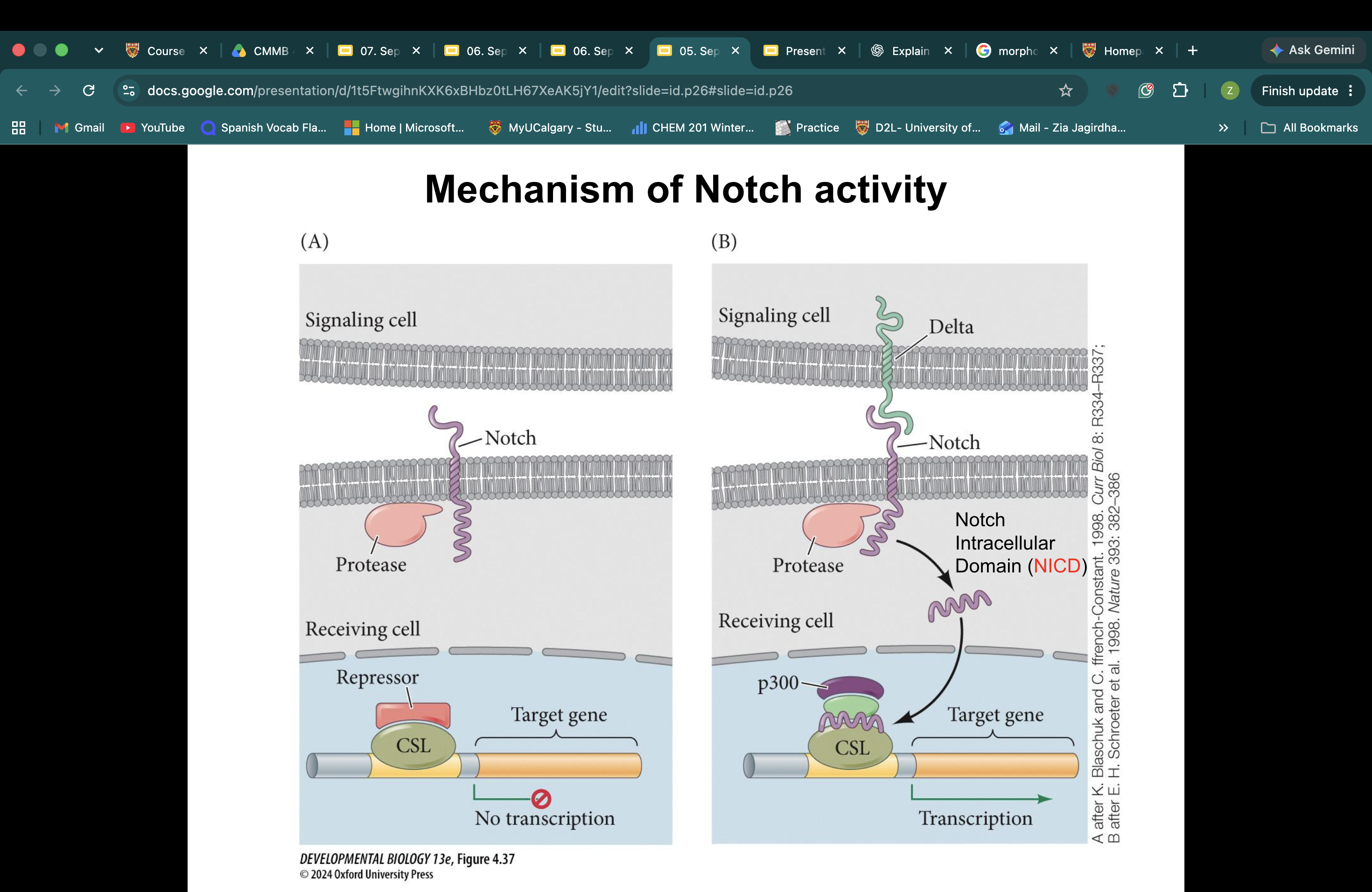
what is a stem cell?
The stem cell concept is based on asymmetric cell divisions. The fundamental notion of a stem cell is that it can make more stem cells while also producing cells committed to undergoing differentiation. This process is called asymmetric cell division. A population of stem cells can also be maintained through population asymmetry. Here a stem cell is shown to have the ability to divide symmetrically to produce either two stem cells (thus increasing the stem cell pool by one) or two committed cells (thus decreasing the pool by one). This is called symmetrical renewing or symmetrical differentiating. (C) In many organs, stem cell lineages pass from a multipotent stem cell (capable of forming numerous types of cells) to a committed stem cell that makes one or very few types of cells to a progenitor cell (also known as a transit-amplifying cell) that can proliferate for multiple rounds of divisions but is transient in its life and is committed to becoming a particular type of differentiated cell.
TOTIPOTENT STEM CELLS
can generate ALL the cell types of the embryo and extra-embryonic tissues (part of placenta, or chorion, the amnion and the yolk sac)
PLURIPOTENT STEM CELLS
can generate all the cell types of the embryo, but NOT the extra-embryonic tissues.
MULTIPOTENT or UNIPOTENT STEM CELLS
can generate many cell types or a single cell type, respectively, in a tissue-specific manner.
How does cell potency change during early human embryonic development, and what is the difference between totipotent blastomeres and the pluripotent inner cell mass (ICM)?
Human blastomeres upon 4-cell stage have been shown to maintain totipotency. During early human embryonic development, blastomeres at approximately the 4- to 8-cell stage can maintain totipotent potential, meaning they can give rise to both embryonic and extra-embryonic tissues. Around 5–6 days after fertilization, the embryo develops into a blastocyst containing approximately 200–300 cells. Within the blastocyst, the inner cell mass (ICM) consists of approximately 30–50 pluripotent cells that can give rise to all embryonic cell types but not the extra-embryonic tissues. The surrounding trophectoderm contributes to extra-embryonic structures, including the placenta.

What are hematopoietic stem cells (HSCs), how do they produce different blood cell types, and what regulates their differentiation?
Hematopoietic stem cells (HSCs) are multipotent stem cells found in the bone marrow that continuously produce blood cells throughout an individual's life. They can self-renew to produce more HSCs or differentiate into two major lineages: lymphoid progenitor cells, which produce cells of the adaptive immune system, and myeloid progenitor cells, which produce red blood cells, platelets, and various cells of the innate immune system. The differentiation pathway an HSC follows is regulated by its surrounding microenvironment, or stem-cell niche, which provides signals that influence cell fate.