Common Equine Neuro Diseases
 Call Kai
 Learn
 Practice Test
 Spaced Repetition
 Match
 Flashcards
 Knowt Play
1/63

There's no tags or description
Looks like no tags are added yet.
64 Terms
What are the infectious neuro diseases to be aware of?
Viral: EHV1; rabies; Borna disease; Eastern,
Western and Venezuelan encephalitis; West Nile Virus
Bacterial: meningitis, encephalitis
Protozoal: EPM
Parasitic: aberrant migration
Clostridial toxins: tetanus, botulism
What non infectious neuro diseases are there to be aware of?
Neurodegenerative disorders: EDM; EMND; EGS; cerebellar abiotrophy
Metabolic: HE; intestinal hyperammoniaemia, HYPP, Electrolyte imbalances (Ca, MG, Na…)
CVSM
Trauma
Peripheral nerve pathology: facial paralysis, radial nerve paralysis, sweeney, polyneuritis equi;
Neurotoxins: Avermectins; Lead, Amitraz, Bracken fern; Leukoencephalomalacia (mouldy corn); raygrass staggers
Vascular: Thromboembolic meningoencephalitis
Neoplasia
What syndromes can we split neuro disease in horses into?
Forebrain disease: including seizures and alterations in consciousness
Brainstem abnormalities: Cranial nerve abnormalities (facial, vestibular)
Horner’s syndrome
Urinary incontinence and Cauda Equina Syndrome
Spinal cord disease
Neuromuscular disease
What are the divisions of neuromuscular disease?
Abnormal gait: shivering, stringhalt
Localised weakness: peripheral nerve injuries
Diffuse weakness: EMND, botulism, EGS, HYPP
Excessive activity: Tetatus
How does forebrain disease present?
Disorders of behaviour and personality: aggression, compulsive walking, loss of learnt behaviour, yawning…
Seizures
Blindness
Altered states of consciousness
Head posture (head and neck turn, head pressing)
What are the causes of forebrain disease in horses?
Trauma: ↑↑
Metabolic: HE, intestinal hyperammoniaemia,
Intracarotid injection
Cholesterol granuloma
Infectious meningoencephalomyelitis
Neoplasia
How is forebrain disease due to trauma treated?
Oxygen supplementation
Fluid therapy
NSAIDS
Elevate head
What are some examples of sleep disorders?
True narcolepsia= rare
Sleep deprivation
Due to inability to lie down, MSK pain/ fear
What are the features of seizures in horses?
Rare in adults
High seizure threshold
Tx underlying cause or idiopathic epilepsy PTS due to drug cost and safety
Neonatal seizures and epilepsy
Low seizure threshold
PAS, sepsis trauma
Idiopathic arab epilepsy syndrome
What are the signs of brainstem and cranial nerve disease?
Vestibular disease: peripheral or central
Facial nerve paralysis (VII and VIII frequently affected together)
Pharyngeal/laryngeal deficits: dysphagia
Other cranial nerve deficits
Severe brainstem lesions: gait abnormalities, coma (RAS)
What are the signs of vestibular disease?
Head tilt
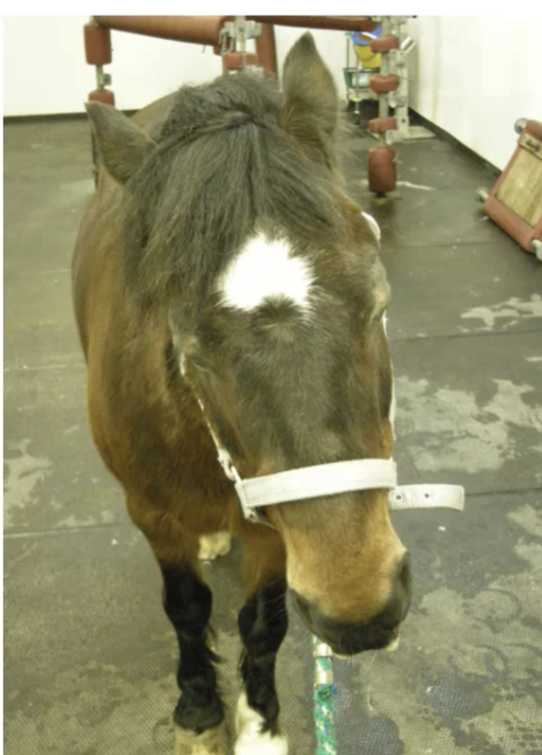
Nystagmus
Ventral strabismus (asymmetry in how much the eye drops)
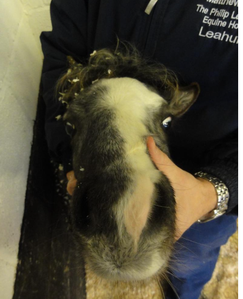
Ataxia
What should you do to ensure that the horse is not compensating for vestibular disease?
Blindfold
Signs may return as eyes are not there to compensate anymore
What can cause vestibular disease?
Trauma: central or peripheral (petrous portion of temporal bone)
Idiopathic
Otitis media/interna (temporohyoid osteoarthropathy)
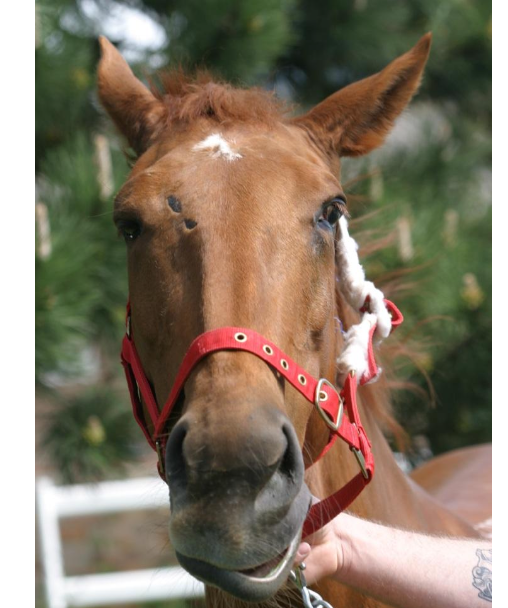
What is being shown here?
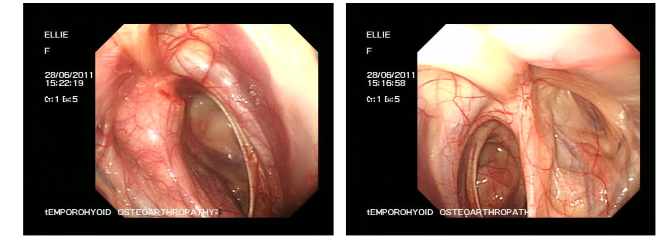
THO
Abnormal left
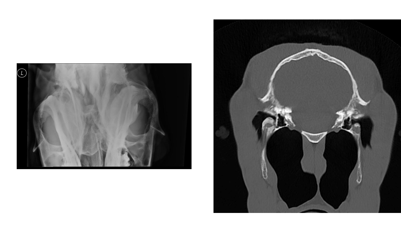
What causes facial nerve injury?
Iatrogenic due to halters left on during field anaesthesia
Trauma (+/- vestibular)
THO (+/- vestibular)
What are the signs of facial nerve injury?
Dropped ear
Ptosis (droopy eye lid)
Nostril deviation (to the other side)
What are the prolonged/ permanent deficits that facial nerve injury can cause?
Exposure keratitis
Dysphagia: feed pouching
Poor performance: nostril collapse
What causes Horner's syndrome?
Interruption of the sympathetic innervation to the head (and neck)
Most common cause iatrogenic due to extravascular injection of irritant substance (PBZ, Buscopan)
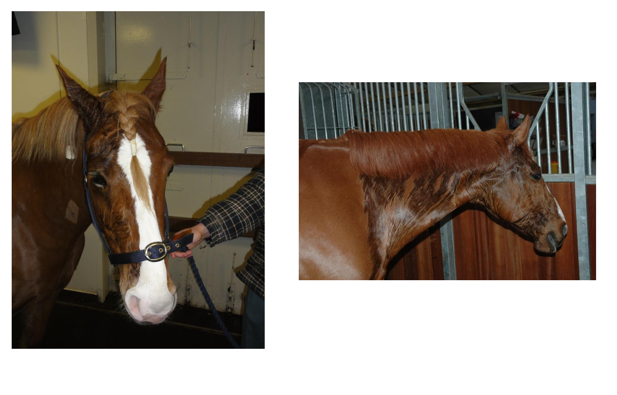
What are the signs of Horner's syndrome?
Miosis, enophthalmos, ptosis, protruding nictitating membrane
Hyperaemic membranes, sweating
MM- half mouth red, half mouth pink
What are the signs cauda equine syndrome?
Degrees of hypotonia, hyporeflexia ad hypalgesia of the tail, anus and perineal region, urinary bladder paralysis, rectal dilation, penile prolapse
May also see LMN weakness and paresis of pelvic limbs
Can be difficult to distinguish UMN disease with urinary retention and 2ary contusion of tail and anus from recumbency
What are some of the causes of cauda equina?
Trauma: sacrococcygeal fracture and luxation, avulsion of the cauda equina
Infectious, inflammatory, immune:
Inflammatory: polyneuritis equi (± cranial nerve signs)
Viral / immune: EHV-1 (± ataxia, ± cranial nerve signs)
Verminous: EPM
How does EHV-1 myeloencephalopathy present?
Uncommon manifestation
May occur sporadically or as outbreak
Recent history of resp disease or abortion
Sudden onset and early stabilisation of signs including ataxia, paresis, urinary incontinence
What causes the CS in EHV-1 myeloencephalopathy?
Result of vasculitis and thrombosis of arterioles in spinal cord (and brain)
Viral endotheliotrophism
How is EHV-1 myeloencephalopathy diagnosed?
Lesion location
Rule out other causes
History of respiratory disease and/or abortion
CSF sample: xanthochromic ± Abs to EHV-1
NP swab and PCR (or buffy coat)
High antibody titres
How is EHV-1 myeloencephalopathy managed?
Isolate
Reasonable prognosis if not recumbent
Give anti inflammatories, steroids, aspirin
Antivirals: acyclovir, valacyclovir
What are the different causes of spinal cord disease?
Spinal cord Trauma
Cervical vertebral malformation (CVM) / Cervical vertebral stenotic myelopathy (CVSM) / Wobbler syndrome
How does spinal cord trauma present?
Sudden onset ataxia or recumbency
No progression, freq improve or may later progress due to callus formation
Ataxia and paresis to recumbency
How is spinal cord trauma managed?
Early anti inflammatory therapy
Surgical therapy? Brace?
Box rest if standing
Nursing care if recumbent
Time
What is the most common cause of ataxia in horses in the UK?
Cervical vertebral malformation
What causes cervical verterbral malformation?
Narrowing of the cervical vertebral canal causing compression of the spinal cord, often in combination with malalignment and malformation of the cervical vertebrae
What are the two broad categories of cervical vertebral malformation?
Type I
Young horses
Compression as result of developmental abnormalities
Dynamic stenosis
Any site
Type II
Older horses
OA of articular processes
Static stenosis
C5-C6 and C6-C7 most common
How is CVM diagnosed?
Lesion location: ataxia all 4 limbs, hindlimbs more severely affected than forelimbs (by a maximum of 2 degres of ataxia)
Neck pain rarely seen
Radiography
What can be seen on a radiograph for CVM?
Stenosis of vertebral canal
Abnormal articular processes
Subluxation of vertebrae on flexion or extension of neck
Enlarged vertebral physeal growth regions
Overriding of the vertebral arch and next caudal vertebral body causing dynamic stenosis during flexion or extension
Proliferation of articular or periarticular soft tissues
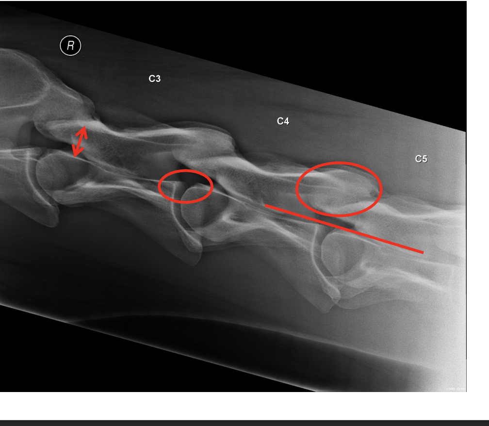
NB- not uncommon to find no Rx abnoramlities, does not rule out CVM
Can do myelography but risky, CT myelogram>
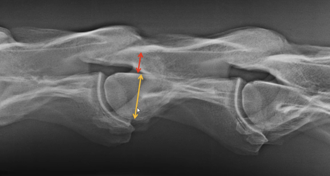
What measurement can be done to diagnose cervical vertebral malformation?
Intravertebral sagittal diameter
Narrowest part of spinal canal: Widest part of spinal body
Intervertebral sagittal diameter
Narrowest part between two vertebrae/ widest part of vertebrae
50% ratio (red line 50% or more of yellow- do on every vertebrae)
How is CVM managed?
Surgery possible but costly and not much improvement
Don't ride ataxic horse
Type I- early detection and dietary restriction can result in resolution
Type II- intraarticular corticosteroids (US guided)
What are some other spinal cord diseases to be aware of?
Occipitoatlantoaxial malformation
Neoplasia
Vertebral body osteomyelitis
What diseases can cause abnormal gait?
Shivering
Stringhalt
What are shivers?
Reflex hypertonia of flexor muscles of pelvic limbs
How does shivers present?
Horse snatches up HL when picked
Accentuated when turhing or backing horse and if excited
HL flexed and held in spastic space for some time
Draft breeds
Always progressive
NB- shivering not that common
What is stringhalt?
Sudden, involuntary, exaggerated flexion of one or both hindlimbs during attempted movement
What are the two types of stringhalt?
Australian or plant associated; dandelion/ flat weed
Remove from pasture
Sporadic stringhalt: aetiology not understood
Tx= myotenectomy of lateral digital extensor
What can cause localised weakness in horses?
Peripheral nerve injuries
How does radial nerve injury present? (most common)
Most common from external blows or following lateral recumbency in anaesthesia
Inability to flex shoulder, extend limb and fix elbow
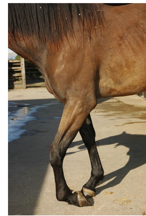
How does suprascapular nerve injury present?
Sweeny (shoulder slip)
Most common from ext blows or poorly fitted collars in draft horses
Atrophy of supra and infraspinatus, abduction of limb and inability to advance shoulder
How are peripheral nerve injuries treated?
Antiinflammatories
Physio
Return of function may take days to months (or never return)
What can cause diffuse weakness in horses?
Equine motor neurone disease
Botulism
Grass sickness
Hyperkalemic Periodic Paralysis
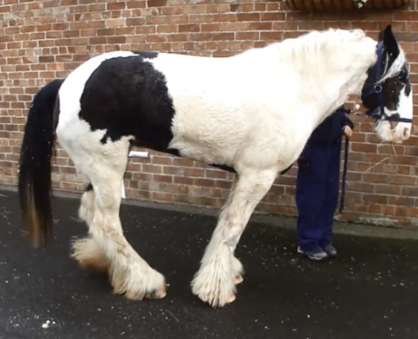
What causes equine MND and what are the signs?
Degen of motor neurons in spinal cord due to low vit E conc
Normally history of stabled with no access to pasture for long time
Signs: weight loss, muscle fasciculations, prolonged recumbency, tail elevation, legs close together when stood
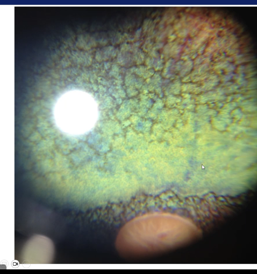
Ocular signs 30-40% of cases: brown pigment accumulation in retina
How is equine MND diagnosed and treated?
Vit E/ Sel levels
Muscle biopsy (tail head)
Tx= vit E supplementation
What causes botulism?
Clostridium botulinum
Ingested toxin from source
Contaminated feed, water, poultry litter and carcasses
Toxico-infectious route
Type B grows in ingesta of suckling foals, infected wound (rare)
Describe the pathogenesis of botulinism
Blocks Ach release at presynpatic membrane of n-m junction
Failure of contraction= weak
Abrupt progressive onset flaccid paralysis of skeletal muscle
What are the signs of botulism?
Symmetrical weakness (muscle tremors)
Stumble, ataxia
Tongue weakness, dysphagia
Hang head, nose on ground
Eventually recumbent
Dyspnoea: intercostal and diaphragm paralysis
GI stasis, urine dribbling
Shaker foal syndrome: 1-3 months, initial episodes of trembling
Die in 10 days (resp muscle paralysis)
How is botulism diagnosed?
ID toxin/ spores
Stomach content (+/- faeces or feed)
A lot of the time diagnosis of exclusion
How is botulism treated?
Antitoxin (USA, expensive)- expensive and not that helpful
Penicillin (not procaine!)
Supportive, fluids
What is the prognosis and prevention of botulism?
Poor prognosis esp if rapid onset tetraplegia
Vaccine -US
What are the features of clostridium tetani? What are the different toxins it produces?
From soil, GI flora
Gram +, anaerobic: sporulates with right conditions (wounds!) and releases toxins:
Tetanospasmin: neurotoxin
Migrates retrograde along the motor neurone to the spinal cord and brainstem
Tetanolysin: increases tissue necrosis at site
How does tetanspasmin affect the horse?
Inhibits release of glycine
Inhibits presynaptic inhibitory neurons in spinal cord
Muscles continuously contract
Tetany elicited by sound, light, touch
Horses very sensitive
What are the signs of tetanus?
Elevated tail head
Stiff gait
Anxious: ears back, eyelids wide open, nostrils flared, head extended
Lock jaw: drool, can’t swallow
Prolapse 3rd eyelid
Recumbency, rigidity
Secondary ulcers, trauma
How is tetanus treated?
Support- quiet, ,dark room, hydration
Eliminate source- flush wound, treat with metronidazole
Antitoxin - if unvaccinated
If horse was vaccinated in past but not up to date use toxoid (separate site from antitoxin)
Muscle relaxation- ACP, diazepam
How is tetanus prevented?
Vaccination
2,3,5 months
Yearly booster
What is headshaking?
Spontaneous occurring disorder consisting of violent head flicks of the head
In severe cases horses may sustain significant self-inflicted trauma and compromise welfare
Likely represents trigeminal neuropathic pain
Shaking head ≠ Headshaking
What branch of the trigeminal nerve is most commonly affected
Maxillary (opthlamic possible)
What are the signs of headshaking?
Violent, usually vertical, shakes or flicks of the head
Snorting
Nostril clamping
Anxious expression
Muzzle rubbing / licking
Striking at nose with thoracic limbs
Self-trauma
Intermittent / continuous
Rest / exercise
Stable / field
How is headshaking diagnosed?
Diagnosis of exclusion:
Ophthalmic exam
Neurological exam
Dental exam
Upper airway endoscopy
Head CT (Radiography?)
How is headshaking treated?
Most neither specific or curative
Environmental management
Avoid known triggers
Can be seasonal, response to weather, sound induced, exercise
Nose nets
Magnesium
PENS- percutaneous electric nerve stimulation