Vascular Exam 3 - Physiologic Testing (Notes)
 Call Kai
 Learn
 Practice Test
 Spaced Repetition
 Match
 Flashcards
 Knowt Play
1/89

There's no tags or description
Looks like no tags are added yet.
90 Terms
Introduction to Physiologic Testing
Physiologic testing is the _____ assessment of _____
Used to detect hemodynamically significant arterial _____ disease
Assessing the degree of _____, not the degree of _____
indirect, arteries, occlusion, disease, stenosis
Goals of Physiologic Testing:
Determine if there is _____ (factual) evidence of _____ disease
Determine if _____ disease is causing the patient’s _____
Assess increasing or decreasing _____ _____ during serial follow-up exams
objective, arterial, arterial, symptoms, limb perfusion
Methods of Physiologic testing:
_____ _____ _____ (PVR)
_____ _____ _____ analysis or _____ analysis
_____ pressures (calculate _____)
_____ stress testing
Pulse volume recording, Doppler analog waveforms, spectral, Segmental, ABI, Exercise
Physiologic Testing Baseline Exam:
Wrap blood pressure cuffs on the upper _____ and _____, obtain _____ _____ pressures __laterally
Obtain _____ _____ _____ from the same sites OR acquire _____ doppler waveform tracings from the axillary, brachial, radial, and ulnar _____
Obtain _____ (PPG) waveforms from both _____ fingers
Interpret _____ and _____ obtained
arms, forearms, systolic segmental, bi, pulse volume recordings, CW, arteries, photoplethysmography, index, pressures, waveforms
Compression of the subclavian artery and vein and the nerves of the brachial plexus. However, the majority of cases are neurogenic in origin.
Thoracic Outlet Syndrome (TOS)
Thoracic Outlet Syndrome (TOS): compression of the _____ artery and vein and the nerves of the _____ _____. However, the majority of TOS cases are _____ in origin.
subclavian, brachial plexus, neurogenic
Thoracic Outlet Testing:
After performing the _____ exam, obtain waveforms with one of the following methods to serve as a baseline reference for the positional study
_____
_____
_____ _____ tracings
Obtain _____ waveforms __laterally
Obtain tracings from the _____ arm using the following maneuvers:
__ducted _____ degrees
_____ _____ – abduct the arm 30 degrees at the shoulder while the arm is manually extended, then have the patient extend their neck and turn their head towards the affected arm
_____-_____ _____ – place arms in “surrender” position, shoulders abducted 90 degrees with elbows flexed at 90 degrees; patient slowly opens and closes their hands
_____ _____ – have patient move their arm to whatever position elicits (provokes) the symptoms most
baseline, PPG, PVR, CW Doppler, resting, bi, symptomatic, Ab, 90, Adson’s maneuver, Stick-up position, Symptomatic position

_____ maneuver – __duct the arm _____ degrees at the _____ while the arm is manually _____, then have the patient extend their _____ and turn their _____ towards the _____ arm
Adson’s, ab, 30, shoulder, extended, neck, head, affected


_____-_____ position – shoulders __ducted _____ degrees with elbows _____ at _____ degrees; patient slowly opens and closes their _____
Stick-up, ab, 90, flexed, 90, hands
The stick-up position for thoracic outlet testing is AKA:
Costoclavicular
Indirect or non-imaging assessment of blood vessels, typically arteries
Physiologic testing
Physiologic Testing:
_____ or non-_____ assessment of blood vessels, typically _____
Indirect, imaging, arteries
What vessels are we assessing when performing physiologic testing?
The _____ can technically also be assessed with physiologic testing (but we don’t focus on this)
Arteries, veins
Physiologic Testing:
Provides information about _____ _____, which _____ alters – we call it “_____” information
Only detects _____ disease
Tells us:
Presence of _____ disease
Relative _____ of disease
_____ impairment
Reliably tells us if symptoms are due to vascular _____
Useful for _____ patients
_____ (toe, finger) measurements
_____ potential
blood flow, disease, functional, significant, significant, severity, Functional, insufficiency, diabetic, Digital, Healing
What are the four methods of physiologic testing?
Doppler waveform analysis, segmental pressure assessment, exercise testing, plethysmography
True of false: Typically you won’t see just one method of physiologic testing done but rather a combination of more than one method
True
_____ level evaluation
Generally the ankle
ABI
Quick method to determine presence and relative severity of disease
Single
_____ evaluation
Two or more levels
Segmental pressures or PVR
Determines the presence and relative severity as well as the region (location) of disease
Multilevel
Single level evaluation
Generally the _____
_____
Quick method to determine _____ and relative _____ of disease
ankle, ABI, presence, severity
Multilevel evaluation
_____ or more levels
_____ pressures or _____
Determines the _____ and relative _____ as well as the _____ (location) of disease
Two, Segmental, PVR, presence, severity, region
Indications for Physiologic Testing
***Basically the same indications as arterial _____ imaging
_____ (always)
_____ _____ pain
Arterial _____
_____ _____ _____ (PAD)
_____-procedure assessment – angioplasty, stent, bypass graft
Follow-up _____ procedure – angioplasty, stent
Prognosis for _____ – ulcerations, amputations
duplex, Claudication, Ischemic rest, ulceration, Peripheral arterial disease, Pre, endovascular, healing
Limitations of Physiologic Testing
Detects only hemodynamically _____ disease
So if someone has _____ disease, physiologic testing will likely show _____ results
Usually cannot distinguish between a _____ or _____
If the exam is abnormal, typically the patient will get an actual _____ exam anyways
Provides the region, but not the precise _____ of disease
Duplex scanning provides _____ AND _____ information
significant, mild, normal, stenosis, occlusion, duplex, location, anatomic, physiologic
Doppler Waveform Analysis
_____ wave Doppler probe
Two _____ crystals – one that constantly _____, one that constantly _____
Two types:
_____ – waveforms on a graph-like presentation; averages the frequencies obtained into a single line tracing
_____ – this is your fast fourier transform (FFT), what you’re used to seeing as a spectral Doppler waveform; displays ALL the frequencies moving through a sample area
Continuous, piezoelectric, transmits, receives, Analog, Digital

Type of Doppler waveform analysis
Waveforms on a graph-like presentation; averages the frequencies obtained into a single line tracing
Analog

Type of Doppler waveform analysis
This is your fast fourier transform (FFT), what you’re used to seeing as a spectral Doppler waveform; displays ALL the frequencies moving through a sample area
Digital
Limitations of Doppler Waveform Analysis:
Cannot differentiate _____ from _____ disease or _____ from _____
Patient must be able to cooperate/tolerate or remain _____ through the exam
Potential for _____ quality signals
Takes a lot of _____/skill to perform correctly
normal, mild, stenosis, occlusion, still, poor, practice
Think of this exam as duplex imaging…without the imaging. We are obtaining waveforms ONLY!
Doppler Waveform Analysis
How a Doppler Waveform Analysis is performed:
_____ wave Doppler (_____) probe, patient is positioned _____
Use _____ for appropriate contact
Doppler _____ still applies!
Tilt your transducer on the patient’s skin to create a Doppler _____ between __-__ degrees
Obtain signals (_____ and _____) from:
_____ extremity – CFA, SFA, Pop A, PTA, ATA, Pero A
_____ extremity – Subclavian A, Axillary A, Brachial A, Radial A, Ulnar A
Continuous, pencil, supine, gel, angle, angle, 40, 60, audible, visible, Lower, Upper

What vessel is being interrogated here?
PTA
True or False: Interpretation of a Doppler Waveform Analysis is quantitative
False (QUALITATIVE)
Doppler Waveform Analysis Interpretation: QUALITATIVE! (How does it look?)
Compare signals from one location to another – was there _____?
When saying _____ in this case it means _____ up the leg or up the arm
Ex: The signal in the PTA compared to the signal in the Pop A compared to the signal in the SFA
Look at the overall appearance of a waveform; indicates _____ vs. _____ and relative _____
A vessel that is well collateralized even with a significant stenosis or occlusion may appear _____ – this is another limitation
Original descriptors: triphasic, biphasic, monophasic…
HOWEVER…there are discrepancies specifically with the _____ waveform
Biphasic waveforms can be _____
New terminology: multiphasic or monophasic
Triphasic and biphasic are considered _____ → _____
HOWEVER, biphasic waveforms can also be _____
Monophasic is still monophasic → _____
deterioration, location, location, normal, abnormal, severity, NORMAL, biphasic, NORMAL, multiphasic, NORMAL, ABNORMAL, ABNORMAL
What type of waveform is being described?
Rapid systolic upstroke
Sharp peak
Rapid mid systolic downstroke
Late systolic flow reversal
Projection below baseline
Forward flow in diastole
Triphasic
What type of waveform is being described?
Rapid upstroke
Sharp peak
Slightly slowed mid systolic downstroke
Late systolic flow reversal
No final forward diastolic flow component
Biphasic
What type of waveform is being described?
Delayed systolic upstroke
Delayed acceleration time
Rounded peak
Slowed mid systolic downstroke
Absence of systolic flow reversal and forward diastolic flow component
Monophasic
True or False: A biphasic waveform is not considered multiphasic
False (is IS considered multiphasic)
True or False: A biphasic waveform can be normal or abnormal
True

Normal – Multiphasic (Triphasic)
Rapid _____ upstroke
_____ peak
Rapid mid systolic _____
Late systolic flow _____
Projection _____ baseline
Forward flow in _____
systolic, Sharp, downstroke, reversal, below, diastole

Normal/Abnormal – Multiphasic (Biphasic)
Rapid _____
_____ peak
Slightly _____ mid systolic downstroke
Late systolic flow _____
_____ baseline
No final forward _____ flow component
upstroke, Sharp, slowed, reversal, below, diastolic

Abnormal – Monophasic (Monophasic)
Delayed systolic _____
Delayed _____ time
_____ peak
Slowed mid systolic _____
Absence of systolic flow _____ and _____ diastolic flow component
upstroke, acceleration, Rounded, downstroke, reversal, forward

Peak to peak frequency difference divided by the average frequency
(P1-P2)/average frequency
Typically calculated by the machine
Normal = >4.0
Abnormal = <4.0
Pulsatility Index
What is the range for a normal pulsatility index?
> 4.0
What is the range for an abnormal pulsatility index?
< 4.0
Time from onset of systole to peak systole – how long it takes to get to the max velocity
Normal = <133 milliseconds
Abnormal = >133 milliseconds
Acceleration Time
What is the normal range for acceleration time?
< 133 milliseconds

What is the abnormal range for acceleration time?
> 133 milliseconds
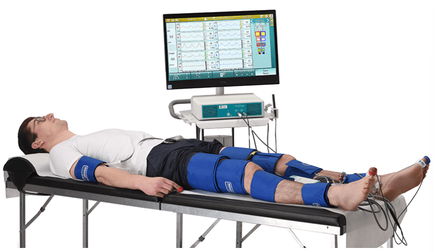
Segmental Pressure Evaluation
Blood pressure cuffs are used to obtain _____ pressures throughout the limbs
Cuff is inflated to _____ flow, then slowly deflated…the _____ Doppler signal heard is recorded as the _____ pressure at that location
Flow is detected using a _____ wave probe
Pressure drops or significant differences between cuffs (segments) indicate _____ _____ _____
systolic, cease, first, systolic, continuous, flow limiting stenosis
Segmental Pressure Evaluation Limitations:
Only detects _____ disease
Patient limiting factors – _____, _____ areas of the limb
_____
Cold → vaso_____
Hot → vaso_____
_____ _____
Very common in diabetic patients
The vessel walls are calcified to a point where they are non-compressible
Stiff to the point where we cannot completely close them off to briefly cease blood flow
_____ _____ _____ (DVT), fistula or grafts, _____ (swelling due to lymphatic system damage), post _____ _____ (BPG) procedure
If ordered at the same time, a DVT study should be done _____ a physiologic testing study.
significant, tolerance, inaccessible, Temperature, constriction, dilation, Medial calcinosis, Deep vein thrombosis, lymphedema, bypass graft, before
Medial calcinosis
Very common in _____ patients
The vessel walls are _____ to a point where they are non-_____
Stiff to the point where we cannot completely close them off to briefly _____ blood flow
diabetic, calcified, compressible, cease
A serious medical condition where a blood clot (thrombus) forms in a deep vein, most commonly in the lower leg, thigh, or pelvis, though it can also occur in the arms.
Deep vein thrombosis (DVT)
What position should patients be lying in for all physiologic testing exams?
Supine
NOTE – _____ position for all physiologic testing exams…WHY?
To eliminate _____ _____ caused by _____
Lying flat removes this _____ variable, allowing baseline pressures and blood flow to be measured accurately and consistently
supine, hydrostatic pressure, gravity, gravitational
What are the two methods of segmental pressure evaluation?
4 cuff and 3 cuff
***4 and 3 refer to the number of blood pressure cuffs that are on the lower extremity (excludes the brachial cuffs)
Segmental Pressure Evaluation:
4 Cuff
__lateral brachial cuffs
Two _____ cuffs – high _____ (1) & low _____/above knee (2)
_____ cuff (3)
_____ cuff (4)
3 Cuff
__lateral brachial cuffs
One (larger) _____ cuff (1)
_____ cuff (2)
_____ cuff (3)
Bi, thigh, thigh, thigh, Calf, Ankle, Bi, thigh, Calf, Ankle
The width of the cuff should be 20% greater than the diameter of the limb (or 40% of the limb circumference)
20% Rule
20% Rule – the width of the cuff should be __% greater than the _____ of the limb (or __% of the limb _____)
20, diameter, 40, circumference
Using too small of a cuff in a segmental pressure evaluation results in artifactually _____ limb pressure
high
4 Cuff Method:
Two thigh cuffs provide _____ and _____ thigh measurements
Can differentiate between _____-_____ (inflow) disease and _____ _____ _____ (outflow) disease
Two thigh cuffs violate the _____ _____; results in artifactually _____ thigh pressures, approximately _____ mmHg higher
3 Cuff Method:
Provides _____ thigh pressure measurements
If abnormal, cannot differentiate _____-_____ (inflow) disease from _____ _____ _____ (outflow) disease
proximal, distal, aorto-iliac, superficial femoral artery, 20% rule, elevated, 30, accurate, aorto-iliac, superficial femoral artery
Aorto-iliac disease is what type of disease?
Inflow
Superficial femoral artery disease is what type of disease?
Outflow
Obtain __lateral _____ pressures – do NOT take an _____ pressure if the patient has a shunt or a dialysis graft!
Obtain leg pressures starting at the _____ and work your way _____ the leg
_____ and _____ pressures first
Calculate _____ – use highest _____ pressure (Lt or Rt) and highest _____ pressure (DPA/PTA)
_____ divided by _____
Example: If you are calculating the right sided ABI you would use – HIGHEST _____ REGARDLESS OF _____ (Lt or Rt), and highest _____ pressure between the right _____ and _____
Obtain _____ pressures
Obtain _____ pressures
Notes Box:
Multiple inflations of a cuff beyond _____ pressure may result in transient ischemic-induced _____ affecting the pressure – this is why it is recommended to start at the _____ and work your way _____ the leg. HOWEVER, I will be honest…I have not seen a clinical site perform it this way.
bi, brachial, arm, ankle, up, DPA, PTA, ABI, brachial, ankle, ANKLE, ARM, ARM, LATERALITY, ankle, DPA, PTA, calf, thigh, systolic, vasodilation, ankle, up
Important to keep in mind: Segmental Pressures
Complete _____ of blood flow is required – inflate the cuff __ to __ mmHg beyond the _____ audible signal
Record the _____ from the _____ audible signal upon deflating the cuff
If you need to repeat a measurement, fully _____ the cuff, wait a minute, then try again. Give the body time to _____ before trying again.
_____ _____ can result in “non-compressible” vessels
The pressure being measured represents the pressure directly _____ the cuff
If you inflate the thigh cuff but use an ankle vessel to record your signal, your pressure represents the _____
cessation, 20, 30, last, pressure, first, deflate, autoregulate, Medial calcinosis, UNDER, thigh
How do you solve for an Ankle Brachial Index (ABI)?
Highest ankle pressure / highest arm (brachial) pressure
***Longer word on top!

Solve for the right and left ABIs in this example:
Right Brachial Pressure = 147 mmHg, Left Brachial Pressure = 140 mmHg
Right DPA = 142, Right PTA = 148
Left DPA = 126, Left PTA = 130
Right ABI: 148/147 = 1.01
Left ABI: 130/147 = 0.88
What is the normal ABI range?
0.9-1.35
***Greater than 1.35 is abnormal
List the ABI values that correspond to the following interpretations:
Normal…usually
Normal, consider exercising the patient
Probable claudication, single level disease
Multi-level disease
Ischemic rest pain
>1.0, >0.9 to <1.0, <0.8, <0.5, <0.3
An ABI value >_____ is USUALLY normal because someone with _____ _____ can have a really high pressure and a high ABI of for example 1.8, which is pretty high and abnormal.
1.0, medial calcinosis
ABI Values:
< 0.8 = _____ disease, < 0.5 = _____ disease, < 0.3 = _____ disease (gangrene, ulcerations, etc.)
mild, moderate, severe
Interpretation: Segmental Pressures
4 Cuff
NORMAL
High _____ pressure should be at least _____ mmHg _____ than the _____ pressure
***With the 4 cuff method, the _____ pressures are going to be artificially _____ because we are not obeying the _____ _____. So that is why the _____ pressure should be at least _____ mmHg greater than the _____ pressure.
ABNORMAL
Thigh pressure _____ than the brachial pressure
_____ mmHg or greater drop in pressure between segments
thigh, 20, greater, brachial, thigh, elevated, 20% rule, thigh, 20, brachial, less, 30
Interpretation: Segmental Pressures
3 Cuff
NORMAL
All segmental pressures should be _____ to or slightly _____ than the _____ pressure
ABNORMAL
_____ mmHg or greater drop in pressure between segments
equal, greater, brachial, 30
Interpretation: Segmental Pressures
*Arm pressures – gradient of 15-20 mmHg (typically 20) between brachial pressures indicates _____ _____ or _____/_____
Horizontal pressures – _____ mmHg pressure drop from one side to the other in the legs is _____ (left vs. right side)
subclavian steel, stenosis, occlusion, 30, abnormal
Interpretation Tips for Segmental Pressures
Step #1: Look at the _____ used – _____ _____ vs. _____ _____, what type of _____
Step #2: Look at/compare the _____
Step #3: Look at the _____, start by comparing the (high) thigh to the arm (brachial) pressures, then go down the leg. Determine where (if at all) things become abnormal (location). → Abnormal if there is more than a _____ mmHg difference from one segment to the next
Step #4: Look at the _____. Determine where (if at all) things become abnormal.
Step #5: Make a conclusion on disease _____ and _____
method, 3 cuff, 4 cuff, waveforms, ABIs, pressures, 30, waveforms, location, severity

When interpreting segmental pressures you compare _____ to _____ and _____ and _____
side, side, up, down

Segmental Pressure Example:
Step #1: What method was used? What type of waveforms?
Step #2: Look at/compare the ABIs
Step #3: Look at/compare the pressures
Step #4: Look at/compare the waveforms
Step #5: Make a conclusion on disease location and severity
4 cuff, no waveforms
Both ABIs are normal
Normal high thigh and brachial pressure differences; normal segmental pressure differences except from 153 to 119 on the left leg
Remember that in the 4 cuff method, high thigh pressures are going to be at least 20 greater than the brachial pressures
From 153 to 119, the pressure difference is 34 but since the ABI is still normal, this could be due to artifact or incorrect recording. We are still going to consider it NORMAL.
No waveforms
Normal, no disease

Segmental Pressure Example:
Step #1: What method was used? What type of waveforms?
Step #2: Look at/compare the ABIs
Step #3: Look at/compare the pressures
Step #4: Look at/compare the waveforms
Step #5: Make a conclusion on disease location and severity
3 cuff, no waveforms
Right ABI is abnormal (0.74), left ABI is normal (1.06)
With the ABI being 0.74, this is likely a mild disease (<0.8).
Abnormal right thigh (89) and brachial (126) pressure difference; all other pressure differences are normal
Remember that in the 3 cuff method, the thigh pressure should be equal to, or potentially slightly greater than the brachial pressure
No waveforms
Right-sided proximal disease
Remember that with the 3 cuff method, because we are only using one thigh cuff, we cannot differentiate between aorto-iliac (inflow) disease and SFA (outflow) disease.
Exercise Testing Evaluation
Metabolic demand _____ during exercise, requiring _____ blood flow to supply oxygen (vaso_____)
And what do we know is the most common symptom of PAD???
_____ → pain with _____
Patients with mild to moderate disease may have normal blood flow at _____
This is why in the ABI chart > _____ to < _____, the interpretation was “normal but consider _____ the patient.”
The patient may have a normal ABI value while at _____ and in the _____ position but if they exercise, an _____ ABI can be visualized!
Exercise testing allows for the differentiation of vasculogenic claudication from _____-_____
_____-_____ is going to be either _____ or _____ in nature
increases, increased, dilation, Claudication, exertion, rest, 0.9, 1.0, exercising, rest, supine, abnormal, pseudo-claudication, Pseudo-claudication, neurogenic, orthopedic
Limitations of Exercise Testing:
_____ _____ (COPD, congestive heart failure), _____ (HTN), _____ _____ _____ (SOB) → patients that physically cannot exercise long enough for the exam
ABI < _____ – patients with evident disease
Resting _____ (ABI < 0.3)
Poor _____ status → patients who cannot walk well
Cardiac status, hypertension, shortness of breath, 0.5, ischemia, ambulatory
True or False: There is little to no benefit of exercise testing for patients with severe PAD, resting ischemia, ulceration, and who cannot walk well enough to elicit (provoke) claudication
True
***We already know that disease is present anyways
How To: Exercise Testing
Complete resting _____
Patient will be _____
Obtain brachial, PTA, and DPA _____
There are several ways to exercise your patient (calf raises, walking, treadmill…)
Treadmill – easily _____
Patient walks on treadmill at < _____% grade (incline)
Speed _____ mph for max of _____ minutes or until the patient complains of symptoms
Document the duration and speed that the patient walked
Immediately obtain _____ pressures (PTA, DPA); don’t have to do the brachial pressures again
Normal = ankle pressures are the _____ or slightly _____
Abnormal = pressure _____
< _____% drop = _____ level disease
> _____% drop = _____ level disease
ABI, supine, pressures, reproducible, 12, 1.5, 5, ankle, same, higher, drop, 50, single, 50, multi
Arterial Plethysmography
Volume/Air (PVR)
*PVR operates under the physical principle that blood flowing through any given limb segment results in changes in the limb’s _____ → How does the limb get bigger or smaller based on the blood flowing underneath that cuff
_____ cuffs are placed around the limb at specific levels (segments)
Cuffs are inflated with a measured amount of _____ (_____ to _____ mmHg) → We are _____ cutting off circulation like we did in a _____ pressure
As arterial flow moves through the vessel, the limb gets slightly _____
The increase in limb volume displaces the _____ in the cuff
Pressure transducer converts this change into an _____ waveform
volume, Pneumatic, air, 40, 65, NOT, segmental, bigger, air, analog
Arterial Plethysmography
Photo (PPG)
Detects _____ blood flow
LED transmits light into the tissue and the _____ light is measured by the adjacent photo-sensor
Blood in the _____ tissue _____ the amount of light reflected back to the sensor → _____ is the gradual loss of amplitude and intensity as a wave propagates through a medium
Increased blood content in the tissue results in increased _____ (more scattering, less being sent back to the photosensor (decreased reflected signal)) and displays as a _____ upstroke _____ waveform
cutaneous, reflected, subcutaneous, attenuates, Attenuation, attenuation, positive, analog
Both PVRs and PPGs display _____ waveforms
analog
PPG =
Photoplethysmography
PVR =
Pulse Volume Recording (Volume Plethysmography)
True or False: The interpretation of Pulse Volume Recording (Volume Plethysmography) is QUALITATIVE
True

Pulsatile Flow Waveform:
Early _____ (A) – _____-velocity ejection of blood; aortic valve _____
Late _____/Early _____ (B)
Rapid deceleration and aortic valve _____ (_____ _____)
Results in temporary flow _____
Late _____ (C)
Flow depends upon vascular _____
Systole, High, opens, Systole, Diastole, closes, dicrotic notch, reversal, Diastole, resistance
PVR Waveform Interpretation: QUALITATIVE!
An abnormal waveform reflects hemodynamically significant disease _____ to the level of the waveform
If you are getting an abnormal waveform at the ankle, the disease is _____ to the ankle (_____ in the leg)
Severity is often _____estimated
Someone might have moderate disease but we’re getting a _____ abnormal waveform
Reduced _____ with no changes in the _____ (outline) of the waveforms MAY represent insignificant disease unless there is a notable difference between levels (up and down) or sides (laterality)
This is saying that a waveform may have a normal _____ but a reduced _____ and therefore look normal-ish (insignificant disease) but if the levels or sides have notable differences, then you know that there is disease
PVR should be done with _____ measurements to get accurate diagnostic information
Reflected wave = _____ _____
proximal, proximal, higher, under, mildly, amplitude, contour, shape, amplitude, pressure, dicrotic notch

PVR Interpretation:
Normal: Rapid systolic _____ with reflected wave (_____ _____)
Mildly abnormal: _____ peak, absent _____ wave, downslope “_____” away from the baseline
Moderately abnormal: _____ systolic peak, upslope and downslope are _____, _____ _____ absent
Severely abnormal: low _____ throughout
acceleration, dicrotic notch, Sharp, reflected, bowed, Flattened, delayed, reflected wave, amplitude
The _____ _____ (_____ _____) is the single most important feature of a normal PVR waveform.
reflected wave, dicrotic notch
Photoplethysmography (PPG)
Limitations:
_____ skin for good contact of sensor
Patient’s resting _____ state
Cuff or sensor _____
_____
Patient Position:
_____ position
Place sensors on _____ and _____
_____ room
Intact, basal, placement, Tremors, Supine, fingers, toes, Warm

Summary