sensory system ( afferent )
 Call Kai
 Learn
Learn Practice Test
 Spaced Repetition
 Match
Match Flashcards
 Knowt Play
1/171

There's no tags or description
Looks like no tags are added yet.
Name | Mastery | Learn | Test | Matching | Spaced | Call with Kai | Chat |
|---|
No analytics yet
Send a link to your students to track their progress
172 Terms
sensory system
this includes sensory receptors, neural pathway from receptors to CNS & portion of the brain that processes sensory information
afferent pathways carry sensory information from the …
PNS to the CNS
sensory receptors
Peripheral ends of afferent neurons that generate graded potentials ( graded potentials are at the receptor, aka receptor potentials)
visceral afferent pathways
carry internal information we are not aware of
Visceral receptors detect stimuli from the internal organs and structures (chemoreceptors and mechanoreceptors)
adequate stimulus
The type of energy to which a certain type of receptor responds best; however, the receptor may also respond to other types of energy (e.g photoreceptors respond best to light but also respond to pressure. If you get hit in the eye, you will see a flash of light)
sensation
conscious awareness of a stimulus
being aware or becoming aware
we are limited to what we are aware off bc of our receptors ( e.g., animals & insects can see more colors than us)
perception
conscious interpretation of sensation
Understanding that the pain is throbbing due to a cut
Perception of stimuli can vary among individuals based on their past experiences
mechanoreceptors
a type of receptor that respond to pressure or stretch
thermoreceptors
respond to diff temp
photoreceptors ( cones)
responds to light of different wavelengths
chemoreceptors
specific chemicals
nociceptors
receptors that respond to pain ( and detect it)
sensory transduction
Sensory receptors convert a physical or chemical stimulus from the environment into an electrical signal
receptor potential
the change we see in a membrane potential in a sensory receptor, resulting in an action potential
The stronger the stimulus, the more action potentials a neuron is going to generate
Weaker stimulus, the fewer action potentials, or less likely to make an action potential

Explain the specialized afferent ending of sensor neurons
Specialized dendrites, or a cell that acts like a receptor and responds to stimuli
1) A stimulus is applied to the specialized ending of an afferent neuron
2) causing a cation channel to open, which depolarizes the receptor
When a sensory neuron acts like the receptor, they open Na channel, causing a change in the MP ( aka RP bc it technically occurs at the receptor now )

Explain when a cell acts like a receptor
1) the cell responds to the stimulus
2) causing the cation to open, changing the MP in a receptor cell ( aka RP)
3) The receptor cell depolarizes to all Ca2+ channels to open
Signals vesicles within the neuron to release NT
4) NT binds to the afferent neuron, causing an ion channel to open and convey a signal ( either inhibitory or excitatory )
5) The stronger the stimulus → NT release by receptor cell is more frequent, and action potentials are generated by the afferent neuron

for both cells that have a receptor and specialized afferent neuron endings, a stimulus is causing a change in the permeability of the receptor ions → causing a generation of an AP

Explain this photo
The greater the stimulus → more APs
stimulus must be transduced into an electrical signal that the neuron can understand & carry into the CNS
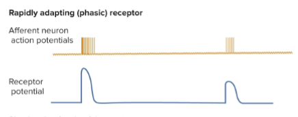
receptor adaptation
a decrease in the strength of the RP over time in the presence of a continuous stimulus
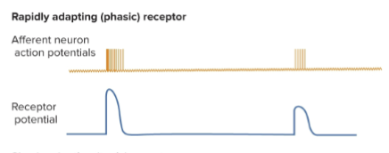
Phasic receptors
a stimulus occurs on an afferent neuron, causing an action potential. It adapts quickly, meaning that even though the stimulus remains, the neuron stops firing APs.
What happens?
It sends one big AP to signal the start of a stimulus and one small AP to mark the end of a stimulus (when you feel the weight of the ring after you take it off)

tonic receptor
1) a stimulus begins flipping the on switch, causing a rise in the receptor potential, which eventually causes an AP to fire
2) Compared to phasic receptors, this one doesn’t adapt. it has a starting stimulus and keeps a high RP for AP
e.g., photoreceptors; they don’t stop sending signals even if you have been looking at something for a long time
proprioceptors: they must always be aware of where your body is in the environment

the brain is often bombarded with irrelevant and continuous info and responds by ignoring that info bc it needs to focus on new info
neuron habituation
neurons reduce their response by depressing activity at the synapse; basically, they close some Ca channels, causing there to be less NT to be released
Change at the axon terminal, affecting voltage-gated Ca channels
neuron sensitivity
( aka sensitization ) A neuron increases its responsiveness to a strong stimulus, the opposite of neuronal adaptation; the neuron sends more AP to bc sensitive to an out-of-place stimulus
e.g in a loud area, you will still be able to hear a loud bang bc the neurons will be sensitive to out-of-place sounds ( construction site, hypersensitive to danger)
sensory coding
how the brain codes a stimulus to understand it, the brain codes it based on location, type, and strength
peripheral ending of sensory neurons can have many branches acting as receptors innervating the same region of the skin
sensory unit
a afferent neuron and all of its receptors

motor unit
a motor neuron and all the muscle fibers it innervates

receptive field
a region of our skin that is activated by a sensory unit and produces a response
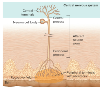
dense receptive fields =
more receptive units, which means high acuity ( e.g finger tips)
sensory unit with smaller receptive fields have more acuity compared to sensory unit with larger fields bc
small fields are easier to “search” compared to larger ones
all the afferent neurons in a receptive field can resolve when a stimulus is strong enough
cerebral cortex
the outer 4 mm of the brain
All sensory receptors have a path to the cerebral cortex
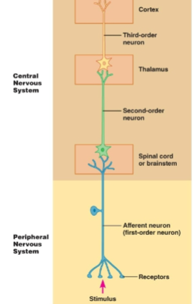
explain the sensory receptor pathway to the cortex
1st order neuron: the afferent neuron that first detects a stimulus and sends it to the spinal cord
2nd order neuron: an interneuron that sends info from the spinal cord → thalamus
3rd order neuron: synapses with the second, where it will take the signal to the right region of the cortex to determine: type, strength, and location

all sensory info goes through the thalamus, then the cortex, except olfaction (smell), which goes directly to the temporal lobe, where the olfactory cortex is located
vestibular cortex
balance
gustatory cortex is in the
temporal lobe
when sensory info reaches the cortex, that is when you become aware of it
stimulus modalities
The ability of the brain to distinguish the modalities it receives, e.g heat, cold, pressure, light
Sensory units will only respond to their specific modality
How does the brain ID stimulus modality
the receptor it activates
the pathway it takes to the brain ( there are two )

dorsal column lemniscal pathway
involves mostly mechanoreceptors, proprioceptors, touch, vibration, two-point discrimination
1st-order neuron: enters the spinal cord in the dorsal horn on the same side of the stimulus and synapses with the 2nd-order neuron
2nd-order neuron: crosses over to the other side ( decussation) to the thalamus to synapse with the 3rd-order neuron
3rd-order neuron: goes to the specific region of the brain where that stimulus belongs to

decussation
the crossing over of signal in the brain stem
spinothalamic pathway ( aka anterolateral pathway)
carry info: nociceptors & thermoreceptors ( pain and temp )
1st order: pain or temp receptors carry info to the spinal cord and synapse with the 2nd neuron, right as it enters the spinal cord, bc of the reflex involved ( pulling your hand off the stove )
2nd order: descends, then goes all the way up to the thalamus, then the 3rd order neuron
3rd order neuron: takes it into the part of the cortex that associates with that part of the body
Pain also activates the limbic system, which deals with emotion, meaning pain will not just cause a physical response but an emotional response ( making you angry or cry )
e.g chronic pain can cause depression

the brain codes the strength of a stimulus based on
how many sensory units are activated
How many APs are sent

stimulus strength is also coded by the recruitment of more sensory neurons or more afferent neurons
more neurons are needed to send one signal
more neurons = more AP
population coding
the recruitment of more receptors

acuity
How precisely a stimulus is perceived
depends :
size of receptor field
how many of the receptor units (density) → two-point discrimination

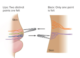
the lips have more smaller sensory units, making you more likely to hit the two different fields compared to the back, where you have a larger field, so you are more likely to hit one field with two points
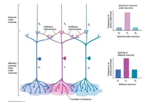
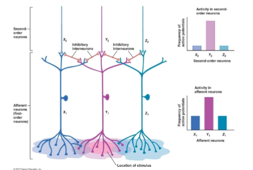
lateral inhibition
occurs in the field that overlaps with each other, increasing the density and acuity of receptive fields
increasing sensory discrimination & loc
works by exciting one receptive field and inhibiting others at the 2nd-order neuron, causing them to send fewer APs ( eliminating the excess APs to be more precise)


explain the process of lateral inhibition
1) a group of sensory units activates
2) Out of that group, the sensory unit that receives the strongest stimulus uses its lateral inhibitory neuron to weaken the AP of the other units lateral to it
3) They make the lateral receptive fields send weak enough APs to the brain that it doesn’t notice, making the strong receptive field stand out, without changing the intensity or AP of the original neuron
somatic sesation
sensation associated with the skin
mechanoreceptors
… response to pressure touch, sound ( vibration), any mechanical distortion
thermoreceptors
temp receptors specifically on the skin
nocireceptors
pain through chemical detection
proprioceptors
a type of mechanoreceptor found in muscles, tendons, & ligament tell the brain where your body is in space and how stretched your muscles & tendons are, and what angle your limbs are in
photoreceptors
respond to diffent wavelengths of light
chemoreceptors
detects specific chemical associated with taste and smell

meissner’s corpuscle
tonic mechanoreceptors detect distortion in the skin, when someone move the skin these structures do too and send AP to the brain about the sensation

merkel’s corpuscle
like meissner corpuscle but slow reacting, detect distortion in the skin, when someone move the skin these structures do too and send AP to the brain about the sensation

free nerve endings
basically Merkel’s corpuscle but included nociceptors, itch receptors, thermoreceptors, and mechanoreceptors

pacinian corpuscles
loc in the dermis ( deeper than the Meissner’s), not covered in connective tissue, but will rapidly adapt. Important for vibration & deep pressure

Ruffini corpuscles
slow adapting mechanoreceptors, that detect stretch
thermoreceptors & nocireceptor are all free nerve endings
warm receptors
adapt rapidly ( phasic receptors) send frequent AP to let you know about the extreme temp (response to temps between 30 - 45 °C)
cold receptors
adapt rapidly ( phasic receptors ) send frequent APs to let you know about the extreme temp ( responds to temp around 35 - 20o C)
nociceptor
respond to chemicals released by damaged tissue, they don’t adapt, respond to noxious stimuli ( dangerous stimuli), any chemical that is related to tissue damage

Paradoxical hot /cold
when the temp receptors of one temp stop firing, and nociceptors and the opp start firing due to recruitment bc there is so much stimulation

capsaicin
a chemical found in hot peppers that warm receptors response to
methanol
bind to receptors and give the sensation of cold
NT released by damaged cells
prostaglandin, cytokines, bradykinins, histamine, potassium
pain perception
Helps to prevent further damage; makes us aware that something is wrong
Causes sweating, an increase in blood pressure, and emotional responses of anxiety or even fear
two types: fast and slow pain
fast pain
Carried by A-delta fibers, transmitted very quickly because these fibers are myelinated
Initial pain, aka acute pain, is localized (sharp pain)
slow pain
the pain that comes after fast pain is going to last longer because it is carried on C-fibers, which are unmyelinated, making the propagation of pain slower
Slow pain is fast in transmission but slow compared to fast pain
Localized and poorly defined region
guarantees you are aware that your body needs to heal ( throbbing)
3 types of nociceptors bc there is more than one way to get hurt
mechanical nociceptors
travel on A-delta fibers, making them fast-responding
Detects crushing, pinching, cutting, and tearing; any intense mechanical stimulation
thermal nociceptors
travel on A -delta fibers, making them fast, responding to extreme temperatures
polymodal nociceptors
travels on c fibers, making them slow reacting, they respond to all types of damage stimuli
endogenous analgesic system
the brains pain blocking system
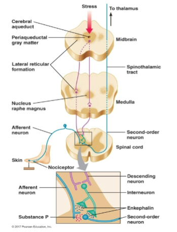
how does the endogenous analgesic system work?
1) 1st order neurons: nociceptors will receive the pain signal and start releasing substance P, which will bind to the 2nd order neuron
2) Afferent neurons (substance P) from the brainstem are going to synapse with inhibitory neurons (interneurons) to release endogenous opioids, which are NTs (enkephalin & endorphins, substances similar to morphine; they are powerful analgesics )
3) Instead of substance P binding to the 2nd-order neuron to create a damping effect by preventing the second-order neuron from sending an action potential
4) Analgesics are painkillers, including ibuprofen, aspirin, narcotics, and NSAIDs


referred pain
visceral pain in the organ you can’t actually feel it is instead felt on the surface of the body
This happens bc the nociceptors on the skin & nociceptors on the organ both synapse on the same second-order neuron in the spinal cord; they also share the same neural pathway
Someone experiencing a heart attack will feel pain in their chest but also in their shoulder and arm


reflection
when light waves bounce off an object and enter our eyes and become the color we see
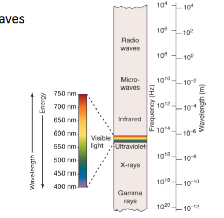

fibrous tunic ( outermost layer)
sclera + cornea; extraocular muscles insert into the sclera, making the eyeball
has collagen fibers
cornea is the clear part of the eye that allows light into the eye


vascular tunic ( middle layer)
includes the choroid, ciliary body, iris, and contains a lot of blood vessels
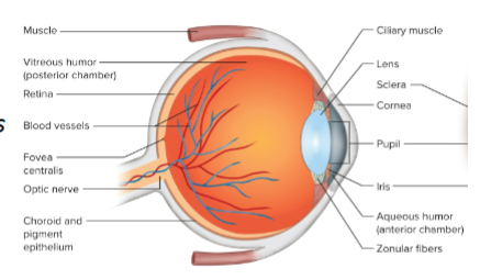

neural tunic ( inner tunic)
a layer that includes the retina
Contains the blind spot, where there are no photoreceptors


iris
colored part of the eye
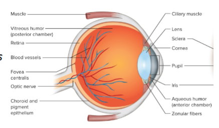

anterior chamber
the part of the eye that is filled with a “ fluidity” humor
loc in front of the lens → to the cornea
aqueous humor is nourishing the lens


posterior chamber
Everything posterior to the lens is much larger and filled with a gel-like humor called the vitreous humor
Vitreous humor functions to keep the retina in place and support the eye as well


how light enter the eye
1) goes through the cornea, then the pupil, which is loc in the center of the iris
2) hits the lens, then passes through the vitreous humor to eventually hit the retina, where the photoreceptors are

the functions of tears
to cleanse the eye, also contains a lysosome to prevent bacteria from growing over the eye
The iris is the part of the eye that contains color and is unique to every person, more unique than fingerprints
Has 2 sets of smooth muscles: circular and radial muscles

circular muscles
muscles that surround the pupil of the eye
When these muscles contract, they cause the pupil to constrict
The parasympathetic NS innervates here; this is why doctors shine a light in your eye ( parascope)


radial muscles
the outermost ring of muscles around the pupil; when they contract , they cause the iris to dilate
The sympathetic NS innervates here; it dilates if there is not enough light
I am sympathetic when people fall in love


refraction
the bending of light
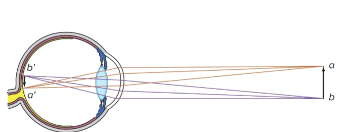

when light enters the eye …
It hits curved surfaces: the cornea & the lens, changing the light waves to refract
The lens can change shape, changing the refractive power of the light waves. Light hits your retina upside down, but when it hits your brain, it’s right side up again ( the visual cortex reverses it back to the original image)


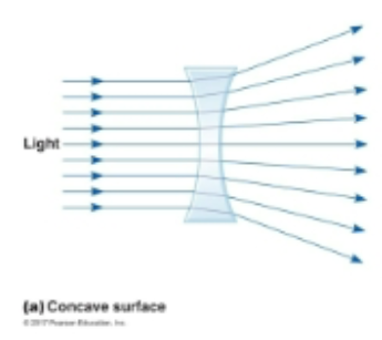
concave surfaces
surface curves in ( caves in ); light spreads out
When light passes through this surface, the edges of the refracted light move away from each other ( aka divergence of light )
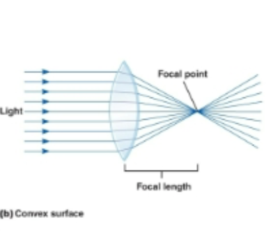
convex surfaces
surface curves out; light spreads in
The light bends inward toward a single point ( focal point) that must be projected onto the retina to see the image clearly


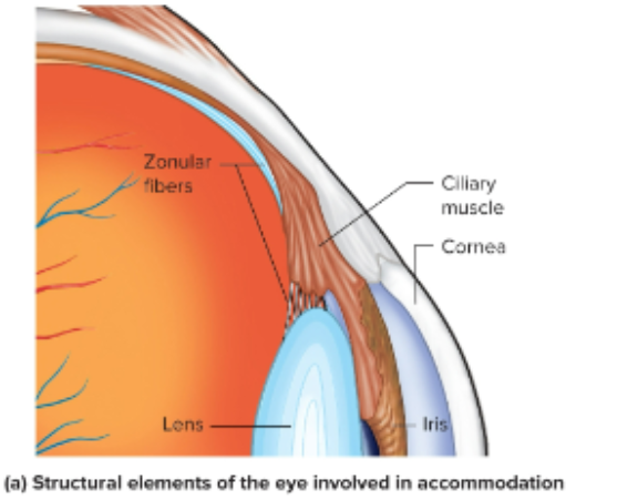
zonular fibers ( aka suspensory fibers)
attach and circle the lens. The contraction of the ciliary muscle will either tighten or loosen the suspensory fibers, causing the thickness of the lens to change

looking at distant objects
1) light rays come into the eye at most parallel, close to each other
2) When they hit the lens, they refract

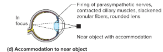
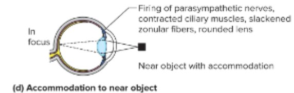
looking at object close up
1) light will come in at a wider angle
2) Light will hit the lens and refract into a focal point behind the retina
3) the lens will accommodate and move the focal point onto the retina