Molecular Bio Exam 1 Review
 Call Kai
 Learn
Learn Practice Test
 Spaced Repetition
 Match
Match Flashcards
 Knowt Play
1/29

There's no tags or description
Looks like no tags are added yet.
Name | Mastery | Learn | Test | Matching | Spaced | Call with Kai | Chat |
|---|
No analytics yet
Send a link to your students to track their progress
30 Terms
4 main macromolecules and their monomers
1) Nucleic acids - nucleotides
2) Proteins - amino acids
3) Carbohydrates - monosaccharides
4) Lipids - fatty acids
Elements that make up most of living matter
C(Carbon), H(Hydrogen), N(Nitrogen), O(Oxygen), P(Phosphorous), S(Sulfur)
What levels of protein structure involve the side chains(R) of an amino acid?
Tertiary and quaternary
What structure is mostly stabilized by INTRAchain hydrogen bonds?
Alpha helices
Protein denaturation
Loss of secondary, tertiary, and or quaternary structure WITHOUT breaking peptide bonds(reverts back to primary structure)
Reasons to isolate a protein
1) You want to study the function of a known protein
2) You want to recapitulate a cellular process in a test tube and need to assemble the protein components
3) You want to isolate a protein in pure form for structural biology(X-ray crystallography)
4) You are trying to identify a novel protein responsible for cell function
Differential centrifugation
Sequential/stepwise centrifugation at increasing speeds to fractionate the extract into constituent cellular organelles(large sediments=lower speed; small organelles=higher speed)
Stationary phase
Resin/matrix packed in a column
Mobile phase
Buffer containing the protein mixture moves through the matrix
Equilibration
Preparing the column with an appropriate buffer before loading the sample
Flow through
Material that passes through the column WITHOUT binding
Washing
Step used to remove unbound/weakly bound material by using different pH/ionic strengths
Elution
Releasing the protein that is bound to the column
Fraction
Small volume collected as material exits the column
Eluate
A collected fraction containing the target protein released from the column
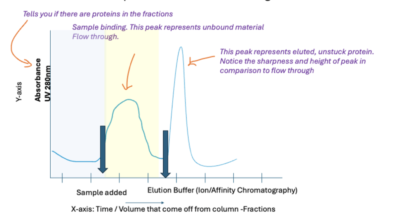
Sharpness of peak = purity of protein
Height of peak = how much protein is in the sample
In size exclusion chromatography, why do large proteins elute first?
They cannot enter the pores and go between the porous beads so they take a shorter path through the column to elute first
Smaller proteins fit in these porous beads and therefore take a longer path through the column to elute later
Unit of molecular weight
1 kDa = 1000 Da
Average weight of an amino acid
110 Da(amino acids x 110 Da = average size of the protein)
How does ion exchange chromatography work?
The + or - charged proteins will stick to the opposite charged groups on the beads while proteins that are neutral/same charge as groups on beads will pass through column quickly
What can you do to elute the stuck proteins?
Alter the affinity of the protein for the column by…
1) Increasing the salt concentration in the buffer which weaken ionic interactions as salt ions compete with the proteins for binding sites
2) Changing the pH of the buffer which alters the proteins net charge and possibly reducing its affinity for the resin in the column
You have two purified samples of protein Y: the wild-type (nonmutated) protein and a mutant version with a single amino acid substitution. When washed through the same gel-filtration column, mutant protein Y runs through the column more slowly than the normal protein. Which of the following change in the mutant protein is most likely to explain this result?
The single amino acid substitution results in the loss of a binding site on the mutant-protein surface through which protein Y normally forms dimers
Gel filtration separates proteins by size
The single amino acid substitution diminished the interaction between the 2 molecules of the Protein Y DImer
The resulting mutant protein is now a MONOmer
Monomer=Smaller=Fits in porous beads and take longer path=Slower
What pH would you use if Protein X has a pI of 7.5 and is using Cation Exchange Chromatography?
CATION exchange chromatography means protein=+ and resin=-
To have Protein X elute from this, you want it to be - so it doesn't stick to the resin
In order to have a net - charge, you need the pI<pH
pH=8.5 works best
Immunoprecipitation
Specialized form of affinity purification that uses an ANTIBODY to isolate a SPECIFIC protein from a complex mixture
What are the limitations of assays?
They establish if a specific protein is present within a sample but can not tell you if there are other contaminants
What is SDS-PAGE used for and what are the steps?
Used to separate/visualize ALL the proteins within a sample by molecular weight
1) Prepare samples by adding SDS and heating to fully denature proteins
2) Load the gel, apply electrical current where - molecules move to + electrode, and add molecular weight-marker to separate wells
3) Run electrophoresis where proteins migrate through gel(smaller=further/faster; larger=slower/smaller distance)
4) Visualize the proteins by staining the gel to reveal the separated protein bands
What does adding SDS do in SDS-PAGE?
Proteins naturally differ in shape and charge so proteins are first treated with SDS to make migration depend primarily on molecular weight
What is a limitation to the SDS-PAGE method?
Proteins that have multiple similarly sized subunits(in their quaternary structure) cannot be identified; they will blend together in one protein band
Therefore it cannot PROVE the identity of a protein
Protein molecular weight marker(AKA Ladder)
Proteins of KNOWN sizes used to estimate the molecular weights of unknown proteins by comparing migration distances
What is western blotting used for and what are the steps?
Uses antibodies to detect a SPECIFIC protein
1) Electrophoretic transfer of proteins from an SDS-PAGE gel to a paper-like membrane
2) Blocking of nonspecific protein binding sites on transfer membranes
3) Wash then incubation of the membrane with a primary antibody specific for the epitope of interest
4) Wash then incubation with a secondary antibody that recognizes primary antibodies
5) Wash then visualization of bound antibodies