BGSU BIOL 3310 Lecture Exam 2- Muscles
 Call Kai
 Learn
 Practice Test
 Spaced Repetition
 Match
 Flashcards
 Knowt Play
1/173

There's no tags or description
Looks like no tags are added yet.
174 Terms
skeletal, cardiac, smooth
three types of muscular tissue
skeletal muscle
long, cylindrical, multinucleate (located peripherally), striated
voluntary
most commonly attaches tendons to bones
cardiac muscle
branched cylindrical fiber with one centrally located nucleus; intercalated discs joing neighboring fibers, striated
smooth muscle
fiber is thickest in the middle, tapered at each end, has one centrally positioned nucleus; not striated
-excitability
-contractility
-extensibility
-elasticity
Properties that enable muscle to functionand contribute to homeostasis
excitability
ability to respond to stimuli
contractility
ability to contract forcibly when stimulated
extensibility
ability to stretch without being damaged
elasticity
ability to return to an original length
cells, connective tissue, blood, nerves
skeletal muscle components
anchor and structure
role of connective tissue in skeletal muscle
signals and nutrients
why do muscles need extensive nerve and blood supply
epimysium
Outermost layer
Surrounds numerous bundles of fascicles
fibrous sheath surrounding the entire muscle
outer surface grades into the fascia
inner surface sends projections between fascicles to form perimysium
Perimysium
Separates 10-100 muscle fibers into bundles called fascicles
slightly thicker layer of connective tissue
fascicles - bundles of muscle fibers wrapped in perimysium
carry larger nerves and blood vessels, and stretch receptors
endomysium, perimysium, epimysium, fascia
connective tissues of a muscle
Endomysium
thin sleeve of loose connective tissue surrounding each muscle fiber
allows room for capillaries and nerve fibers to reach each muscle fiber
fascia
sheet of connective tissue that separates neighboring muscles or muscle groups from each other and the subcutaneous tissue
lines body wall, limbs
occupies space between muscles
rich in blood and nerves
myoblasts
skeletal muscle cells arise from
hypertrophy
increase in cell size
same
_________ number of muscle cells at birth as the rest of your life time (usually)
wraps around all muscles and body parts
difference between fascia and other connective tissue
ligaments
bone to bone
does not
heavily damaged muscle _______ repair
tendon
Connects muscle to bone
fascicle
bundle of muscle fibers
muscle fiber
a single muscle cell
satellite cell
can fuse to make new fibers (but heavily damaged muscle does not repair)
aponeurosis
A broad, flat tendon
somatic motor neurons
Neurons that stimulate skeletal muscle to contract are _________________
fusiform
spindle-like shape that is wide in the middle and tapers toward both ends

biceps brachii
example of fusiform muscle
parallel
muscle fascicles (bundles of fibers) that run in the same direction as the long axis of the muscle
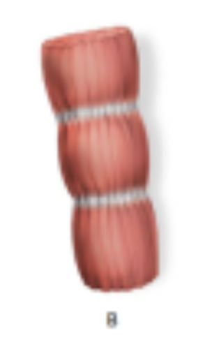
rectus abdominis
ex of parallel muscle
triangular
muscle fibers that spread widely across a broad origin (base) and pull together toward a single, narrow insertion point (apex)
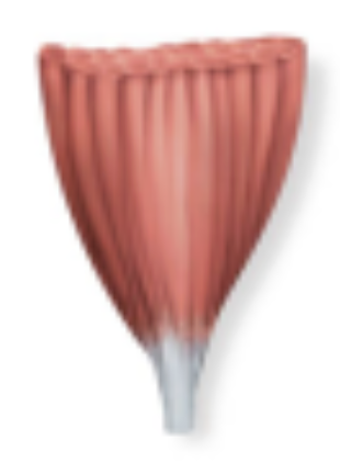
pectoralis major
ex of triangular muscle
Unipennate
the muscle fascicles are positioned on only one side of a central tendon

palmar interosseous
example of unipennate muscle
bipennate
muscle fibers (fascicles) are arranged on both sides of a central tendon, resembling the structure of a regular feather

rectus femoris
example of bipennate muscle
multipennate
features muscle fibers that approach a central tendon from multiple angles, branching out around or within the tendon to resemble a complex feather or multi-headed structure

deltoid
ex of multipennate
circular
muscle fibers arranged in concentric rings that act as sphincters to control body openings

orbicularis oris
example of circular muscle
orientation of its facicles
strength of a muscle and the direction of its pull are determined partly by the __________________________________________
origin
bony attachment at stationary end of muscle
belly
thicker, middle region of muscle between origin and insertion
insertion
bony attachment to mobile end of muscle
indirect attachment to muscle
tendons bridge the gap between muscle ends and bony attachment
direct attachment to bone
little separation between muscle and bone muscle seems to emerge directly from bone
prime mover
muscle that produces most of force during a joint action
ex. brachialis
synergist
Muscle that assists a prime mover
stabilizes the nearby joint
modifies the direction of movement
ex. biceps brachii
antagonist
opposes the prime mover
relaxes to give prime mover control over an action
preventing excessive movement and injury
antagonistic pairs - muscles that act on opposite sides of a joint
ex. triceps brachii
fixator
muscle that prevents movement of bone
ex. rhomboids
intrinsic muscles
contained within a region, such as the hand
both origin and insertion in the same region
extrinsic muscles
acton a designated region, but has its origin elsewhere
fingers - in the forearm
myofibrils
Muscle fibers are full of _____________, but also contain a number of notable chemicals and structures
sarcolemma
plasma membrane of muscle fiber
transverse tubules
tunnel-like indentions of the sacrolemma
sarcoplasm
cytoplasm of a muscle fiber
myoglobin
a cytoplasmic protein that binds oxygen in a muscle fiber
glycogen
Storage form of glucose
sarcoplasmic reticulum
network of membranous sacs around the myofibrils.
calcium
the sarcoplasmic reticulum stores ___________ ions
myofibril
contractile organelle
contraction
in the SR, release of Ca++ triggers myofibril ___________
nucleus
1

sarcomere
2

z disc
3

m line
4

h band
5

a band
6

i band
7

contractile proteins, regulatory proteins, structural proteins
the three kinds of proteins that build a myofibril
contractile proteins
generate force during contraction
actin and myosin
the 2 contractile proteins
myosin
thick filaments
actin
thin filaments
regulatory proteins
switch the contraction process on and off
tropomin and tropomyosin
the two types of regulatory proteins
structural proteins
align the thick and thin filaments properly
provide elasticity and extensibility
link the myofibrils to the sarcolemma
myosin, actin
the 2 contractile proteins
myosin
Functions as a motor protein which can achieve motion
Convert ATP to energy of motion
Projections of each myosin molecule protrude outward (myosin head)
actin
______ molecules provide a site where a myosin head can attach
Tropomyosin and troponin are also part of the thin filament
In relaxed muscle
titin, dystrophin
2 structural proteins
titin
Stabilize the position of myosin
accounts for much of the elasticity and extensibility of myofibrils
Extends from Z disc to M line
where does ca++ bind in a muscle contraction
sarcolemma
dystrophin links thin filaments (actin) to this
Tropomyosin
Covers myosin-binding sites, keeps myosin from engaging actin
troponin
Bind to tropomyosin
In the presence of Ca++, causes tropomyosin to uncover myosin binding sites, allowing contraction to begin.
ca++
if there is a muscle contraction what needs to bind to troponin
action potential
what causes ca++ to release in a muscle contraction
transverse tubule
action potential travels along the______
thin filament, myosin
when ca++ binds to troponin on the __________ it exposes the binding sites for ___________
thin filaments, actin
where do myosin heads hook on
troponin
when a muscle wants to contract where does ca++ bind
skeletal muscle
in _______ almost all ca++ is stored in the SR
cardiac muscle
in __________ most ca++ is stored in the SR and some comes from outside the cell
smooth muscle
in ________ almost all ca++ comes from outside the cell
tropomyosin, troponin
2 regulatory proteins
rigor mortis
hardening of muscles and stiffening of the body beginning 3 to 4 hours after death