Musculoskeletal System
 Call Kai
 Learn
Learn Practice Test
 Spaced Repetition
 Match
Match Flashcards
 Knowt Play
1/138

There's no tags or description
Looks like no tags are added yet.
Name | Mastery | Learn | Test | Matching | Spaced | Call with Kai | Chat |
|---|
No analytics yet
Send a link to your students to track their progress
139 Terms
Skeletal muscle
Striated, voluntary muscle; can also be cruise controlled (e.g. breathing, swallowing)
Cardiac muscle
Striated, involuntary muscle; contracts rhythmically, adopt the contraction rate of the most rapid cell
Smooth muscle
Nonstriated, involuntary muscle; forms of this muscle include visceral and multiunit
Visceral smooth muscle
Large sheet organ walls made of cells (wall the stomach, intestine, uterus, bladder, blood vessels, respiration); responds to the stretching of the lumen; reacts in rhythmic waves
Multiunit smooth muscle
Small and delicate movements are required for this muscle; examples include the iris, ciliary body of the eye, small blood vessels and small air passages; require specific impulses from the nervous system to contract (e.g. light changes)
Aponeuroses
Connect muscle to bone or to another muscle
Primary mover (agonist)
Muscle group that directly produces a desired movement
Antagonist
Directly opposes the action of a muscle group
Synergist
Muscles that contracts at the same time as a prime mover and assists in the action
Fixator
Stabilize joints to allow other movements to take place
Extension
Act of not being bent
Flexion
Act of being bent
Flexor surface
Decreases the resting angle of a muscle
Extensor surface
Increases the resting angle of a muscle
Extrinsic muscles
Muscles that attach the limb to the body; originates outside the structure it moves and inserts itself into it
Superficial pectoral
Pectoralis major
Craniolateral
The descending portion of the superficial pectorial that arise from the first two sternebrae and runs in a _____ direction
Transversely
The transverse portion of the superficial pectorial that originates from first three sternebrae and runs…
Sternum (cranial sternum)
Superficial and deep pectoral origin
Humerus (great tubercle)
Superficial and deep pectoral insertion
Adducts thoracic limb
Superficial and deep pectoral action
Deep pectoral
Pectoralis minor
Trapezius
Thin triangular muscle that lies over the dorsal part of the scapula; divided into cervical and thoracic portions
Neck and thoracic vertebrae (dorsal aspect)
Trapezius origin
Scapular spine
Trapezius insertion
Elevates and abducts the forelimb
Trapezius action
Latissimus Dorsi
Large triangular shaped muscle lies caudal to the scapula and covers much of the thoracic wall
Draws thoracic limb caudally and flexes shoulder joint
Latissimus Dorsi action
Cervical vertebrae
Serratus Ventralis (cervical) origin
Ribs
Serratus Ventralis (thoracic) origin
Scapula (serrated face)
Serratus Ventralis insertion
Supports the trunk and depresses the scapula
Serratus Ventralis action
Intrinsic muscle
Muscle that attaches to the bones within the limb; have both their origin and insertion entirely within the specific body region they move
Infraspinatus
Muscle fills the infraspinous fossa caudal to the spine of the scapula
Infraspinous fossa (scapula)
Infraspinatus origin
Humerus (greater tubercle)
Infraspinatus insertion
Flexes the shoulder joint and abducts shoulder limb
Infraspinatus action
Supraspinatus
Fills the supraspinous fossa cranial to the spine of the scapula, wider and longer than its counterpart
Supraspinous fossa (scapula)
Supraspinatus origin
Humerus (greater tubercle)
Supraspinatus insertion
Extends glenohumeral joint
Supraspinatus action
Subscapularis
Muscle occupies the subscapular fossa on the medial aspect of the scapula
Subscapular fossa (scapula)
Subscapularis origin
Humerus (lesser tubercle)
Subscapularis insertion
Adducts shoulder limb and rotates forelimb medially
Subscapularis action
Biceps Brachii
Located on the craniomedial aspect of the humerus; only has one head in domestic animals
Scapula (supraglenoid tubercle)
Biceps Brachii origin
Ulna and radius (proximal)
Biceps Brachii insertion
Flexes the elbow and extends the shoulder
Biceps Brachii action
Triceps brachii
Antagonist of the biceps brachii
Supraspinatus
Synergist of the biceps brachii
Triceps Brachii
Makes up the large muscle mass on the caudal aspect of the humerus; commonly has 3 heads, but carnivores have a fourth head (long, medial, lateral, accessory)
Scapula (caudal border) and humerus (proximal border)
Triceps Brachii origin
Ulna (olecranon)
Triceps Brachii insertion
Extends the elbow and flexes the shoulder
Triceps Brachii action
Extensor reticulum
Thick fascial band, near carpal joint; holds the tendons of these muscles in place

Extensor Carpi Radialis
Largest craniolateral muscle of the antebrachium
Humerus (distal)
Extensor Carpi Radialis origin
Metacarpals (proximal aspect II and III)
Extensor Carpi Radialis insertion
Extends the carpus
Extensor Carpi Radialis action
Common Digital Extensor
Lies caudal to the extensor carpi radialis on the lateral side of the forelimb; tendon of insertion branches into four separate branches and each inserts on the dorsal surface of the proximal phalanges of the major digits; craniolateral muscle of the antebrachium
Humerus (lateral epicondyle)
Common Digital Extensor origin
Digits II-V (dorsal surface)
Common Digital Extensor insertion
Extends the carpus and digits
Common Digital Extensor action
Superficial Digital Flexor (SDF)(thoracic)
Lies caudomedial to the ulna; tendon of insertion divides into four major branches and each branch inserts on one digit
Humerus (medial epicondyle)
Superficial Digital Flexor (thoracic) origin
Digits II-V (middle phalanx)
Superficial Digital Flexor (thoracic) insertion
Flexes the carpal and digital joints
Superficial Digital Flexor (thoracic) action
Deep Digital Flexor (DDF)(thoracic)
Muscle has 3 heads of origin (humeral, ulnar, and radial); tendons of all three heads fuse at the carpus to form a single tendon and then subdivide to attach to the palmar aspect of each digit

Superficial Gluteal
Small muscle that overlies the caudal part of the middle gluteal muscle; fibers extend laterally from its origin to the insertion on the femur
Sacrum and coccygeal vertebrae
Superficial Gluteal origin
Femur (greater trochanter)
Superficial Gluteal insertion
Abducts the pelvic limb and extends the hip
Superficial Gluteal action
Middle Gluteal
Large egg-shaped muscle that lies cranial to the superficial gluteal muscle and caudal to the tensor fascia late; fibers are directed caudoventrally to its insertion on the femur
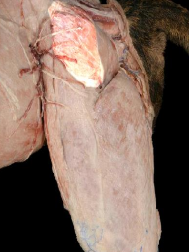
Lateral ilium
Middle Gluteal origin
Femur (greater trochanter)
Middle Gluteal insertion
Abducts the limb and extends the hip
Middle Gluteal action
Biceps Femoris
Longest and widest muscle of the thigh and covers its entire caudolateral aspect; muscle spans over 3 joints and has actions at all 3 (coxofemoral, stifle, tarsus); contributes to common calcanean tendon
Pelvis (ischiatic tuberosity)
Biceps Femoris origin
Patella, proximal tibia, and tuber calcanei
Biceps Femoris insertion
Extends the hip, flexes the stifle, and extends the tarsal joint
Biceps Femoris action
Semitendinosus
Strap-like muscle lies medial to the Biceps femoris and lateral to the semimembranosus; member of hamstring group
Extends the hip, flexes the stifle, and extends the tarsal joint
Semitendinosus action
Semimembranosus
Muscle is not as long semitendinosus, but is slightly thicker; wedged between the biceps femoris (laterally) and gracilis (medially); member of hamstring group
Extends the hip and flexes the stifle
Semimembranosus action
Semitendinosus, semimembranosus, and biceps femoris
The hamstrings consist of…
Pectineus
Small spindle shaped muscle on the medial aspect

Adducts the pelvic limb
Pectineus action
Adductor
Large pyramid shaped muscle in the leg

Adducts the pelvic limb and extends the hip
Adductor action
Sartorius, pectineus, adductor, and body wall
The femoral triangle hallow triangular space that contains the femoral nerve, artery, and vein created by the borders of the…
Rectus femoris, Vastus lateralis, Vastus intermedius, Vastus medialis
The quadriceps femoris s most powerful extensor of the stifle that consists of four heads that fuse distally called the ____________ to form a common tendon

Ilium and proximal femur
Quadriceps Femoris origin
Tibial tuberosity
Quadriceps Femoris insertion
Extends the stifle and flexes the hip
Quadriceps Femoris action
Biceps femoris
Antagonist of the quadriceps femoris
Cranial Tibial
Most cranial muscle of the lower leg; group of cranial muscles are held in place by the extensor retinaculum; strong band of fascia

Flexes the tarsal joint
Cranial Tibial action
Long Digital Extensor
Spindle shaped muscle that lies lateral to the cranial tibial muscle and is partly covered by it; tendons of this muscle run under the proximal extensor retinaculum and divide into the four tendons of insertion

Extends digital joints, flexes tarsal joint and extends the stifle joint
Long Digital Extensor action