4 - Surgical Management of Temporomandibular Disorders (Dr. Chang)
 Call Kai
 Learn
 Practice Test
 Spaced Repetition
 Match
 Flashcards
 Knowt Play
1/99

There's no tags or description
Looks like no tags are added yet.
100 Terms
What should you include in your HPI for a patient that is complaining of a TMJ disorder?
- Description of symptoms
- Location, quality, severity, quantity, timing, remitting or exacerbating factors, associated manifestations
What should you include in your exam for a patient that is complaining of a TMJ disorder?
- Differentiate between joint and muscular pain
- Evaluate clicking, crepitus, trigger points, muscle tenderness
- Deviation on opening, trismus, limited protrusion, lateral movements
- Dental exam to evaluate for clenching or grinding
T/F: Muscular TMJ problems is a surgical problem
False
T/F: Joint TMJ problems is a surgical problem
True
What are the components of an Evaluation of the Muscles of Mastication:
- Muscle tenderness
- Fasciculations
- Trigger points

What are the components of an Evaluation of the TMJ?
- Tenderness
- Clicking/crepitus
- Deviation on function
- Range of motion (opening, lateral excursions, protrusion)
Which muscle attaches to the lateral surface of the lateral pterygoid plate?
Lateral pterygoid (inferior head)
Where does the lateral pterygoid insert?
Neck of mandibular condyle (pterygoid fovea), TMJ articular disc, and joint capsule
The first 20-23mm of jaw opening is a purely ________ movement
A) Rotational
B) Sliding
C) Mixed
A) Rotational
If the right TMJ has restricted movement due to injury, which direction does the mandible deviate during opening?
Right (toward injured side)
If the right TMJ is injured, which lateral movement is restricted?
Left lateral excursion (away from the injured side)
A patient presents with mandibular deviation to the right upon opening, normal right lateral excursion, and restricted left lateral excursion. Which TMJ is injured?
Right TMJ
All of the following are requirements of TMJ surgery EXCEPT:
A) Limited movement: MIO
C - DISCOMFORT
What is the 1st line of imaging for evaluating a TMJ issue?
Pano
What two structures do you look for an a panoramic when evaluating a TMJ issue?
- Condyle
- FOssa
Your patient complains of TMJ pain. After you obtain a pano and determine it is a hard tissue/bony problem, What is you next line of imaging?
CT
(More detailed view of boney anatomy)
Your patient complains of TMJ pain. After you obtain a pano and determine it is a soft tissue problem, What is you next line of imaging?
MRI
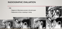
What image allows for the following analyses?
- Diagnosis of inflammatory process in the joint space
- Displacement of disc on opening or closing
MRI
If you suspect an issue with the meniscus, what imaging is indicated?
MRI
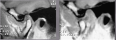
ID the problem with the TMJ:
anterior disk displacement with reduction

ID the problem with the TMJ:
anterior disk displacement without reduction
T/F: Joint issues is an acute condition
False, it is a CHRONIC condition and patient who undergo TMJ surgery will require future intervention
What image allows for the following analysis?
- Use IV T-99 to evaluate areas of active bone metabolism
Nuclear imaging
For nuclear imaging, take one radiograph every ______ months to see growth trend
Six
You can use a ______ scan to evaluate areas of active bone metabolism
IV T-99
____of pts suffer from depression at time of initial presentation of TMJ disorder
1/3
____ of TMD pts have a history of severe depressive episode
2/3
What are the 8 Classifications of TMD?
- Myofascial Pain and Dysfunction (MPD)
- Internal Derangements
- Degenerative Joint Disease
- Systemic Arthritic Conditions
- Chronic Recurrent Dislocation
- Ankylosis (intracapsular, extracapsular)
- Neoplasia
- Infections
What classification of TMD?
- TMD from muscular pain from clenching or grinding
Myofascial Pain and Dysfunction (MPD)
What classification of TMD?
- TMD from Anterior disk displacement with reduction or
- Anterior disk displacement without reduction
Internal Derangements

What classification of TMD?
TMD from Anterior disk displacement with reduction

What classification of TMD?
Anterior disk displacement without reduction
Internal Derangements are categorized by severity using the ________ system
Wilke's classification
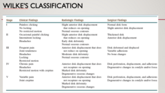
What Wilke's Classification has the following clinical findings?
- Painless clicking
- No locking
- No restricted motion
Stage I
What Wilke's Classification has the following clinical findings?
- Frequent pain
- Joint tenderness
- Headaches
- Locking
- Restricted motion
Stage III
What Wilke's Classification has the following clinical findings?
- Variable pain
- Joint crepitus
Stage V
What Wilke's Classification has the following radiographic findings?
- Slight anterior disk displacement that reduces on opening
- Normal osseous contours
Stage I
What Wilke's Classification has the following radiographic findings?
- Anterior disk displacement that does not reduce on opening
- Moderate disk deformity
- Normal osseous contours
Stage III
What Wilke's Classification has the following radiographic findings?
- Anterior disk displacement that does not recapture on opening
- Marked disk deformity
- Degenerative osseous changes
Stage V
What Wilke's Classification has the following surgical findings?
- Normal disk form
- Slight anterior disk displacement
Stage I
What Wilke's Classification has the following surgical findings?
- Disk deformed and displaced
- Variable adhesions
- No bone changes
Stage III
What Wilke's Classification has the following surgical findings?
- Disk perforation, displacement, and adhesions
- Degenerative changes in condyle and/or fossa
Stage V
Degenerative Joint Disease (Arthrosis, Osteoarthritis) can be due to one of what three possible mechanisms of injury?
- Direct mechanical trauma
- Hypoxia reperfusion injury
- Neurologic inflammation
What is the most common Systemic Arthritic Condition that can cause TMD?
Rheumatoid arthritis
What are all of the following?
- Rheumatoid Factor --> Rheumatoid Arthritis
- ANA --> Autoimmune (e.g. RA, SLE)
- CCP --> helps dx RA and its prognosis
- HLA-B27 --> Reactive arthritis, ankylosing spondylitis, Juvenile reactive arthritis
- Vit D
- 17B-Estradiol
Serologies for classification of TMD
As a general dentist, you can do what 3 things for patients that experience TMD?
- Analgesics
- Physical therapy
- Rx night guard
T/F: You should treat the click of TMJ issues
False
What are three options for patient education for TMD non-surgical management?
- Modification of diet
- Jaw exercises
- Avoiding aggravating factors
What type of medications can be used for TMD non-surgical management?
- NSAIDs (celebrex, or ibuprofen) to tx inflm and pain (Ibuprofen 800mg q8h x 2 weeks)
- Other analgesics for pain (no longer than 2 weeks)
- Muscle relaxants (Flexeril 5mg qhs, Soma) to relieve masticatory pain
- Antidepressants (esp. TCAs-Elavil) improve sleep, decrease bruxism, decrease joint and muscle pain
- Botox
When it comes to management of TMD, what should you always do first?
Try non-surgical management for 4-6 weeks first
T/F: Botox is used to treat both muscular and TMJ problems
False
What type of treatment?
A mouthguard that acts like crutches to prevent clenching
Autorepositioning splints
What type of treatment?
A guard that pull the jaw forward to recapture the meniscus and then slowly pull the jaw back to reposition the meniscus
Anterior repositioning splints
If you prescribe NSAIDs for a patient for part of their TMD non-surgical management, how long do they need to take them at MINIMUM to see any benefit?
2 weeks
What are 2 minimally invasive surgeries used to manage TMD?
- Arthorcentesis
- Arthroscopy
What type of surgery?
- Uses 1-2 canulas to access the superior joint space
- Lactated ringer solution to wash out the joint to eliminate inflammatory products, release fine adhesions
- Sterioids can be placed to decrease inflm
Arthorcentesis
What type of surgery?
- Same access as arthrocentesis but with an arthroscope
- Joint and disc can be recontured, adhesions can be removed
- Small tears in the disc can be repaired
Arthroscopy
What is the success rate for minimally invasive surgeries like Arthroscopy?
80% (higher for Wilkes II and III)
Which surgery is known as a blind technique: Arthorcentesis or Arthroscopy?
Arthorcentesis
At which Wilke's stage would we begin to consider surgical management to help with TMJ function?
Wilkes 3
What are four open joint surgeries used to manage TMD?
- Disk repositioning surgery
- Disk repair or removal
- Condylotomy
- Total Joint Replacement
What nerve do you have to be cautious of with preauricular access?
Temporal branch of facial nerve

Why do you only want to do about 3-4 open joint surgeries in a patient's life time?
Results in decrease blood supply to the joint
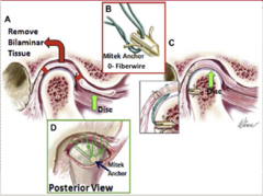
ID the procedure:
Disk repositioning surgery

ID the procedure:
Disk repositioning surgery
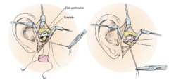
ID the procedure:
Disk Repair or Removal
Disk repair surgeries are falling out of favor due to their short term success rate before regression back to the original problem. When replacement is done, what is the preferred material for replacement due to its ability to become flexible after it scars?
A) Piece of septal cartilage
B) Piece of skin
C) Piece of belly fat
D) Temporalis muscle
C) Piece of belly fat
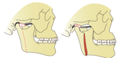
ID the procedure:
Modified Condylotomy
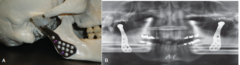
ID the procedure:
Total Joint Replacement
What is the last line of surgical treatment for patients with severe TMD?
Total Joint Replacement
In a Total Joint Replacement of the TMJ, the materials used for the prosthetic condyle on joint should be:
A) Metal on metal
B) Metal on plastic
C) Plastic on plastic
B) Metal on plastic
If a patient jaw deviates to the left on opening, which side is not moving?
Left
If a patient jaw deviates to the right on opening, which side is not moving?
Right
If a patients moves their jaw in lateral movements and has 3mm to right, 8mm to left, Which side has the problem?
Left
What occlusal bite most commonly presents with TMD?
Class II
If a patient has anterior disk displacement without reduction, what is sitting between the eminence and the condyle?
Retrodiscal tissue
you should limit open TMJ surgery to only about ___-___ times
3-4
When were x-rays discovered?
1895 by Wilhelm Roentgen

When was Linear tomography developed?
1930s

When wer computed tomography (CT) and magnetic resonance imaging (MRI) developed?
1970s and 80s

When was nuclear medicine developled?
1990s

What is the use of computed tomography for the head and neck? (4)
- Facial trauma
- Infection
- Neoplasms
- Surgical Planning
Computed tomography (CT) uses __________ values to measure attenuation of radiation through tissue, assigns a gray scaled image
- Depending on the range of values, an image can be "windowed" to the anatomy of interest (e.g. bone, brain, liver, soft tissue)
Hounsfield

ID the plane of view:
Axial

ID the plane of view:
Coronal

ID the plane of view:
Saggital
What is the most accurate plane of view for a CT?
Axial (bc that is how image is taken)
Soft tissue CTs can be enhanced with ________
Contrast
(Usually iodine-based contrast media (either ionic or non-ionic): Can be used intraarterially, intrathecally, intraabdominally, and intravenously)
What has the following pros/cons?
Pros:
- Fast
- Good soft tissue and bone imaging
Cons:
- High radiation
- Cost
- Requires iodine based contrast for soft tissue (can not be done for iodine allergic or pts with poor kidney function)
Computed tomography
What image...
- Use a cone-shaped beam
- Processed to generate 3D volumetric data
- Reconstructed to all 3 planes of view
Cone beam computed tomography
What has the following pros/cons?
Pros:
- Lower radiation compared to standard CTs
- Less artifact scatter
Cons:
- For evaluation of osseous structure and NOT soft tissue structures
- Not covered by medical insurance
Cone beam computed tomography
Which imaging modality typically has a lower radiation dose: CBCT or CT?
CBCT
What is the difference in X-ray beams between a CBCT and CT?
- CBCT: Cone-shaped beam
- CT: Fan-shaped beam (or cone-like geometry in some multidetector CT systems)
What image...
Applies a magnetic field to tissue, picks up signals produced by magnetized protons, signals are processed using Fourier transform to generate an image
Magnetic resonance imaging (MRI)
What imaging has the following uses?
- Good for delineating soft tissue pathology
- Evaluation of suspected osteomyelitis
- Perineural spread of malignancy
- Head and neck infections and neoplasms (requires gadolinium based IV contrast
- TMJ pathology (closed and open mouth position)
Magnetic resonance imaging (MRI)
What has the following pros/cons?
Pros:
- No radiation
Cons
- More time consuming
- More expensive
- Less accessible
- Gadolinium may cause nephrogenic systemic fibrosis in patients with renal insufficiency
Magnetic resonance imaging (MRI)
You cannot obtain MRIs in patients with what conditions? (3)
- Pacemakers
- Neurostimulators
- Any feromagnetic implants/fragments near vital structures

What is A to B showing?
Anterior disc displacement with reduction
What image...
- Technetium 99 bone scans
- Determine bone metabolic activity
Nuclear medicine
What image...
- Uses 18-fluorodeoxyglucose (18FDG)
- Neoplasms has increased glucose metabolism than normal tissue
- Can be fused with a diagnostic CT imaging (PET/CT)
- System detects pairs of gamma rays emitted indirectly by a positron-emitting radionuclide tracer (18 FDG)
- Used for staging and surveillance of head and neck Ca
Positron emissions tomography (PET)