Chp 3: Abdomen
 Call Kai
 Learn
 Practice Test
 Spaced Repetition
 Match
 Flashcards
 Knowt Play
1/36

There's no tags or description
Looks like no tags are added yet.
37 Terms
the 2 large muscles found in the posterior abdomen adjacent to lumbar vertebra that are usually visible in AP radiograph are
psoas muscles

3 parts of small intestine are
duodenum (shortest & widest)
jejunum
ileum (longest)
Don’t Jump In
which of the following organs is part of lymphatic system?
a. liver
b. spleen
c. pancreas
d. gallbladder
b. spleen
3 accessory digestive organs
liver
gallbladder
pancreas
why is right kidney found in a more inferior position than left kidney?
bc of presence of liver
which endocrine glands are superomedial to each kidney?
adrenal glands
peritoneum
double walled membrane that lines abdominopelvic cavity
what are the 4 abdominal quadrants?
right upper quadrant (RUQ)
left upper quadrant (LUQ)
right lower quadrant (RLQ)
left lower quadrant (LLQ)
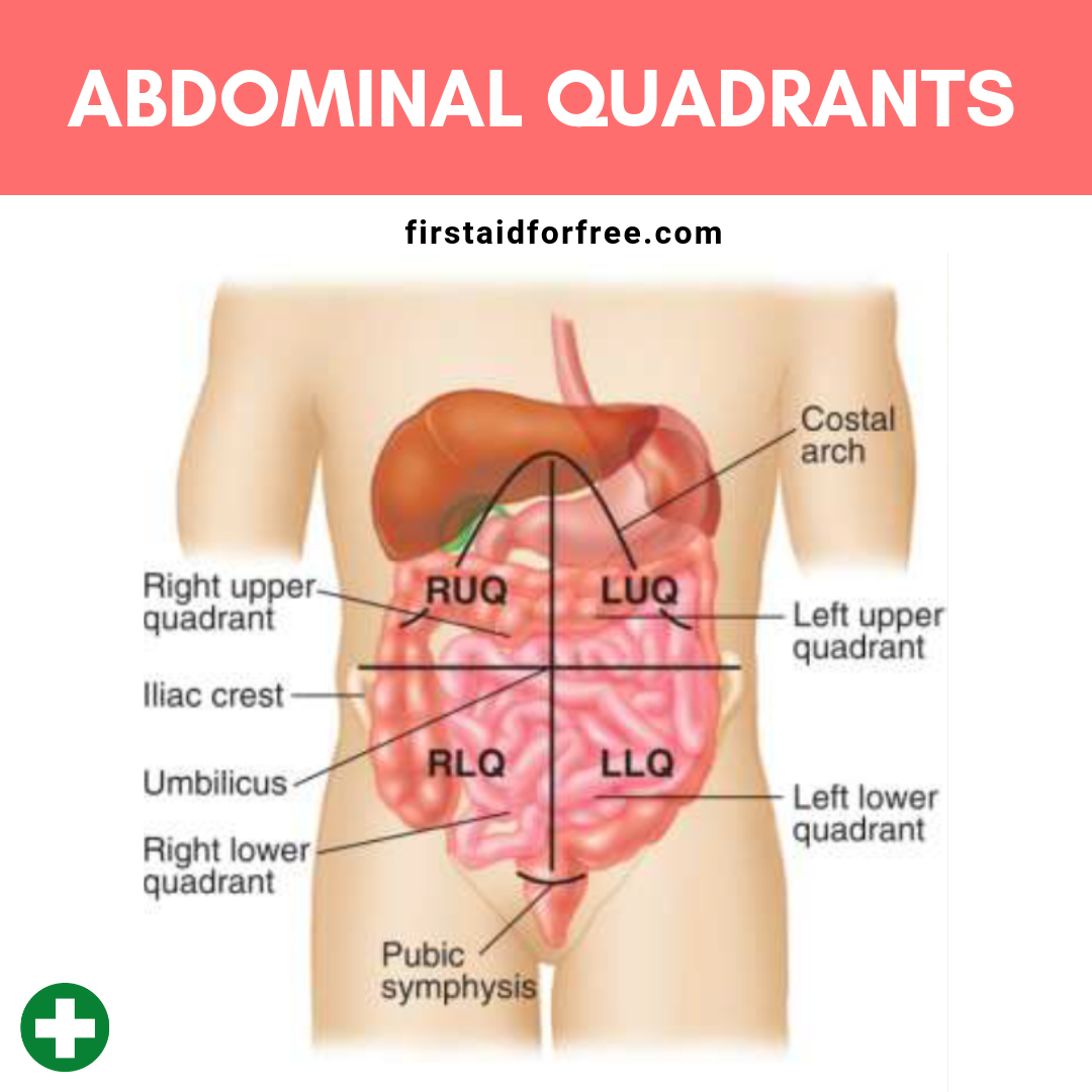
what are the 9 abdominal regions?
right hypochondriac
epigastric
left hypochondriac
right lateral (lumbar)
umbilical
left lateral (lumbar)
right inguinal (iliac)
pubic (hypogastric)
left inguinal (iliac)

topographic landmarks for abdomen xray?
xiphoid process (T9-T10)
ischial tuberosity
symphysis pubis
iliac crests (L4-L5)
ASIS
greater trochanter
inferior coastal margins (L2-L3)
2 way acute abdomen series includes
AP supine KUB
erect AP abdomen OR left lateral decub if pt can’t stand
3 way acute abdomen series includes
AP supine KUB
PA chest
erect AP abdomen
why is left lateral decub preferred over right lateral?
bc stomach is on left side. thus by having pt lay on left it prevents gastric bubbles from mixing with abnormal air bubbles pt may have that rise and might make it more difficult to tell air bubbles from gastric bubbles apart. by lying on left side only bubbles that rise will be due to pathology
ascites
abnormal accumulation of fluids in peritoneal cavity

pneumoperitomeum
free air or gas in peritoneal cavity
best seen with erect abdomen or chest image where even small amount of air will be seen under diaphragm

small bowel obstruction
blockage of flow of intestinal content
types of small bowel obstruction
intussusception
volvulus
ileus
intussusception
section of bowel slides into another loop

volvulus
twisting of a loop of intestine
ileus
non mechanical bowel obstruction caused by lack of intestinal motility
what organs are in the RUQ?
liver
gallbladder
duodenum
head of pancreas
right kidney
right adrenal gland
right hepatic (colic) flexure
what organs are in LUQ?
spleen
stomach
left colic (splenic) flexure
tail of pancreas
left kidney
left adrenal gland
2/3 of jejunum
what organs are in RLQ?
appendix
cecum
ascending colon
2/3 of ileum
ileocecal valve
what organs are in LLQ?
descending colon
sigmoid colon
more jejunum
more ileum
what are 2 causes of voluntary movement?
movement
breathing
voluntary movement can be prevented by
careful breathing instructions to pt
what’s the primary cause for involuntary motion in abdomen?
peristalsis
what’s best mechanism to control involuntary motion?
short exposure time
exposure for an AP projection of abdomen should be taken on
expiration
parts of large intestine
appendix
cecum
ileocecal valve
ascending colon
right colic (hepatic) flexure
transverse colon
left colic (splenic) flexure
descending colon
sigmoid colon
rectum
anus

the kidneys are connected to the bladder by way of the
a. urethra
b. renal artery
c. ureter
d. renal vein
c. ureter
which structure stores & releases bile?
a. liver
b. spleen
c. pancreas
d. gallbladder
d. gallbladder
which of the following structures connects the small intestine to the posterior abdominal wall?
a. greater omentum
b. peritoneum
c. lesser omentum
d. mesentary
d. mesentery
when is dorsal decubitus AP abdomen done?
replaces AP erect & left lateral decub if pt cannot stand nor turn to their side
evaluates air/fluid levels, hernias, aneurysms, calcification of aorta, etc
usually for severe trauma pts
in what position will seeing the psoas muscles be of greater importance?
supine AP abdomen (KUB)
in what position will seeing the diaphragm be of most importance?
erect AP abdomen, PA chest, & left lateral decub
for a left lateral decubitus how much time does the patient need to be in this position prior to the exposure and why?
5 min
to give enough time so that the air can rise and the fluid can fall and level