4. Cells and Tissues 2
 Call Kai
 Learn
 Practice Test
 Spaced Repetition
 Match
 Flashcards
 Knowt Play
1/54

There's no tags or description
Looks like no tags are added yet.
55 Terms
what is epithelium
Epithelia are coherent sheets of cells that cover body surfaces and line cavities. They are supported by a basement membrane that separates them from underlying connective tissue
functions of epithelium
protection
absorption
secretion
selective diffusion/transport
how are epithelia classified
number of layers
cell shape
surface specialisations
how are epithelia cells classified by number of layers
Type | Description |
|---|---|
Simple | Single cell layer |
Stratified | Multiple cell layers |
how are epithelia cells classified by cell shape
Shape | Appearance |
|---|---|
Squamous | Flat |
Cuboidal | Cube-shaped |
Columnar | Tall cells |
how are epithelia classified by their surface specialisations
cilia
microvilli
general features of simple epithelia
Single layer of cells.
Found where exchange occurs between different environments.
Involved in diffusion, absorption and secretion
features of simple squamous epithelial cells
flat, thin cells
minimal diffusion distance

function of simple squamous epithelia cells
rapid diffusion and exchange
example of simple squamous epithelial cells
blood vessel lining (endothelium)
alveoli of lungs
features of simple cuboidal epithelium
cube-shaped cells
central round nuclei
more cytoplasm than squamous cells

functions of simple cuboidal epithelium
secretion and absorption
example of simple cuboidal epithelium
kidney tubules, small glad ducts
features of simple columnar epithelium
tall cells
nuclei near basement membrane
may possess microvilli or cilia

functions of simple columnar epithelium
absorption
secretion
transport
example of simple columnar epithelium
intestinal epithelium
parts of respiratory tract
features of stratified epithelia
more than one cell layer
primarily protective
resistant to physical and chemical damage
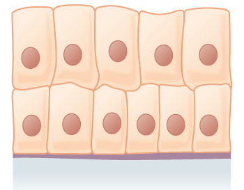
features of stratified squamous epithelium
multiple layers of cells
surface cells are squamous

function of stratified squamous epithelia
protection against abrasion
examples of stratified squamous epithelia
oral cavity
vagina
anal canal
skin (keratinised form)
three key epithelial cell specialisations
junctions
polarity
glands
how are cell-extracellular matrix junctions enabled
by integrins
which are transmembrane adhesion proteins that link the cell's cytoskeleton to the components of the extracellular matrix (such as collagen, laminin, and fibronectin)
cell-extracellular matrix junctions: how do cells attach to the ECM
through integrins
intern-mediated adhesion
cells attach to the ECM through integrins
functions of integrin-mediated adhesion
anchor cells to ECM
connect ECM to intracellular cytoskeleton
allow cells to sense and respond to their environment
example of integrin-mediated adhesions
focal adhesions (actin)
hemidesmosomes (intermediate filaments)
focal adhesions (actin)
sites where a cell attaches to the ECM and links the ECM to the actin cytoskeleton
structure of focal adhesions
Outside cell:
ECM proteins (e.g. fibronectin, collagen, laminin)
Cell membrane:
Integrins (transmembrane receptors)
Inside cell:
Adaptor proteins such as talin, vinculin, paxillin, and α-actinin
Connected to actin filaments (microfilaments)
functions of focal adhesion sites
Cell adhesion to the ECM
Cell migration (formation and breakdown of focal adhesions during movement)
Mechanotransduction (sensing and responding to mechanical forces)
Signal transduction affecting growth, survival, and differentiation
hemidesmosomes
cell-ECM junctions that anchor epithelial cells to the basement membrane
structure of hemidesmosomes
Extracellular side:
Laminin in the basement membrane
Transmembrane proteins:
Integrin α6β4 (main receptor)
BP180 (collagen XVII)
Intracellular side:
Plaque proteins (e.g. plectin, BP230)
Connected to keratin intermediate filaments
function of hemidesmosomes
Provide strong adhesion between epithelial cells and the basement membrane.
Resist mechanical stress (important in skin and other epithelia).
Prevent epithelial layers from detaching from underlying tissues.
3 major classes of cell-cell junctions and their functions
Junction Type | Function |
|---|---|
Occluding | Seal spaces between cells |
Communicating | Allow communication |
Anchoring | Provide mechanical strength |
functions of occluding/tight junctions
Seal adjacent cell membranes.
Prevent leakage between epithelial cells.
Form selective barriers.
Separate apical and basolateral membrane domains
e.g. bladder epithelium preventing urine leakage
major proteins in occluding/tight junctions
claudins and occludins
structure of communicating/gap junctions
channels connect neighbouring cells
build from proteins called connexins
function of communicating/gap junctions
allow movement of:
ions
small metabolites
signalling molecules (<1000 Da)
importance of communicating/gap junctions
cell-cell communication
electrical coupling in some tissues
function of anchoring junctions
link cytoskeletons of neighbouring cells into a mechanically strong sheet
2 major types of anchoring junctions
adherens junctions
desmosomes
components of adherens junctions
cadherins
catenins
actin cytoskeleton
function of adherens junctions
maintain tissue architecture
how do adherens junctions structure support their function
Cadherins (transmembrane proteins) | Cadherins on adjacent cells bind to each other, creating direct cell-cell adhesion. |
|---|---|
Calcium-dependent binding | Ca²⁺ stabilizes cadherins, allowing strong, specific adhesion between neighbouring cells. |
Catenins and other adaptor proteins | Link cadherins to the actin cytoskeleton inside the cell. |
Actin filament attachment | Forms a continuous actin network across many cells, allowing forces to be transmitted through a tissue. |
Belt-like arrangement (zonula adherens) | Encircles the cell, distributing tension evenly and helping maintain tissue architecture. |
example of adherens junctions in epithelia
In epithelial sheets, adherens junctions form an adhesion belt just below tight junctions. Because they are connected to contractile actin filaments, they can change cell shape during processes such as:
Tissue folding
Wound healing
Embryonic morphogenesis
components of desmosomes
cadherins
desmoplakin
intermediate filaments
function of desmosomes
resist mechanical stress
particularly important in:
skin
cardiac muscle
how does desmosomes structure support its function
Structural feature | How it supports function |
|---|---|
Cadherins (desmoglein and desmocollin) span the membrane | Bind to cadherins on neighbouring cells, creating strong cell-cell adhesion. |
Dense intracellular plaque (desmoplakin, plakoglobin, plakophilins) | Forms a robust anchoring structure beneath the membrane. |
Attachment to intermediate filaments (keratin) | Distributes mechanical forces throughout the cell and tissue, preventing cells from being pulled apart. |
Numerous desmosomes between cells | Creates a network of strong connections across the tissue. |
why are intermediate filaments important
Unlike actin filaments, keratin intermediate filaments are very good at resisting tensile forces. When a tissue is stretched, the force is transmitted from:
Keratin in one cell
→ Desmosome
→ Keratin in the neighbouring cell
This spreads the stress across many cells, preventing damage.
where are desmosomes found
They are especially abundant in tissues exposed to mechanical stress:
Epidermis (skin)
Cardiac muscle (intercalated discs)
clinical relevance of desmosomes
autoantibodies against desmosomes cause pemphigus, a severe blistering disease
what polarity do epithelial cells exist
pico-basal polarity
3 domains in epithelial cells for epithelial polarity
Apical surface → faces the lumen/exterior
Lateral surface → contacts neighbouring cells (contains tight junctions, adherens junctions, desmosomes)
Basal surface → contacts the basement membrane (contains hemidesmosomes)
apical surface
the free surface of an epithelial cell that faces the lumen or external environment
specialises in microvilli and cilia
how is the apical surface specialised to perform the cells specific role (structure → function)
Apical specialization | Function |
|---|---|
Microvilli | Increase surface area for absorption (e.g. intestinal epithelial cells). |
Stereocilia | Long microvilli that aid absorption or sensory functions (e.g. epididymis, inner ear). |
Cilia | Move fluid or particles across the cell surface (e.g. respiratory tract). |
Glycocalyx | Protective carbohydrate-rich coating involved in protection and cell recognition. |
examples of apical surfaces
Small intestine: apical microvilli increase nutrient absorption.
Trachea: apical cilia move mucus and trapped pathogens towards the throat.
Kidney tubules: apical membrane contains transport proteins for reabsorption.