pacing, capture, and threshold
 Call Kai
 Learn
Learn Practice Test
 Spaced Repetition
 Match
Match Flashcards
 Knowt Play
1/64

There's no tags or description
Looks like no tags are added yet.
Name | Mastery | Learn | Test | Matching | Spaced | Call with Kai | Chat |
|---|
No analytics yet
Send a link to your students to track their progress
65 Terms
pacemaker battery
lithium iodine, first implanted in 1972, unsurpassed reliability and predictability
why do polarities reverse between battery and tissue
tissue has electrical charge (+anode and -cathode)
battery anode
donates or losses electrons through oxidation
battery cathode
receives or gains electrons through reduction
depolarization
electrons sent from pulse generator to -cathode tip; attract +Na
phase 0
rapid depolarization
phase 1
initial repolarization
phase 2
plateau
phase 3
final repolarization
phase 4
alert period or resting phase
depolarization action potential for someone with CHB
isoelectric line because nothing to depolarize
action potential x and y axis
time (msec) and transmembrane potential (mV)
threshold
have enough energy to trigger depolarization; without, give impulse with no reaction
time zero
phase 4: resting phase pacing impulse begins
0.3 mSec
impulse delivered: electrons sent to lead tip (cathode)
0.6msec
phase 0: capture/depolarization
0.7 msec (after potential)
phase 1-3: ions return to repolarization
what drives polarization to depolarization
battery
amplitude
how many volts are delivered per pacing impulse; how much power
pulse width
duration of the impulse; how long amplitude delivered; pacing output setting
goal
effective and safe output setting while maximizing battery longevity
safety margin
2:1
why do you want to pace with minimum power
to save battery
pulse width
mseconds; how long impulse is delivered usually 0.4 ms or 0.5
strength-duration curve
amplitude (V) and pulsewidth (ms); lowest threshold for best battery
rheobase
smallest amplitude that stimulates myocardium at infinitely long pulse duration; double to get reference
chronaxie
threshold pulse duration at 2x rheobase voltage; approximates point of minimum threshold energy
3 lines on strength duration curve
rheobase, 2. chronaxie, 3. energy
center of strength-duration curve
knee of the curve; defines lowest amount of energy to reliably capture the heart
Ohm’s law
Voltage = I (current-amps) x R (resistance/impedence in Ohm’s)
units of Ohm’s law
amps but deliver in milliamps
energy equation
E=V²/R x t; E=energy in microjoules, V=voltage, R=impedance, t= Pulse Width
when threshold elevates
increase output/volts, or increase/extend pulse width
doubling voltage
quadruple energy
doubling pulse width
double energy
energy equation units
want microjoule, convert from millijouleto
to depolarize
run a threshold test; control the heart rate
when to not run threshold test
AFib or VT
mode for PPM dependent patient
DDD never AAI
Dual chamber device testing
DDD only one chamber decremented at a time
ventricular threshold test mode
VVI
overdrive pace
power (adequate starting amplitude) and speed (faster than underlying rhythm)
decrement power
decrease amplitude incrementaly and watch the rhythm
when to stop threshold test
loss of capture, verify rhythm again
important when running a threshold test
know the presenting rhythm and know the underlying rhythm
final result of threshold test
previous output with successful depolarization, not loss of capture
factors affecting threshold
lead integrity/location, medications, electrolyte imbalance, arrhythmia (flutter, fib, tach, slow VT)
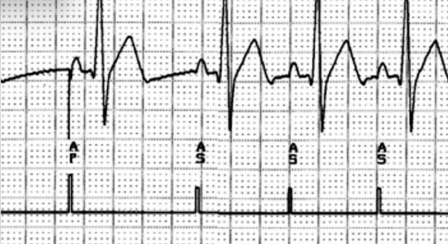
atrial capture
AAI

ventricular capture
VVI

dual chamber capture
DDD

atrial failure to capture

ventricular failure to capture
failure to capture- short term
physician fix: lead dislodgment or poor connection at connector block
failure to capture- long term
hyprid fix; lead maturation (exit block)or lead failure
failure to capture you can fix
low output; pulse width and amplitude

intrinsic r-wave

fusion beat

paced ventricular beat

pseudo fusion beat
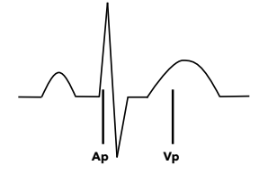
pseudo pseudofusion beat
why use automatic capture algorithms
scheduled frequency and prolong battery life
ACA scheduled frequency
•More frequent testing compared to in-office visits
•Every 8hrs, 24hrs, etc
•Some beat-to-beat
•Typically set to 1:00/2:00 am (programmable)
ACA safety margin
•Pacing Amplitude reprogramming (if necessary)
•By not applying a 2:1 safety margin and instead use 1.0V safety margin,
you prolong battery life
ACA temporary programming and evoked response
•Ventricular morphology discriminator – creates a higher output template for a captured beat. When LOC occurs, the LOC morphology is confirmed against the reference template
ACA backup pulse
protects patient and minimizes symptoms at LOC