protein sorting
 Call Kai
 Learn
 Practice Test
 Spaced Repetition
 Match
 Flashcards
 Knowt Play
1/100

There's no tags or description
Looks like no tags are added yet.
101 Terms
Stages from gene to functional protein
Transcription, translation, folding and transport
Why is energy needed during protein synthesis?
To regenerate aminoacyl-tRNAs and drive ribosome translocation
Is peptide bond formation energy-requiring?
No, it is spontaneous because energy is stored in charged amino acids
ATP cost per peptide added
2 ATP
GTP cost per peptide added
1 GTP
GTP
Guanosine triphosphate used in signalling, conformational changes and molecular switches, signal transduction mechanisms
ATP
Adenosine triphosphate used for conventional cellular energy costs
Why is protein degradation energetically favourable?
No energy is required to build the protein
Main challenge during protein folding
Hiding hydrophobic residues from water
Cause of protein aggregates
Exposed hydrophobic regions interacting with each other
Why do unfolded proteins aggregate?
Exposed hydrophobic surfaces interact and tangle together
Role of chaperones
Assist protein folding and prevent aggregation
How do chaperones prevent aggregation?
Bind exposed hydrophobic regions
Effect of chaperone binding to hydrophobic regions
Masks hydrophobic surfaces
Unfoldase activity
A chaperone activity that allows proteins to attempt folding again
Does chaperone unfoldase activity require energy?
Yes
What process of chaperone action is ATP-dependent?
Substrate release
Major family of molecular chaperones
Heat shock 70 family
Examples of Hsp70 proteins
Hsp70, Hsc70 and BiP
Location of Hsp70 chaperones
Cytosol and other cellular compartments
Protein-binding domain of Hsp70
Binds unfolded proteins
ATPase domain of Hsp70
Binds and hydrolyses ATP
Is protein folding energetically favourable?
Yes
Is protein unfolding energetically costly?
Yes
Passive function of chaperones
Prevent aggregate formation
Active function of chaperones
Dissolve aggregates and promote refolding
Quality-control role of chaperones
Target proteins for degradation and initiate unfolded protein response
Are chaperones part of the final protein structure?
No

Evidence for chaperone function
Mutant chaperones cause protein aggregation and failed refolding

Folding intermediate
A partially folded protein
Chaperone ligand during folding
A folding intermediate with exposed hydrophobic regions
Monomeric folding intermediate
Usually inactive
Dimerisation
Association of two protein subunits
Effect of dimerisation on chaperone binding
Protein is no longer recognised by chaperones
Why can completed folding still require chaperones?
Some proteins must assemble into complexes before being fully folded
Prokaryotic protein destinations
Cytosol or secretion across the plasma membrane
Relationship between transcription and translation in prokaryotes
Coupled
Why must secreted bacterial proteins unfold?
Folded proteins cannot pass through membrane pores
What places can a protein in a prokaryote end up?
Inner membrane
outer membrane
cytosol
Number of membranes in Gram-positive bacteria
One
Number of membranes in Gram-negative bacteria
Two
what is the protein targeting signal in bacteria known as?
Signal peptide
Function of bacterial signal peptides
Direct proteins for secretion
Fate of bacterial signal peptides
Cleaved after translocation
Example of prokaryote proteins for secretion?
Hydrolases; digest extracellular macromolecules
Relationship between transcription and translation in eukaryotes
Separate processes, not coupled
Fate of eukaryotic mRNA
Transported to cytosol for translation
Possible destinations of eukaryotic proteins
Mitochondria, chloroplasts, peroxisomes and secretory pathway
Port of entry into secretory pathway
Endoplasmic reticulum
Transport vesicle
Protein-coated vesicle moving cargo between compartments
Ribosome
Universal molecular machine that synthesises proteins from mRNA
Signal recognition particle (SRP)
Ribonucleoprotein that targets proteins to membranes
Function of SRP in eukaryotes
Targets proteins to the ER membrane, by recognising a signal peptide; via translocation
Function of SRP in prokaryotes
Targets proteins to the plasma membrane
Two possible fates of translated proteins
Secretory pathway or cytosolic localisation
SRP-dependent pathway
Protein enters secretory pathway
SRP-independent pathway
Protein remains in cytosol
Where does protein synthesis begin?
Cytosol
Exception to cytosolic protein synthesis
Chloroplast and mitochondrial proteins
Fate of proteins synthesised in cytosol
Fold and assemble after translation
Fate of proteins synthesised at rough ER
Fold, assemble and exit in vesicles
—> Exported to ER, translocation pore on ER sealed by ribosome
—> Depend on cytosolic mRNA
Co-translational translocation
Protein enters ER while still being translated
Advantage of co-translational translocation
Protein does not need to be unfolded for transport
Explain process of Co-transnational translocation
-The “signal recognition particle” (SRP) recognises “signal peptides” when they emerge as nascent chains
on the ribosome surface.
--> SRP stops or slows down translation by the ribosome
--> SRP targets the ribosome/mRNA/nascent polypeptide complex to the translocation pore on the ER
--> Engaged SRP's have an increased affinity to the ER Resident SRP receptor, which they bind and hand over ribosome-mRNA complex to translocation pore
--> Nascent protein inserts into aqueous channel of the pore and translation by the ribosome resumes
--> Protein fold up with ER lumenal chaperones (BiP)
--> SRP and the SRP-receptor are being recycled back to cytosol (GTP hydrolysis),
-Cleavage of the signal peptide occurs well before the protein is fully translocated and is never part of the finally folded structure (The mature protein)
what happens after cotranslational translocation?
--> Ribosome seals a translocation pore on the rough ER
--> Protein needs to finish translation and folding within ER
-ER contains a special chaperone: The binding protein (BiP)
Why is BiP especially important in the ER?
High protein concentration increases aggregation risk
Role of BiP when pore is unused
Seals the translocation pore
Role of BiP during protein folding
Masks exposed hydrophobic regions
How long does BiP assist folding?
Until no hydrophobic regions remain exposed
Destination of correctly folded ER proteins
Golgi apparatus
Why do monomeric subunits bind BiP?
Exposed hydrophobic regions remain visible
When are multimeric proteins released from BiP?
After assembly to other proteins hides hydrophobic surfaces
Why do completed multimers escape chaperone binding?
Hydrophobic regions are buried in quaternary structure
Antibody subunits
Light chains and heavy chains
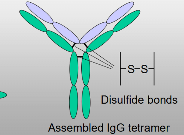
Where are antibody chains synthesised?
In the ER lumen
Why is antibody assembly into tetramers required?
To prevent continued BiP binding and allow secretion
Location of antibody signal peptides
N-terminus
What happens to antibody signal peptides?
They are cleaved after translocation
How do heavy chains interact with BiP?
Bind BiP until light chains assemble and form di-sulphide bridges
What stabilises assembled antibodies?
Disulphide bonds
Complex antibody example
IgM tetramers and pentamers
Destination of secreted antibodies
Bloodstream, soluble in the aqueous lumen of the endoplasmic reticulum
Membrane-anchored antibodies
Remain attached to membranes
Importance of membrane antibodies
Essential for immune system function
Location of antibody domain in membrane antibodies
Faces ER lumen, Golgi lumen, vesicle lumen or extracellular space
How is the light chain membrane-associated?
Through interaction with the heavy chain
Transmembrane domain (Protein insertion to membranes)
Hydrophobic stretch of amino acids
Are transmembrane domains cleaved?
No
Do transmembrane domains remain in mature proteins?
Yes
BiP discovery
Found bound to unassembled antibody heavy chains
GRP78
Protein discovered in glucose-starved cells, where many proteins failed to fold properly due to missing sugar modifications.
Relationship between BiP and GRP78
They are the same protein
Function of BiP/GRP78
Assists protein folding and prevents accumulation of misfolded proteins
Importance of BiP discovery
Showed that cellular stress can reveal important proteins
Proteins destined for membrane insertion contain
A signal peptide and transmembrane domain
Explain process of protein membrane insertion
Final orientation of inserted membrane proteins
N-terminus in ER lumen and C-terminus in cytosol
Location of transmembrane domain
Embedded within the membrane
Why is ER lumen considered topologically outside the cell?
It lies beyond the final membrane barrier before secretion
Order of the secretory pathway
RER → Golgi → Vesicle → Secretion