4. The process of Diagnosis ✅
 Call Kai
 Learn
 Practice Test
 Spaced Repetition
 Match
 Flashcards
 Knowt Play
1/43

There's no tags or description
Looks like no tags are added yet.
44 Terms
DIAGNOSIS
Instant recognition of a disease from previous experience
What process is occurring? - 1st question
What tissue is involved?
Past & present medical & dental histories
Medication history (side effects of medicine)
Radiographic, clinical & pathologic correlations
SIRMIN- origin of disease

Structural Processes
Longstanding and unchanging
May have family history
May be congenital
Young patients often
Slow to develop

Immune Processes
Features may overlap with infections
Classic signs of inflammation
Pain or itching ex herpes
Females > males generally
Allergic or autoimmune
Response to steroids

Reactive Process
Reaction to what?
–Physical, chemical, radiation, trauma?
Should resolve when the causative agent goes away (~10 days)
May mimic neoplasm

Metabolic Processes
Usually a systemic condition
Other medical findings
May be hormonal
May be nutritional
May be genetic
Infectious
Classic signs of inflammation
Often painful
May look similar to tumors
Source of infection?
Radiographic findings
Soft tissue findings
Response to antimicrobials

Neoplastic Processes
Benign neoplasms
Usually encapsulated
Usually slow growing
Localized mass
even if its benign you will biopsy to ensure no malignant cells + it can turn malignant

tori
Malignant Neoplasms
Usually NOT encapsulated
Usually NOT slow growing
More likely to mimic infection
Ill defined mass (so is an infection)
May be painful or cause loss of sensation (so may an infection)
May cause enlargement of lymph nodes (so may an infection)
Types of Oral Tissues: Epithelium
Surface Mucosa: oral cavity has squamous epithelium
Under surface: Salivary, Odontogenic
Types of Oral Tissues: Connective Tissue
Hard - bone or cartilage
Other - fibrous, fatty
Blood vessels or blood cells
Muscle
Nerves
Look for Evidence of Disease
Changes in
Color
Form
Function or sensation
Feel
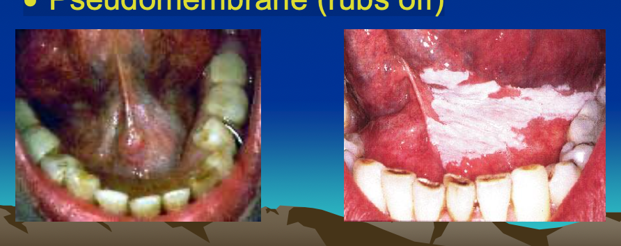
Changes in Color: White
white = Less blood visible
Thick epithelium
Calloused epithelium
Scarred fibrous tissue
Pseudomembrane (rubs off)
Changes in Color: yellow
Fat or xanthoma cells
Sebaceous glands
ex: lipoma (benign)

Changes in Color: Brown/blue/black
Melanin
Foreign sources
Systemic conditions
Physiological
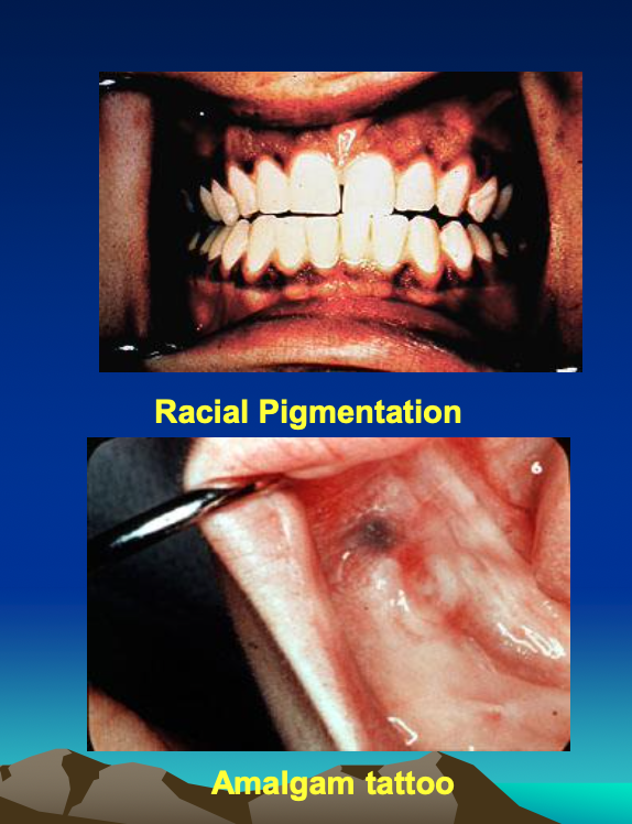

melanoma = most common pigmented lesion
changes in form: When a lesion grows under the epithelium in connective tissue
it creates a smooth-surfaced lesion by stretching the overlying epithelium
Masses that arise under the epithelium are usually smooth

changes in form: When epithelium grows faster than connective tissue
a rough surface results.
corrugated, papillomatous or verrucous

these pictures arise from epithelium because its rough
Flat colored lesions
Local deposits of pigments
Slight local changes in blood vessels
Not significant change in cell number or size
Form
Bilateral lesions, rarely neoplasms – Usually normal anatomic structures
Exceptions i.e. Multifocal canalicular adenoma
Systemic diseases may present multiple lesions
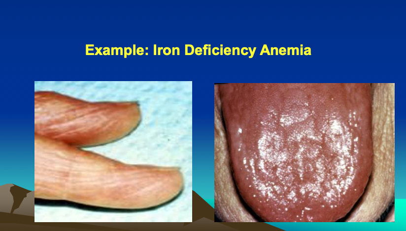
Metabolic - Addison, Iron deficiency
Neoplasms - Lymphoma, Metastases
Infections
Developmental - Syndromes
Loss of Function - sign of Inflammation
Sialadenitis: saliva flow altered
tendinitis: difficulty moving muscles
Loss of Function - sign of Malignancy
Squamous cell carcinoma of the tongue
May produce change in phonation as tumor invades muscle
change in Function or Sensation
Pain due to a tumor
Most tumors painless
Some intrinsically painful - Osteoblastoma
Invade nerve sheath ---Adenoid cystic carcinoma
invade nerve = perineural invasion
Destroys adjacent tissues - Pancreatic adenocarcinoma
change in feel: freely movable
A mass that is freely moveable in all directions is usually benign or encapsulated, originating in the underlying connective tissue.
Example: Lipoma
When the mass is fixed in deep connective tissues, you need to figure out what tissue the mass is bound to - That is likely the tissue of origin

change in feel: attached to skin
A mass that moves freely but together with the overlying epithelium probably arises in the epithelium
Example: Nevus (mole

Induration
may indicate:
• Fibrosis
• Infection
• Malignancy
Spreading from epithelium to deeper tissues
Spreading from deeper structures to epithelium

Differential Diagnosis
A list of diseases with features consistent with the observed signs and symptoms
Arranged in order of probability, as determined by epidemiology and frequency of occurrence
you pick MOST likely
Working Diagnosis
Most likely of the diseases in the differentialdiagnosis
Next steps in managing the patient is based on the working diagnosis
BIOPSY
Surgical removal of living tissue for definitive diagnosis
Must be representative of the lesion
Examined microscopically
Is only performed IF INDICATED BY THE WORKING DIAGNOSIS
Biopsies are fairly costly and stressful for a patient - Use them when you need them! But use them wisely
Problems with biopsy before establishing the working diagnosis
Wrong type of biopsy?
Wrong type of fixative?
Risky biopsy?
Inappropriate biopsy?
Unnecessary biopsy?
When to Do a Biopsy
When signs and symptoms of the lesion do not provide enough information to make a definitive diagnosis
If neoplasia is in the differential diagnosis
Any chronic non-healing ulcer of more than two weeks duration
ulcer
yellow appearance with red border
THINGS TO WATCH OUT FOR
If working diagnosis doesn’t call for a biopsy, don’t do it
e.g
• Herpetic gingivostomatitis
• Hematoma following trauma
• Chronic periodontitis
aphthous ulcers
Risk of trauma, infection, bleeding, and complications
Systemic health of the patient (i.e. bleeding disorder)
Skill of the clinician
Refer to appropriate health care provider - such as OMS
Types of Biopsies: Incisional Biopsy
A representative portion of the lesion is removed
Most of the lesion is left behind
Should try to include normal tissue at edge of lesion
For large lesions or suspected malignant neoplasms
ex: lichen planus (immune related)
Types of Biopsies: Excisional Biopsy
Entire lesion is removed
Also margin of normal tissue
Small lesions
Not expecting malignancy
Other kinds of Biopsy: Punch biopsy

can be incisional or excisional depending on size
Other kinds of Biopsy: Shave biopsy
Esthetic areas such as anterior gingiva
Superficial lesions only, such as lip epithelium
other kinds of Biopsy: needle biopsy
For deep tissues not accessible by direct exploration… e.g. Thyroid gland
Not usually needed for oral lesions
Cytologic Smear Biopsy
Scrape mucosa with spatula
Collect cells on glass slide
Fix cells with cytospray / hairspray containing alcohol
Write patient name on slide
Diagnosis of candidiasis
Pap Smear is a type of cytologic smear
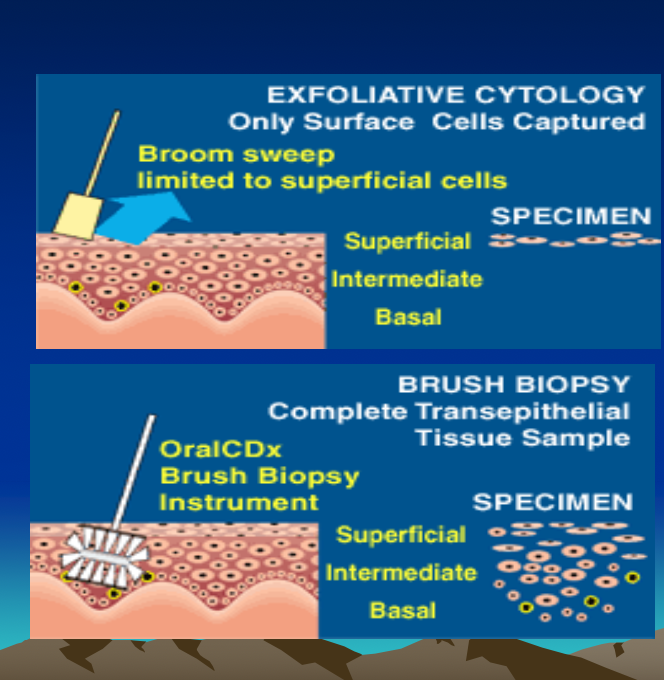
Why not smears to diagnose oral dysplasia?
Cell sample
• Abnormal + normal cells
• Only surface sampled
• Typically 300,000 cells per slide
• “Needle in a haystack”
No architecture is visible
• Cells, but no pattern to them
Improvement in Cell Sampling – Cytobrush Biopsy
Cells from all layers of epithelium if used as directed
Lab will reject biopsy if sample is not optimal
Only tested on white lesions
Toluidine Blue
Stain with affinity for perinuclear cisternae of DNA and RNA
Only + areas retain dye after rinsing with acetic acid
Theoretical risks of a vital dye incorporated into cellular DNA
Rate of false positives
hyperactivity/ Inflammation / ulceration may give false readings
Return visit within 14 days if a test is positive, to repeat the procedure
Misuse in inexperienced hands
Submission of Biopsy
All specimens need to be put in preservative immediately
Make sure you have it before you cut
10% buffered formalin usually
Spray a smear with fixative
Sometimes other special preservatives
Next best is clear ethanol - drinking alcohol like vodka, NOT rubbing alcohol
Fixation for Immune Lesions: Michel’s Solution
Used fresh
Refrigerate for up to 2 weeks
Request in advance
Which lesions?- Vesiculobullous (blister) autoimmune diseases
put ½ lesion in this + ½ in formalin
What’s Next?
Dentist correlates microscopic and clinical findings
Dentist informs patient of her/his diagnosis
Pathologist will not release results to patient
Dentist decides on and arranges further treatment, if any