Physiology Sliding Filament Theory ICE
 Call Kai
 Learn
Learn Practice Test
 Spaced Repetition
 Match
Match Flashcards
 Knowt Play
1/28

There's no tags or description
Looks like no tags are added yet.
Name | Mastery | Learn | Test | Matching | Spaced | Call with Kai | Chat |
|---|
No analytics yet
Send a link to your students to track their progress
29 Terms

What structural component of the sarcomere is associated with arrows in model
1?
Z-line

Based on this, which component of the sarcomere is actually being moved when
the sarcomere contracts and relaxes?
Z-line

Which component of the sarcomere is physically attached to the structure that
gets moved (i.e. the answer to question 2)?
Thin filament

What component of the sarcomere is NOT directly attached to the Z-line?
Thick filament

Based on your answer to question 4, what component of the sarcomere is likely
pulling on the thin filaments to bring the Z-lines closer together?
Thick filament
In the space below, write a short description as a group that explains the role of
the thin filaments, thick filaments, and Z-line in sarcomere contraction.
Thin filament: attaches the Z line, aids in contraction/relaxation
Thick Filament: pulls thin filament and Z line together
Z-line: pulls thin filament to relax
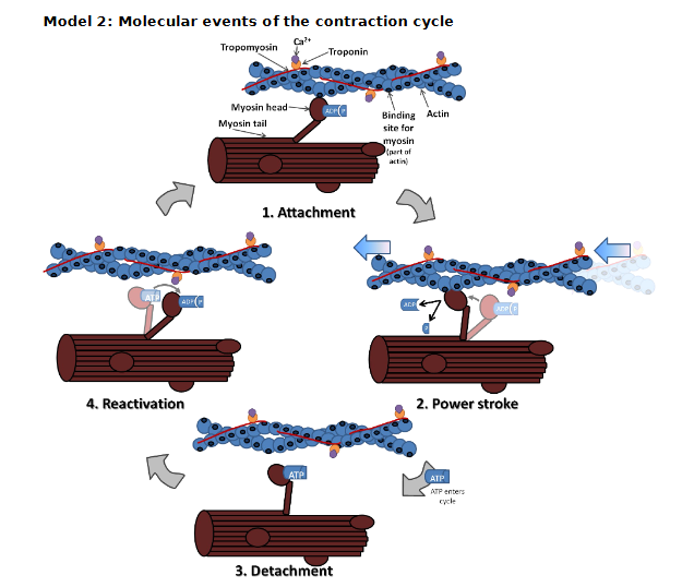
Which component of the thin filament makes the main ‘string-of-pearls’
portion of the filament?
Actin

List the other molecules that are found in/on the thin filament.
Troponin and tropomyosin

The myosin head is bound to some other molecules; what are they? To which specific region of actin is the myosin head bound?
ADP and P/thin filament; on the binding site

In your own words, describe the change that occurs in the myosin molecule
between stage 1 (attachment) and stage 2 (power stroke)?
ATP is hydrolyzed into ADP and a phosphate group, which cocks the myosin head. The myosin then attaches to actin and releases a phosphate group as well as ADP. The myosin head then changes position and pulls actin during the power stroke.

The myosin head doesn’t release from the actin binding site until a new
molecule enters the cycle. According to the model, what molecule allows myosin to
release from actin?
ATP

In stage 4 of this cycle, the myosin molecule moves back into the cocked
position. Where does it get the energy to re-cock itself?
From the hydrolysis of ATP

Look at the left half of the model. Is calcium present here?
No

Is the myosin binding site of actin exposed so that myosin can actually bind to it?
No

What is blocking myosin’s access to the binding site on actin?
Tropomyosin

Now look at the right side of the model. Is calcium present here? To what molecule did it bind?
Yes; troponin

What changed about the troponin molecule after it bound to calcium?
Its position

What effect did this change in troponin have on tropomyosin?
It moved so actin is exposed/pulled it off of the myosin binding site

Is the myosin binding site of actin exposed when calcium is present?
Yes

Describe the role of troponin, tropomyosin, and calcium in muscle cell contraction.
Calcium binds to troponin to move tropomyosin to expose the binding site on actin.
What would happen to a muscle cell if the cell ran out of ATP?
It would stay bound (rigor mortis)
What would happen to a muscle cell if there was an excess amount of calcium in the cell?
Over contraction
What would happen to a muscle cell if there was no free calcium in the cell?
There would no contraction or relaxation

According to model 4, where is calcium stored in the muscle cell? Does this appear to be a part of an organelle? If so, which organelle?
The lateral sac; yes, the sarcoplasmic reticulum

What receptor channel is responsible for calcium release? What causes the activation of this receptor channel?
Ryanodine; DHP receptor

The muscle fiber action potential is carried toward the interior of the cell via what tubule? Does this tubule bring the action potential closer to our storage site for calcium or farther from it?
The transverse/T tubule; brings it closer

Why might this be important for the release of calcium?
Depolarization in the T tubule activates DHP receptor which activate the ryanodine receptor causing the release of calcium

How is calcium taken back up after release from its storage site? What is the source of energy for this uptake?
Through active transport (Ca2+ or ATPase pumps); ATP
Put the following events in order from action potentials in the
skeletal muscle fiber to the contraction of the whole muscle.
Myosin bends in two places, releasing ADP and pulling on the thin filament
Calcium is released from the sarcoplasmic reticulum
An action potential travels down the muscle fiber membrane
The Z-lines are pulled closer together
Calcium ions bind to troponin causing a conformational change
The ryanodine receptor is activated
The action potential is propagated into the transverse tubule
The conformational change in troponin moves tropomyosin off of the myosin
binding site on actin
The myosin head binds the myosin binding domain of actin
The DHP receptor is activated
An action potential travels down the muscle fiber membrane
The action potential is propagated into the transverse tubule
The DHP receptor is activated
The ryanodine receptor is activated
Calcium is released from the sarcoplasmic reticulum
Calcium ions bind to troponin causing a conformational change
The conformational change in troponin moves tropomyosin off of the myosin binding site on actin
The myosin head binds the myosin binding domain of actin
Myosin bends in two places, releasing ADP and pulling on the thin filament
The Z-lines are pulled closer together