Week 5 - Bacteria and Viruses
 Call Kai
 Learn
 Practice Test
 Spaced Repetition
 Match
 Flashcards
 Knowt Play
1/35

There's no tags or description
Looks like no tags are added yet.
36 Terms
The Importance of Bacteria and Viruses in Genetics
Bacteria and viruses have made most important contributions to the science of genetics.
Evidence for the nature of genetic material (DNA)
First characteristics and definitions of a gene
First evidence for gene interactions at the molecular level
Basic principles of inheritance
Why are bacteria valuable organisms for genetic research?
Small size
Rapid reproduction
Selective media (e.g., antibiotics) that can easily identify the presence of an active allele
Simple structures and physiology
Genetic variability
What are bacteriophages?
Viruses that infect bacterial cells.
Several important genetic concepts have been discovered through studies of bacteriophages.
How does bacterial growth differ in liquid versus solid media?
In liquid media, bacteria grow and divide throughout the culture.
On solid agar, individual bacterial cells or small groups can multiply into visible colonies.
Key distinction: Liquid media support growth throughout the culture, while solid media allow researchers to distinguish individual colonies.

What is a bacteriophage plaque, and how does it differ from a bacterial colony?
A plaque is a clear or less-dense area in a bacterial lawn caused by phage infection and bacterial lysis. (Destroyed Bacterial Cells!)
A colony is a visible cluster of growing bacteria.

What are the major structural components of bacteriophage T4?
A protein head containing DNA, tail fibers, a baseplate, tail pins, a contractile tail sheath, and a hollow tail core.
Genome contains 168,800 base pairs and 150 characterized genes
These structures help T4 attach to bacteria and deliver its DNA.

What type of life cycle does bacteriophage T4 undergo?
The phage goes through a lytic phase, meaning it lyses the cell to infect other cells.
This feature provides for quick and simple genetic experiments
The lytic cycle: T4 infects a bacterial cell, produces new phages, and lyses the host cell to release them.

What are the main characteristics of bacteriophage lambda (λ)?
Genome contains, 48,502 base pairs and about 50 genes
May be lytic (lyses the cells like T4) or dormant lysogenic (inserts its DNA in the host cell genome and goes into latency)

What is the main difference between bacteriophage T4 and bacteriophage lambda (λ)?
T4 is presented as a lytic phage, whereas lambda can undergo either a lytic cycle (host-cell lysis) or a lysogenic cycle (viral DNA integrates into the bacterial chromosome and remains dormant).
What four immune-evasion strategies are described for viruses?
Antigenic Variation: viruses can change their surface proteins (antigens) to avoid detection by the immune system.
Example: Influenza viruses frequently mutate their surface proteins, making it difficult for the immune system to recognize and respond to them.
Latency: some viruses can enter a latent state, where they remain dormant within host cells and do not produce new virus particles.
Example: Herpes simplex virus (HSV) can remain latent in nerve cells and reactivate later, avoiding immune detection during dormancy.
Inhibition of Immune Responses: viruses can produce proteins that interfere with the host's immune responses.
Example: HIV produces proteins that inhibit the function of immune cells, weakening the immune response.
Mimicry: some viruses mimic host molecules to avoid detection.
Example: Certain viruses can produce proteins that resemble host proteins, allowing them to evade immune recognition
What is reverse transcriptase, and why is it important for retroviruses?
Reverse transcriptase is a viral enzyme that synthesizes DNA from an RNA template (RNA → DNA).
It allows retroviruses to convert their RNA genome into DNA that can integrate into the host chromosome.
During retroviral replication:
Viral RNA enters the host cell.
Reverse transcriptase produces complementary DNA and then double-stranded viral DNA.
The viral DNA enters the nucleus and integrates into the host chromosome, forming a provirus.
The host cell produces viral RNA and proteins, allowing new viruses to assemble.

What is the difference between a prophage and a provirus?
A prophage is bacteriophage DNA integrated into a bacterial chromosome.
A provirus is viral DNA integrated into a host chromosome, such as during retroviral replication.
Describe the steps of the lytic cycle in bacteriophages.
Attachment & entry: The phage attaches to a bacterium and injects its DNA.
Host DNA degradation: The bacterial DNA is broken down.
Replication: Viral DNA is replicated using host machinery.
Protein synthesis: The host produces viral proteins.
Assembly: New phage particles are assembled.
Lysis: The bacterial cell bursts, releasing new phages to infect other cells.
Outcome: The host bacterium is destroyed.

Describe the steps of the lysogenic cycle in bacteriophage lambda (λ).
Attachment & entry: The phage attaches to a bacterium and injects its DNA.
Integration: Viral DNA integrates into the bacterial chromosome, forming a prophage.
Dormancy: The prophage's lytic genes remain switched off, so new phages are not immediately produced.
Replication: The prophage is copied along with the bacterial chromosome and passed to daughter cells during division.
Induction: Under certain conditions, the prophage can leave the chromosome and enter the lytic cycle.
Outcome: The bacterium survives and reproduces during lysogeny.
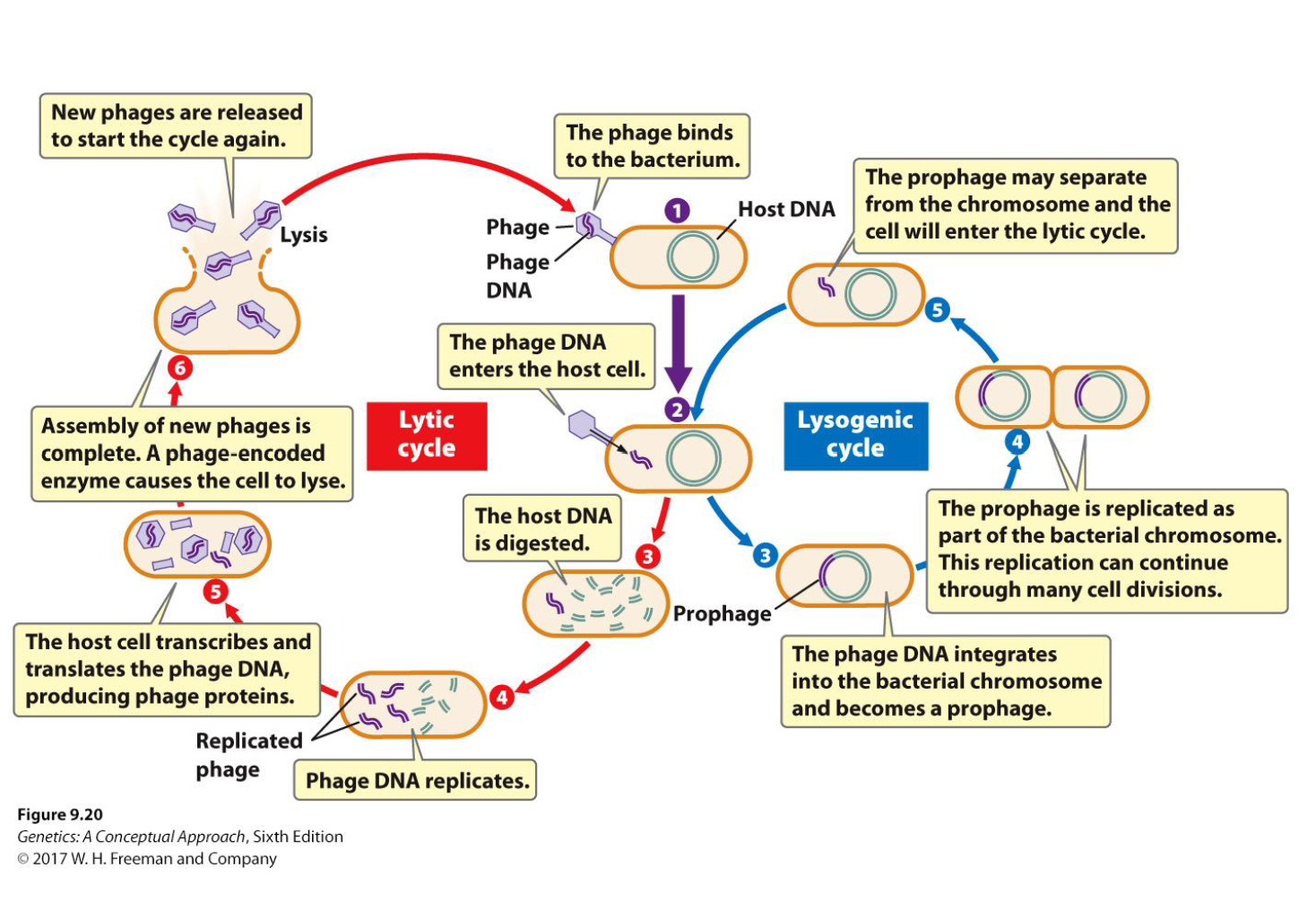
What is the main difference between the lytic and lysogenic cycles?
Lytic: Viral DNA replicates → new phages assemble → host cell lyses (dies).
Lysogenic: Viral DNA integrates into the bacterial chromosome as a prophage → replicates with the host without immediately killing it → may later enter the lytic cycle.

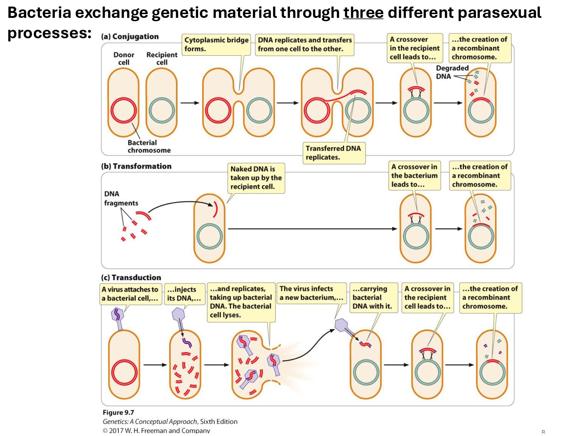
What are the three parasexual processes of bacterial genetic exchange?
Transformation: A bacterium takes in foreign DNA from the
environment and incorporates it into its genomeConjugation: A bacterium transfers genetic material (usually a plasmid) to another via a pilus
Transduction: Viruses (bacteriophages) transfer DNA from one bacterium to another.
These processes allow genetic recombination (cross-over) without meiosis.
Even without crossover, these mechanisms allow bacteria to adapt quickly and evolve resistance to antibiotics or survive in new environments
What is the difference between a bacterial chromosome, a plasmid, and an episome?
Chromosome: Main bacterial DNA/genome containing genes for basic functions (several million base pairs!).
Plasmid: Smaller DNA element that replicates independently of the bacterial chromosome and often carries accessory genes.
Episome: Large circular DNA that can integrate into the bacterial chromosome for replication or remain separate (e.g., F factor).
How can different mutations be observed in bacteria?
Colony colour and morphology
Nutritional mutants (can not metabolise certain sugars, like lactose or galactose)
Prototrophs and auxotrophs (can not make certain amino acids, these need to be added to the growth medium)
Antibiotic resistance
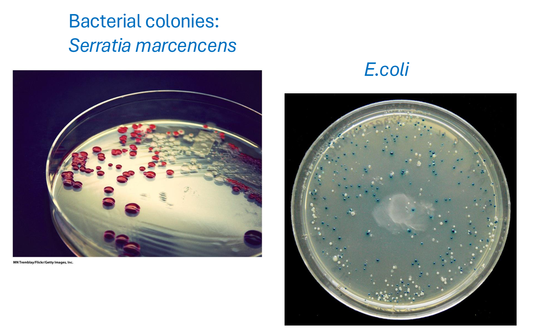
What is the difference between a prototroph and an auxotroph?
Prototroph: Can synthesize the nutrients needed to grow on minimal medium.
Auxotroph: Requires a particular nutrient to be supplied in the growth medium.
Example: A leucine auxotroph needs added leucine.
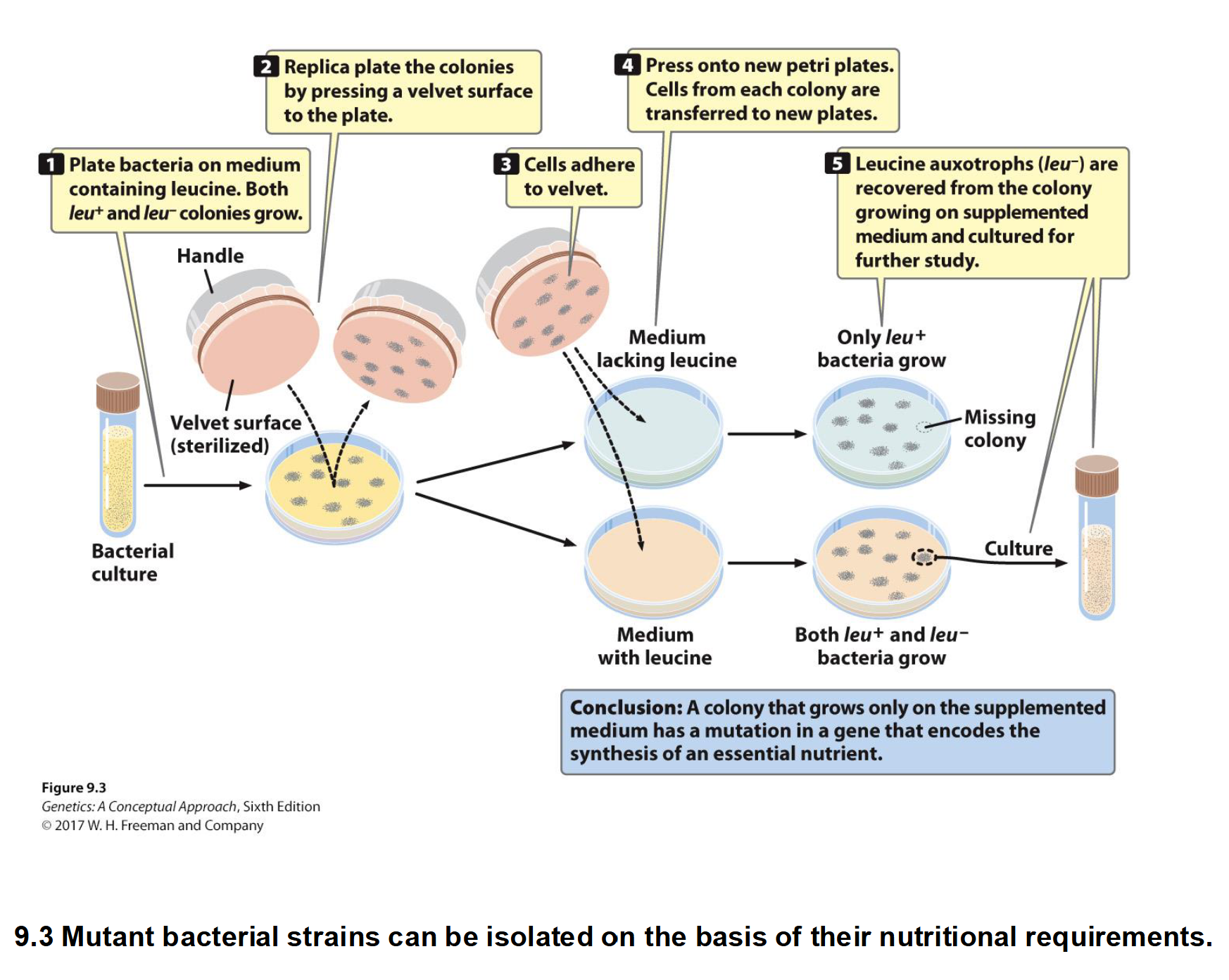
How can replica plating be used to identify bacterial auxotrophs?
Bacterial colonies are first grown on a master plate containing nutrients.
Cells are transferred to complete and minimal media while maintaining the same colony positions.
Prototrophs can grow on minimal medium because they synthesize the necessary nutrients.
Auxotrophs grow on complete medium but fail to grow on minimal medium if their required nutrient is absent.
Comparing colony positions identifies nutritional mutants.
Note: A mutation in a gene required for leucine synthesis makes the bacterium a leucine auxotroph, meaning it cannot produce its own leucine and can only grow on complete medium that supplies it.
How do transformation, conjugation, and transduction differ?
Transformation: Bacteria take up free DNA from their environment.
Conjugation: DNA transfers directly from a donor to a recipient through cell-to-cell contact.
Transduction: A bacteriophage (vector) transfers bacterial DNA between cells.
All three permit bacterial genetic exchange without meiosis.
Quick Contact Summary Table

What does the U-tube experiment demonstrate about bacterial conjugation?
Two bacterial strains are separated by a filter that prevents direct cell contact.
Genetic exchange by conjugation does not occur when the cells are separated.
Conclusion: Conjugation requires direct contact between donor and recipient bacteria.
No prototrophic bacteria were produced, meaning the strains did not exchange genetic material to restore their ability to grow on minimal medium.

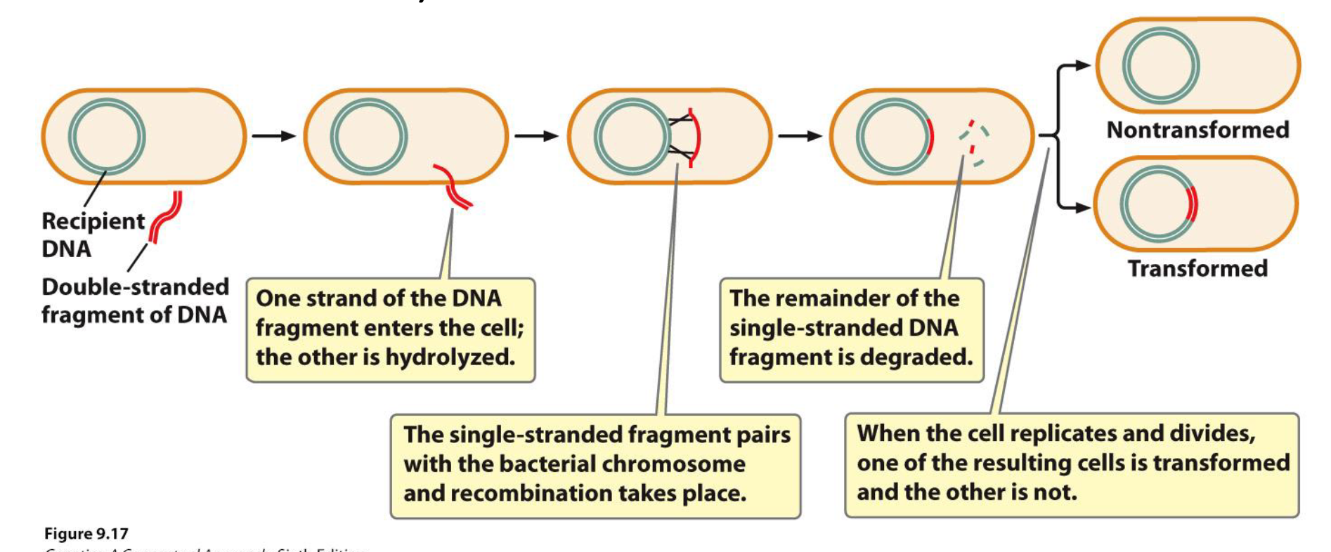
What is bacterial transformation, and what role do competent cells play?
Transformation: Uptake of free DNA from the environment by a bacterial cell.
Competent cells: Bacteria capable of taking up environmental DNA.
A bacterium must be competent to undergo transformation.
The acquired DNA may integrate into the bacterial chromosome.
If integrated, the DNA can be stably inherited by daughter cells and may produce a new phenotype.
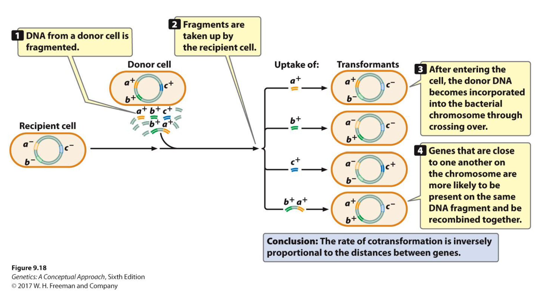
How can bacterial transformation be used to determine the distance between genes?
Only about 0.2–0.5% of the bacterial chromosome undergoes transformation.
Closely linked genes are more likely to be present on the same DNA fragment.
Higher cotransformation frequency = shorter distance between genes.

What is cotransformation, and how does its frequency relate to gene distance?
Cotransformation: Transfer of two or more genes into a recipient bacterium during the same transformation event.
High frequency: Genes are close together.
Low frequency: Genes are farther apart.
Cotransformation frequency is inversely proportional to gene distance.
What is the F factor, and how does it distinguish F⁻, F⁺, and Hfr cells? (Conjugation)
F factor: Fertility factor that mediates bacterial conjugation; it is an episome.
Can exist as an extra-chromosomal DNA OR it can insert into the bacterial chromosome and replicate as part of it
F⁻: No F factor.
F⁺: F factor exists independently of the chromosome.
Hfr: F factor is integrated into the bacterial chromosome.

How does bacterial conjugation occur between F⁺ and F⁻ cells?
F pili bring the F⁺ donor and F⁻ recipient together.
A conjugation bridge forms, allowing one strand of the F factor to transfer.
Rolling circle replication
The F factor is replicated in both cells.
Result: F⁺ × F⁻ → F⁺ + F⁺.

How does conjugation between Hfr and F⁻ cells occur?
The Hfr donor has its F factor integrated into its chromosome.
DNA transfer begins at the integrated F factor, allowing bacterial chromosomal genes to enter the F⁻ recipient.
Crossing over may incorporate donor genes into the recipient chromosome.
Result: The recipient may become genetically recombinant but usually remains F⁻.

What is the main difference between F⁺ × F⁻ and Hfr × F⁻ conjugation?
F⁺ × F⁻: Transfers the F factor, converting the recipient into F⁺.
Hfr × F⁻: Transfers bacterial chromosomal genes that may recombine with the recipient chromosome.
The Hfr recipient usually remains F⁻ because the entire F factor is rarely transferred.
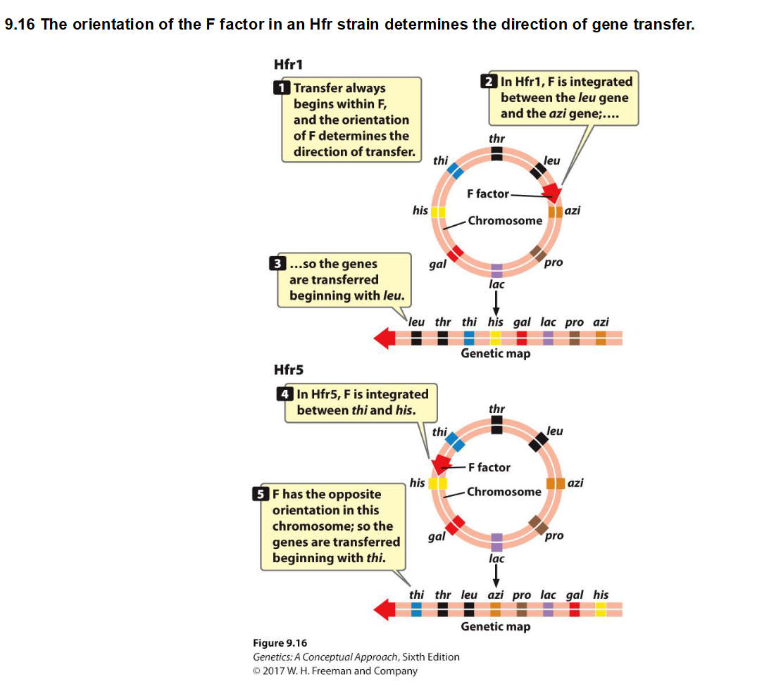
How do the location and orientation of the F factor affect gene transfer during Hfr conjugation?
In Hfr bacteria, the F factor is integrated into the bacterial chromosome, and DNA transfer begins at the F factor.
Location: Determines the starting point of gene transfer and which genes enter the recipient first.
Orientation: Determines the direction of transfer (clockwise or counterclockwise around the circular chromosome).
Example: Hfr1 transfers genes beginning with leu → thr → thi, while Hfr5 begins with thi → thr → leu.
Key point: The genes remain in the same positions on the chromosome; only their order of transfer changes.
How is interrupted conjugation used to map bacterial genes?
Conjugation is interrupted at different times to determine when genes enter the recipient.
Earlier entry: Genes closer to the transfer origin.
Later entry: Genes farther from the transfer origin.
1 minute = 1 map unit in the lecture example.
Review: What are the three types of bacterial genetic elements described in the lecture?
Chromosomal DNA: Contains essential genes for basic bacterial functions.
Plasmids: Smaller DNA elements providing additional traits not essential for survival.
Circular extrachromosomal DNA that replicates independently and often provides additional traits.
F factor: Specialized plasmid involved in conjugation that can integrate into the chromosome.
Are Episomes: Genetic elements that can replicate independently or integrate into the bacterial chromosome.
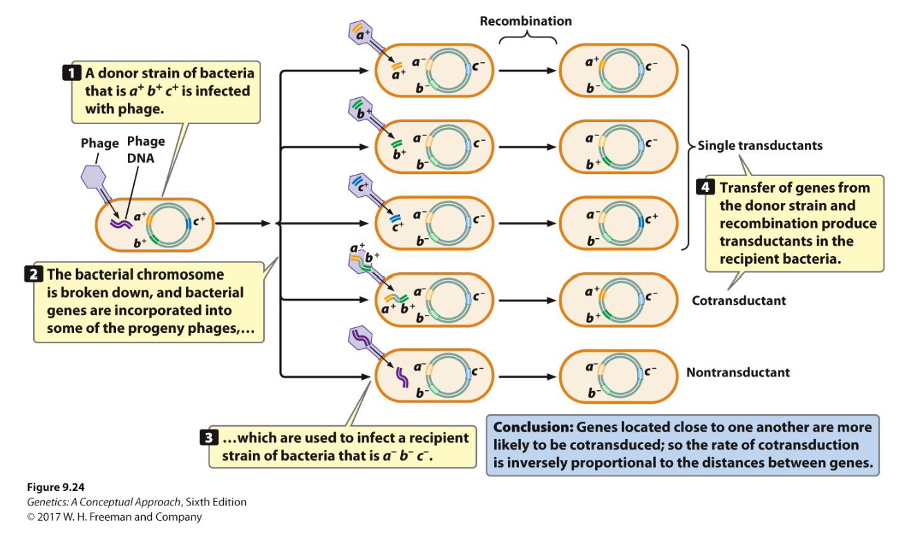
What is bacterial transduction, and how does it occur?
Transduction: Transfer of bacterial genes between cells by a bacteriophage.
A phage infects a donor bacterium, whose chromosome becomes fragmented.
Some bacterial DNA is accidentally packaged into new phages.
These phages infect recipient bacteria, transferring donor DNA that may recombine with the recipient chromosome.

What is cotransduction, and how is it used to determine gene distance?
Cotransduction: Transfer of two or more bacterial genes together by a bacteriophage.
Closely linked genes are more likely to be packaged into the same phage.
High cotransduction frequency: Genes are close together.
Low cotransduction frequency: Genes are farther apart.
Cotransduction frequency is inversely proportional to gene distance.
Review Slide
