module 9: mesodermal differentiation
 Call Kai
 Learn
 Practice Test
 Spaced Repetition
 Match
 Flashcards
 Knowt Play
1/22

There's no tags or description
Looks like no tags are added yet.
23 Terms
gastrulation (chickens)
blastodisk: initial divisions, always tilted-gravity determines orientation,
mesoblastic discoidel
cytokinesis furrow
part of meroblastic discoidel cleavage in chickens
subgerminal cavity
space between the yolk and the epiblast layer where the hypoblast develops
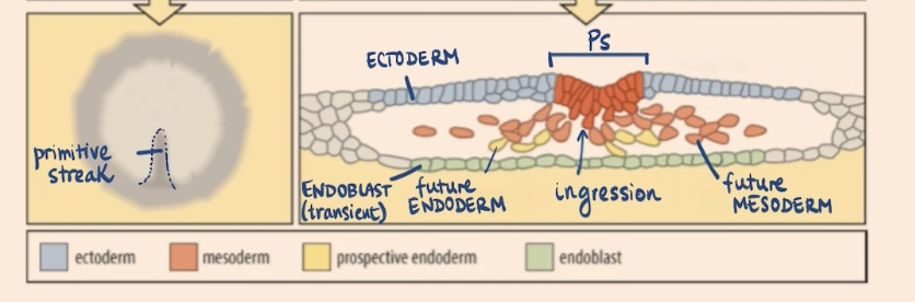
koller’s sickle
more dense area of cells underneath the area pellucida, occurs after laying, where the primitive streak grows from
area opaca
outer layer of the embryo
area pellucida
the inner area of the embryo
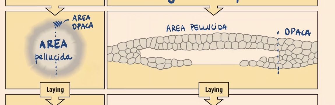
epiblast
ectoderm + mesoderm + endoderm
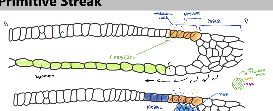
specification of the primitive streak stage 1
wnt8c is expressed around marginal zone
vg1 (TGFb ligand) expressed on posterior side marginal zone
cerberus expressed from hypoblast —> seqeusters and inhibits wnt8c and Vg1
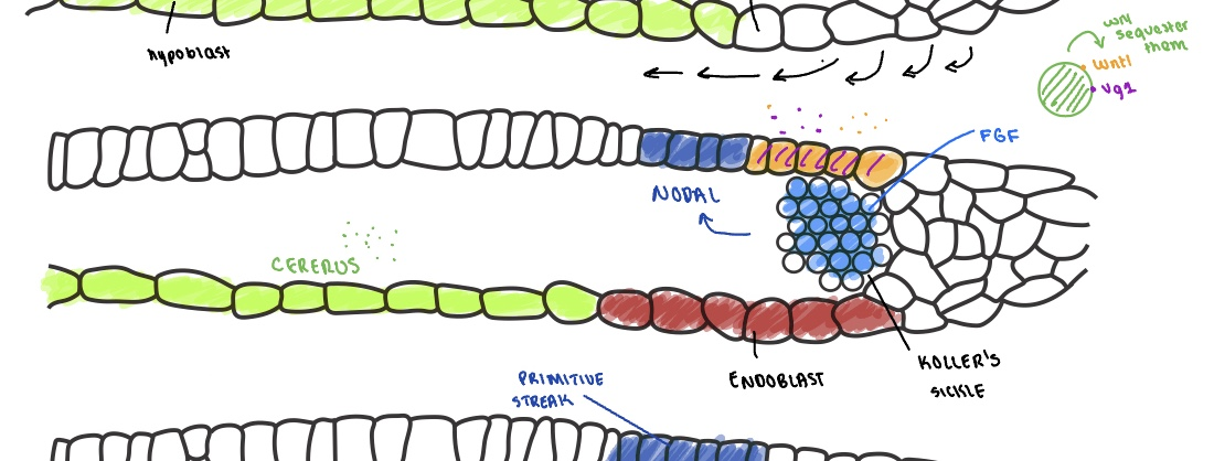
specification of primitive streak stage 2
proliferation of cells from area opaca form the endoblast next to the hypoblast —> pushes cerberus expression farther away allowing wnt8c and Vg1 activation
expressed wnt8c and Vg1 + FGF expressed from the growing Koller’s sickle —> activate NODAL (TGFb)
specification of primitive streak stage 3
NODAL expression defines the primitive streak
NODAL forms the organizer: organizes cells into mesodermal and endodermal cells (needed and sufficient for orientation of embryo)
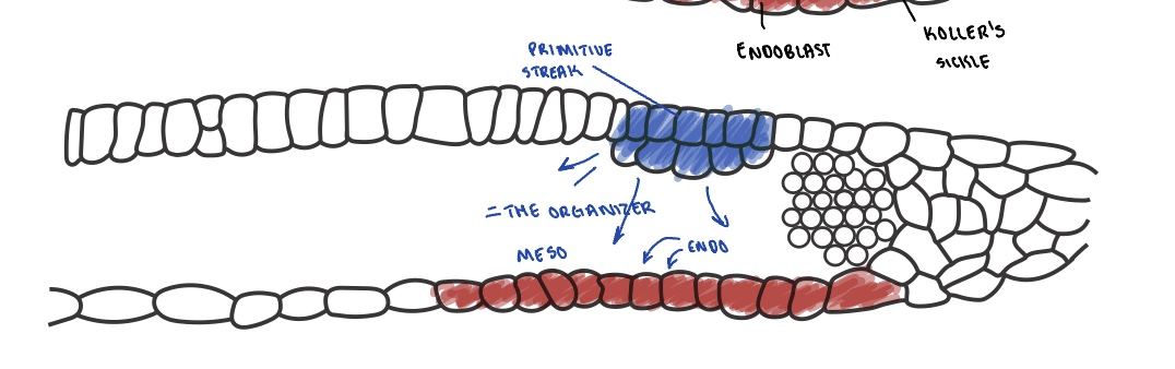
Hensen’s node
signaling center
node between notochord and primitive streak (AP)
the most anterior side of the primitive streak
primitive streak development
migration: streak moves from posterior to anterior side , mesoprogenitors enter through ingression
regression: entire streak regresses from the anterior back to the posterior side
the notochord is left behind — acts as a zipper that leaves behind the dorsal axis
AP border region: early
Anterior: DKK-1 (dickopf-1) = wnt agonist
posterior: Wnt
positive feedback loop: Wnt→activates Fgf8→activates wnt8c→ activates wnt
absence of wnt = head // wnt = posterior
AP border region: later
Anterior:
expressing retanoic acid (morphogen)
RA inhibits Fgf8
posterior:
Wnt majority → activates RALDH2 (releases retanoic acid)
wnt expresses fgf8 -→ inhibits RA

retinoic acid signaling
retinol + retinol binding proteins → retinol transporter into cell
retinol gets turned into retinaldehyde (via retinol dehydrogenase)
retinaldehyde into retinoic acid (via RALDH2)
retinoic acid 2 paths
RA enters neighbor cell → RA binds RAR (RA receptor) + RXR (reinoid receptors) -→ enter nucleus, bind RARE (RA response elements) → activates transcription
RA is degraded (via cyp26, cytochrome P450) → RAR+RXR bind RARE → but no transcription

mesodermal regionalization
axial mesoderm
paraxial mesoderm
intermedial mesoderm
lateral plate mesoderm

axial mesoderm
notochord (transient) —> intervertebral disks
paraxial mesoderm
dermomyota → skeletal elements, dermis, skeletal muscle
sclerotome → tendons, vertebral structures (proximal ribs), endothelial cells, aorta
intermedial mesoderm
kidneys, gonads
lateral plate mesoderm
somatic: pelvis, limb bones
splanchnic: vessels, blood, heart
blastocyst
blastula for mammals. trophoblast + inner cell mass (embryonic stem cells)
trophoblast
syncytiotrophoblast —> placenta, invade endometrium
cytotrophoblast
inner cell mass ICM
epiblast: embryo proper + mesoderm extra
hypoblast: primitive endoderm → yolk sac
