chapter 8 lab-muscular system
 Call Kai
 Learn
 Practice Test
 Spaced Repetition
 Match
 Flashcards
 Knowt Play
1/24

There's no tags or description
Looks like no tags are added yet.
25 Terms

label the figure


label the head


label the figure


rectus femoris
Origin (Where it starts): It originates on the pelvis by two heads:
Straight Head: Aligns at the anterior inferior iliac spine (AIIS) of the pelvis.
Reflected Head: Attaches just above the upper lip of the hip socket (acetabulum).
Insertion (Where it ends): It travels straight down the center of the thigh and inserts into the base of the patella (kneecap) via the quadriceps tendon. From there, the force travels through the patellar ligament to anchor onto the tibial tuberosity (the bony bump on your shin).
Hip Flexion: It helps lift the thigh up toward the torso. (For example, bringing your leg forward when taking a step, sprinting, or kicking a ball).
Knee Extension: It works alongside the other three quadriceps muscles to straighten the knee joint from a bent position. (For example, standing up from a chair, jumping, or the final snap phase of a kick).
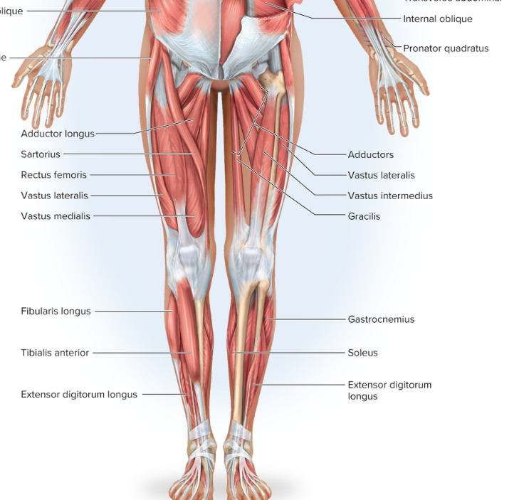

sartorius
The sartorius is the longest muscle in the human body. It is a long, thin, strap-like muscle that runs diagonally down the front and inside of the thigh, crossing both the hip and knee joints.
Origin (Where it starts): It originates on the outer flare of the pelvis at the anterior superior iliac spine (ASIS), which is the prominent bony point at the front of your hip.
Insertion (Where it ends): It snakes across the front of the thigh to insert onto the upper, inner (medial) surface of the tibia (shinbone). It attaches via a shared, fan-shaped tendon footprint called the pes anserinus (which translates to "goose's foot"), alongside the gracilis and semitendinosus muscles.
At the Hip Joint:
Flexion: Bends the hip, lifting the thigh up.
Abduction: Moves the thigh outward away from the midline.
External (Lateral) Rotation: Turns the thigh outward, pointing the knee to the side.
At the Knee Joint:
Flexion: Bends the knee joint.
Internal (Medial) Rotation: Slightly twists the lower leg inward when the knee is bent.

gluteus maximus
Origin (Where it starts): It originates across several structures at the back of the pelvis:
The outer surface of the ilium (the broad blade of the upper hip bone, behind the posterior gluteal line).
The posterior surfaces of the sacrum and coccyx (tailbone).
The sacrotuberous ligament (a strong ligament connecting the sacrum to the pelvis).
Insertion (Where it ends): The muscle fibers run diagonally downward and outward, dividing into two attachment points:
Upper/Superficial Fibers: Insert directly into the iliotibial (IT) band, a thick band of connective tissue running down the outside of the thigh.
Lower/Deep Fibers: Insert onto the gluteal tuberosity, a rough, raised ridge on the upper back portion of the femur (thigh bone).
Hip Extension: This is its main action. It forcefully straightens the hip joint, pulling the thigh backward. It is heavily recruited when generating power against gravity—such as standing up from a deep squat, running, climbing stairs, or leaping.
External (Lateral) Rotation: It twists the thigh outward, turning the knee and toes away from the midline of the body.
Hip Abduction: Its upper fibers assist in pulling the thigh outward to the side.
Trunk Stabilization: When your feet are firmly planted on the ground, it exerts a backward pull on the pelvis, stabilizing your torso and preventing you from tipping forward at the waist while standing or walking.

biceps femoris long head
Origin (Where it starts): The long head originates from the lower, back portion of the pelvis at the ischial tuberosity. This is commonly referred to as the "sit bone" because it is the bony bump you sit directly on. It shares this origin via a common tendon with the semitendinosus muscle.
Insertion (Where it ends): The muscle fibers run down the outer (lateral) backside of the thigh. They merge with the short head to form a thick tendon that inserts onto the lateral side of the head of the fibula (the smaller shin bone on the outside of your lower leg). A small portion of the tendon also blends into the adjacent lateral condyle of the tibia.
Hip Extension: It forcefully pulls the thigh backward relative to the pelvis. This action is heavily used when walking, running, sprinting, or coming up out of a squat.
Knee Flexion: It bends the knee joint, pulling the heel up toward the buttocks.
External Rotation: It helps twist the lower limb outward.
When the hip is extended, it assists in externally rotating the thigh.
When the knee is bent (flexed), it externally rotates the lower leg (tibia)

semimembranosus
The semimembranosus is the deepest of the three muscles that make up the hamstring muscle group on the back of the thigh
Origin (Where it starts): It originates from the lower, back portion of the pelvis at the ischial tuberosity (the "sit bone"). Its origin point sits slightly higher and further outside than the shared origin of the semitendinosus and the long head of the biceps femoris.
Insertion (Where it ends): Its fibers run straight down the inner backside of the thigh and insert via a thick tendon onto the horizontal groove on the back of the medial condyle of the tibia (the inner upper part of the main shinbone).
Hip Extension: It pulls the thigh backward relative to the pelvis. This action is crucial for driving the body forward during walking, running, and climbing stairs.
Knee Flexion: It bends the knee joint, pulling the heel up toward the glutes.
Internal Rotation: It helps twist the lower limb inward.

semitendinosus
The semitendinosus is one of the three muscles that make up the hamstring group on the back of the thigh.
Origin (Where it starts): It originates from the lower, back portion of the pelvis at the ischial tuberosity (the "sit bone"). It shares this exact origin point via a common tendon with the long head of the biceps femoris.
Insertion (Where it ends): It travels down the inner back of the thigh and winds around to insert onto the upper, inner (medial) surface of the shaft of the tibia (shinbone). It anchors here via a shared, fan-shaped tendon footprint called the pes anserinus ("goose's foot"), sharing this site with the sartorius and gracilis muscles.
Movements
Because the semitendinosus crosses both the hip and knee joints, it acts as a two-joint lever system:
Hip Extension: It forcefully pulls the thigh backward relative to the pelvis. This action is critical for forward propulsion during walking, running, sprinting, and driving up from a squat.
Knee Flexion: It bends the knee joint, drawing the heel up toward the buttocks.
Internal Rotation:

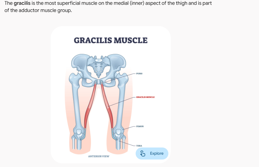
gracilis
Origin:Anterior pelvis: Inferior margin of the pubic symphysis, the body of the pubis, the inferior pubic ramus, and the adjacent ramus of the ischium (ischiopubic ramus). [1, 2, 3]
Insertion
Proximal tibia: The medial surface of the upper tibial shaft (just inferior to the medial condyle), via a shared tendon known as the pes anserinus (alongside the sartorius and semitendinosus muscles).
Hip Joint: Adducts the thigh (draws it inward) and weakly assists in thigh flexion.
Knee Joint: Flexes the leg at the knee and helps medially (internally) rot
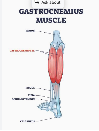
gastrocnemius
Origin
Medial head: The posterior surface of the distal femur, specifically just superior to the medial condyle.
Lateral head: The upper, lateral surface of the lateral condyle of the femur.
Insertion
Calcaneus (heel bone): The two muscle heads converge to form a broad, flat belly that tapers into the Achilles tendon (calcaneal tendon), which inserts into the middle part of the posterior surface of the calcaneus.
Movements / Actions
Ankle Joint: Performs plantarflexion of the foot (pushing down on the gas pedal, raising onto your tiptoes).
Knee Joint: Assists in flexion of the leg
soleus
Origin
Fibula: The posterior surface of the head and upper proximal third of the shaft of the fibula.
Tibia: The soleal line and the middle third of the medial border of the tibia.
Tendinous arch: A fibrous arch stretching between the tibia and fibula.
Insertion
Calcaneus (heel bone): The muscle fibers travel downward and fuse with the tendon of the gastrocnemius to form the Achilles tendon (calcaneal tendon), which inserts onto the posterior surface of the calcaneus.
Movements / Actions
Ankle Joint: Performs plantarflexion of the foot (pushing down on the gas pedal, walking, running, and dancing).
Postural Stability: Crucial for maintaining static posture while standing, as it constantly fires to pull back against gravity and keep the body balanced over the feet.

Posterior Thigh (Hamstrings):
The Biceps femoris, long head, Biceps femoris, short head, Semimembranosus, and Semitendinosus
Anterior Thigh (Quadriceps & Superficial):
Rectus femoris (a quadriceps muscle) and Sartorius.
Medial Thigh (Adductors)
Gracilis
Gluteal Region (Posterior Hip):
GGluteus maximus
Posterior Lower Leg (Calf):
Gastrocnemius and Soleus, which together form the triceps surae.

Quadriceps Tendon
Quadriceps Tendon
The major extensor of the knee, the quadriceps, has four heads: the rectus femoris) the vastus
lateralis, the vastus medialis and the vastus intermedius, They join in a common tendon, the
quadriceps tendon, that extends over the kneecap and becomes the patellar ligament. It inserts
on the tibial tuberosity.


quadriceps tendon

lliotibial Band
lliotibial Band
The iliotibial band is a connective tissue band that the tendons of the gluteus maximus and oth-
er muscles join. The iliotibial band inserts on the lateral condyle of the tibia.
.
Calcaneal Tendon
The calcaneal tendon, or Achilles tendon, attaches three posterior muscles to the calca-
neus bone of the foot. Look at Table 8.1 and name the two muscles you will be creating
that utilize this tendon- gastrocnemius and soleus
where is the insertion of the sartorius?
proximal medial surface of the tibia. [1, 2]
You made two muscles in the posterior, lower leg. Name the two muscles and note which
muscle was the deep muscle.
gastrocnemius and soleus
What is the origin of the rectus femoris?
dual origin consisting of two separate heads that arise from the hip bone (coxal bone):
Straight Head (Anterior Head): Originates from the anterior inferior iliac spine (AIIS) of the ilium.
Reflected Head (Posterior Head): Originates from a groove just superior to the acetabulum (the hip joint socket).
What is the action of the biceps femoris?
Knee Flexion: It bends the knee, bringing the heel toward the glutes.
Knee Lateral Rotation: It externally rotates the lower leg at the knee joint when the knee is already partially bent (flexed).
Hip Extension: Its long head helps extend the hip, pulling the thigh backward (such as during walking or running).
Hip Lateral Rotation: