Anatomy Exam 2 (Heart and Lungs)
 Call Kai
 Learn
 Practice Test
 Spaced Repetition
 Match
 Flashcards
 Knowt Play
1/123

Earn XP
Description and Tags
Heart and Lungs Slides 1, 2, and 3
124 Terms

Heart Anatomy
Superior Vena Cava
Right Atrium
Right Ventricle
Aorta
Pulmonary Trunk
Left Atrium
Left ventricle
Coronary Vessels

Valves of the heart
Semilunar valves
tricuspid valve (right ventricle)
mitral valve
bicuspid valve (left ventricle)
Where does blood returning from the body enter?
the right atrium, then flows through the tricuspid valve into the right ventricle, then pumped through the pulmonary artery to the lungs to pick up oxygen.
How does blood leave the heart to the rest of the body?
oxygen-rich blood returns from the lungs into the left atrium, then passes through the mitral valve into the left ventricle, and then is forcefully pumped through the aorta to distribute oxygen to the rest of the body.
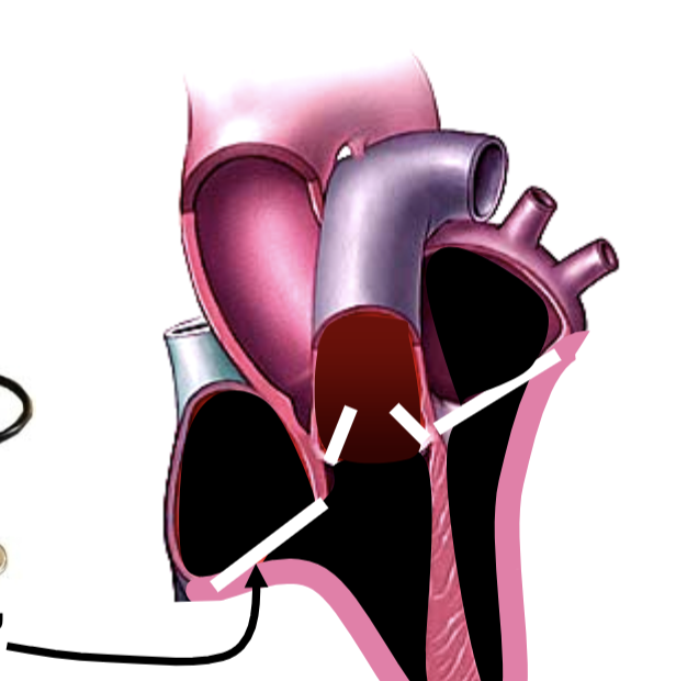
LUB
Atrioventricular valves close

DUP
Aortic and pulmonary valves close

Systole (pumping)
Isovolumetric contraction: atria relaxed, ventricles contract, AV valves closed.
Ejection: atria relaxed, ventricles contract

Diastole (filling)
Isovolumetric relaxation: atria relaxed, ventricles relaxed, semilunar valves closed
Rapid filling: atria relaxed, ventricles relaxed
Atrial contraction: atria contract, ventricles relaxed
Fetal Circulation
Comes from mother’s blood, placenta
Ductus arteriosus
Foramen ovale
Ductus venosus
bypasses lungs

Sinoatrial (SA) Node
Pacemaker
located in the wall of the right atrium
spontaneously generates electrical impulses that set the pace for the heartbeat

Atrioventricular (AV) node
impulse spreads across both atria, causing them to contract and push blood into the ventricles
the signal converges at the AV node, which introduces a brief delay to ensure the ventricles have enough time to fill completely

Atrioventricular Bundle (Bundle of His)
Emerges from the AV node
the signal enters the bundle of His located within the interventricular septum

Right and left bundle branches
The bundle splits into right and left bundle branches, racing rapidly down either side of the septum toward the apex of the heart.

Purkinje Fibers
At the apex, the signal fans out into specialized Purkinje fibers that course upward through the ventricular myocardium. This triggers a powerful, coordinated contraction from the bottom up, efficiently ejecting blood into the aorta and pulmonary artery.

P wave
initial small upward bump
represents the depolarization of the atria
As the electrical impulse travels from the SA node across both atria, it causes them to depolarize and contract, pushing blood down into the ventricles.

QRS complex
Sharp, positive spike
represents the depolarization of the ventricles
ventricles have larger muscle mass than atria, so this electrical sweep creates a large deflection
signal triggers powerful ventricular contraction
atria get repolarized, but that signal is hidden in this large complex

T wave
final, smooth wave
represents the repolarization of the ventricles
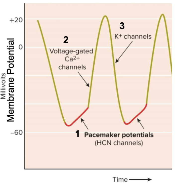
Pacemaker Potential: Membrane continuously polarizes due to what channels for what?
HCN channels for Na+
H (Hyperpolarization) is open at what polarization?
low!
CN (Cyclic Nucleotide) is open more under stimulation of ________ __________ by _________ and ________. what response?
beta-adrenergic receptors
epinephrine and norepinephrine
sympathetic response
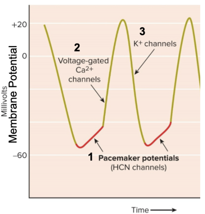
Ca+ channels open at ______, Ca+ floods _____
-40 mV
in

K+ channels open, K+ floods ____
out
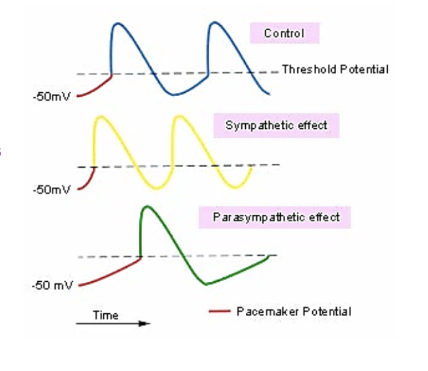
Parasympathetic vagus nerve ______ rate
decreases
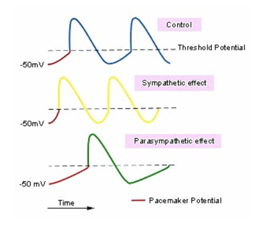
Sympathetic cardiac nerves _____ rate and force
increase

Myocardial action potentials spread from ______ to _______, sweeping across the ______ ________.
cell to cell
entire heart
Cardiac Output (CO) = what?
Heart Rate (HR) * Stroke Volume (SV)
Frank-Starling Law
Increased volume = increased stretch = increased force of contraction
increased preload volume means increased resistance in after load, and therefore increased force of attraction is required to overcome resistance
In arteries, blood can flow ______ ways.
both
In veins, blood can flow ____ way.
one

Artery has a ______, while vein has a ______
Lumen
Valve
The right lung has _____ lobes
3
The left lung has ____ lobes
2

Bronchiole anatomy
Trachea
primary bronchus
secondary (lobar) bronchus
tertiary (segmental) bronchus
bronchiole
terminal bronchiole
respiratory bronchiole
Alveoli are covered with ______
capillaries
Alveolar wall is coated with ______
surfectant

Inflow of oxygen depleted blood is in the _______
arteriole

Outflow of oxygenated blood is in the ________
venule
Liters of O2 per Hour = what?
Surface Area * Change in Partial Pressure / Distance * Resistance
Alveolar wall is _____ ______ and ________ thick!
one cell
0.001 mm
Alveolar surface area is _____ square meters
75 (tennis court size)
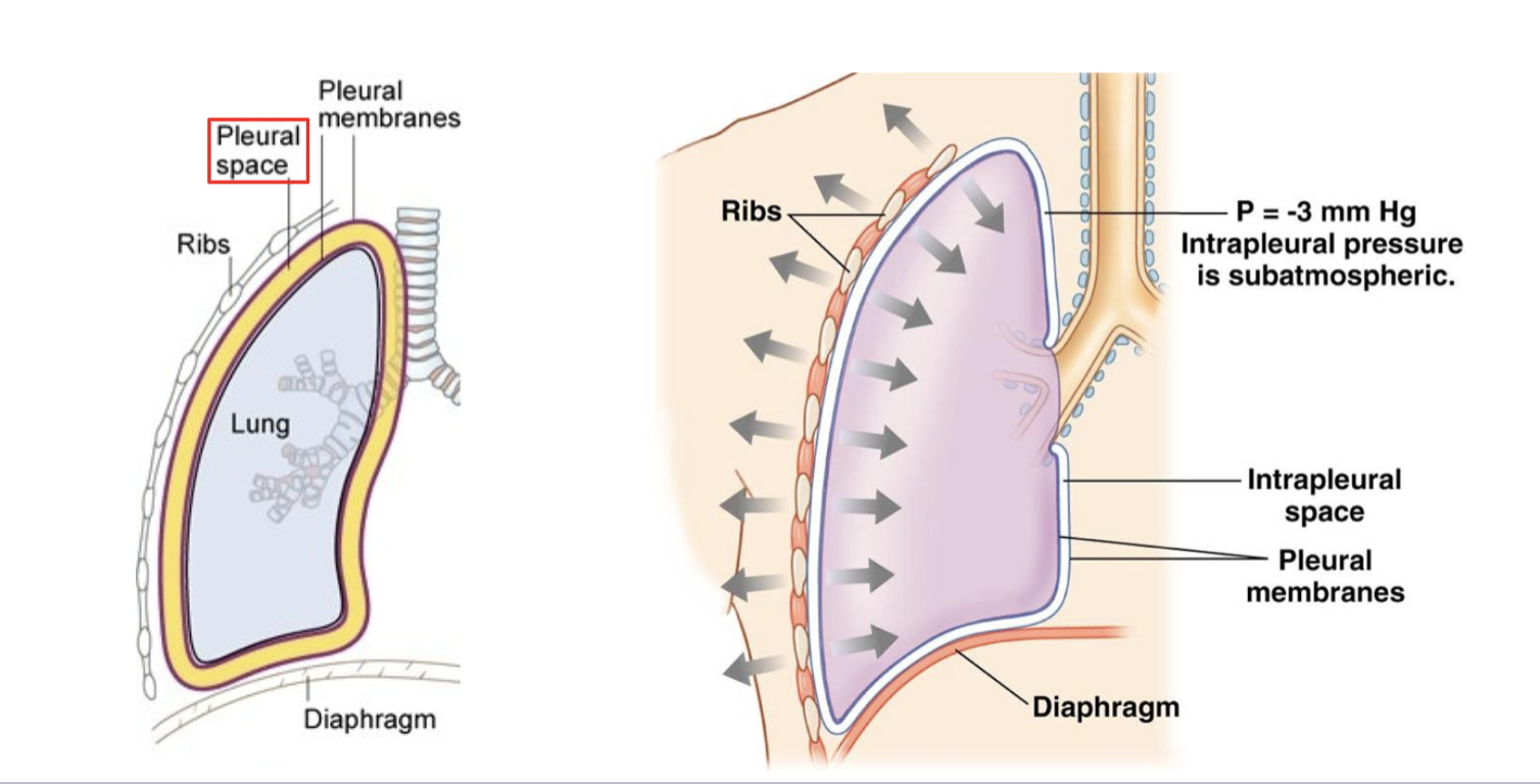
Thoracic Cavity Pressure
-3mm Hg (intrapleural pressure is subatmospheric)
Thorax and Lung expansion creates ______ pressure. Sucks ____ in.
negative
air
Diaphragm is? What nerve innervation?
the primary muscle of breathing
phrenic

Ribcage, what mechanisms?
Pump Handle and Bucket Handle mechanisms
H2O: Inspired Air?
Variable
H2O: Alveolar Air?
47 mmHg
CO2: Inspired Air?
000.3 mmHg
CO2: Alveolar Air?
40 mmHg
O2: Inspired Air?
159 mmHg
O2: Alveolar Air?
10 mmHg
N2: Inspired Air?
601 mmHg
N2: Alveolar Air?
568 mmHg
Total Pressure (Inspired and Alveolar Air)?
760 mmHg
Atmosphere
159 atm
Airway
149 atm
Alveolus
104 atm
Pulmonary Capillary
100 atm
Tissue and Vein
40 atm
In the alveoli, oxygen goes ______ and CO2 goes _____
in
out
In the Alveoli capillaries, PO2 is _____ and PCO2 is ______
104
40
As we transition to pulmonary vein, PO2 is _____ and PCO2 is _____
100
40
(pulmonary vein)
As we go past left heart and into systemic arteries, PO2 is ______ and PCO2 is ______
100
40
(systemic arteries, same as pulmonary vein)
In body cells, PO2 is _____ and PCO2 is _____
40
46
(body cells)
When we get to body cells, oxygen goes ____ and CO2 goes _____
out
in
As we go back to alveoli, PO2 is ______ and PCO2 is ______
40
46
(stays the same as body cell)
PO2 of blood only reflects O2 dissolved in ______
plasma (much more in red blood cells)
Plasma is?
mostly water, proteins like Albumin, Globulins, and Fibrinogen (which is an important clotting factor produced by the liver)
Leukocytes
immune system
Erythrocytes
Red blood cells, carries oxygen
Plasma is what percent of blood?
55%
Buffy coat (leukocytes, platelets) is what percent of blood?
less than 1%
Erythrocytes is what percent of blood?
45%
What is Hematopoiesis?
Making blood cells!
How many blood cells are made per day?
500 billion!
Where are blood cells made?
bone marrow! (liver in fetus)
Red blood cell homeostasis is a ______ feedback loop.
negative.
Kidneys and liver sense low oxygen and produce what hormone (red blood cell homeostasis)?
Erythropoietin (EPO), stimulates red blood cell production in kidneys in adults. liver in fetus
Oxygen levels ______, EPO _______
rise
inhibited
How many hemoglobin per red blood cell, and how many O2 molecules can it carry?
280 million per red blood cell
carries 0 to 4 O2 molecules
Heme group with O2 is…
oxyhemoglobin
heme group without O2 is…
deoxyhemoglobin
Amount of O2 unloaded to tissues


CO2 in 3 forms in the blood:
Dissolved CO2 in plasma
carbaminohemoglobin
bicarbonate
Inside red blood cell…
CO2 + H2O → w/ carbonic anhydrase, becomes H2CO3 → H+ (binds to hemoglobin) + HCO3-, which leaves RBC, Cl enters, chloride shift
Dissolved CO2 is
acidic
bicarbonate is
alkaline (basic)
inadequate ventilation leads to…
decreased pH (acidosis). regulate pH balance (7.4). kidneys help as well, producing bicarbonate and excreteing H+

How does brain use chemoreception to sense blood pH?
with carotid body and aortic bodies in aortic arch
Bohr effect:
O2 affinity changes with pH, makes it easier to load oxygen in the lungs and unload at peripheral tissues
Higher affinity
O2 loading (loads in lungs)
pH 7.6 (lung)
higher pH (more basic)
lower affinity
pH 7.2
O2 unloading (leaves to tissues)
lower pH (more acidic)
Temperature effect
O2 affinity decreases at higher temperature
makes it easier to unload oxygen into exercising muscle

Myoglobin
left-shifted O2 curve
higher affinity for O2

Fetal hemoglobin
left-shifted O2 curve
higher affinity for O2
pulls oxygen from mother’s red blood cells
Evolution as hunter gatherers means…
selection for increased physical activity and increased energy metabolism (kcal/day). Need increased cardiac output!
Human left ventricle is more ____ ______. We have more ______ muscle, more _____, and more efficiency.
Endurance adapted
compact
twist (to “wring” out blood")
What is the most common blood type allele in humans?
O type
Earliest hominins had what blood type?
A type
Blood type B evolved how many years ago?
3.5 mya
Blood type O evolved how many years ago?
1.1 mya