BLOOD AND HEART FLASHCARDS
 Call Kai
 Learn
 Practice Test
 Spaced Repetition
 Match
 Flashcards
 Knowt Play
1/35

There's no tags or description
Looks like no tags are added yet.
36 Terms

Leukocytes
Also known as white blood cells, leukocytes are a key component of the immune system, helping to defend the body against infections and foreign substances.

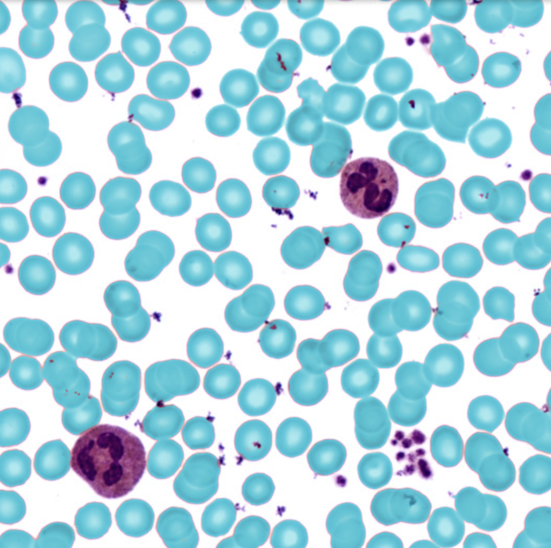
Erythrocytes
Also known as red blood cells, erythrocytes are responsible for transporting oxygen from the lungs to the body's tissues and returning carbon dioxide from the tissues back to the lungs.


Thrombocytes
Also known as platelets, thrombocytes are small cell fragments that play a crucial role in blood clotting and wound healing.


Apex
The apex is the pointed tip of the heart, located at the bottom of the heart chamber. It is oriented towards the left side of the body and plays a crucial role in the heart's overall structure.


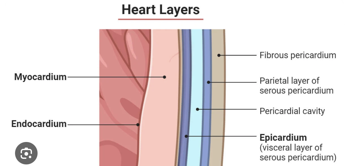
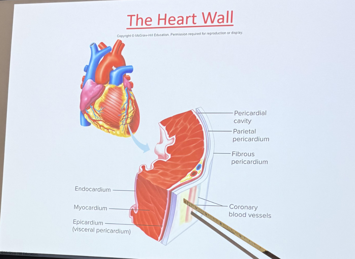
Epicardium
The epicardium is the outermost layer of the heart wall, consisting of a thin layer of connective tissue and fat that protects the heart. It is also part of the pericardium.
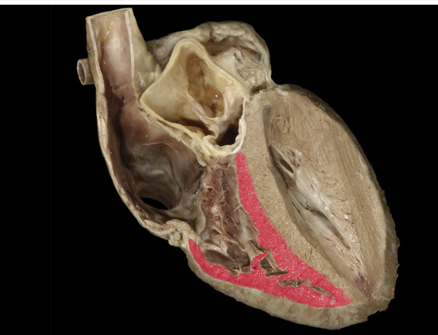
Myocardium
The myocardium is the middle layer of the heart wall, composed of cardiac muscle tissue responsible for contracting and pumping blood throughout the heart.

Endocardium
The endocardium is the innermost layer of the heart wall, composed of a thin layer of endothelial cells that line the heart chambers and valves, providing a smooth surface for blood flow.


Right and Left Atria ( Atrium)
The right and left atria are the two upper chambers of the heart that receive blood from the body and lungs, respectively. They play a crucial role in the heart's function by collecting blood before it is pumped into the ventricles.

Right and Left Ventricles
The right and left ventricles are the two lower chambers of the heart responsible for pumping blood out to the lungs and the rest of the body, respectively. They receive blood from the atria and contract to propel blood into the pulmonary and systemic circulations.

Interventricular septum
The interventricular septum is the muscular wall that separates the right and left ventricles of the heart. It plays an essential role in maintaining proper blood flow and pressure within the heart's chambers.

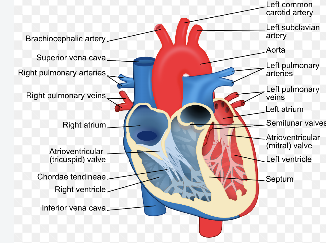
Chordae tendineae
The chordae tendineae are fibrous cords that connect the papillary muscles to the atrioventricular valves in the heart. They help prevent the valves from inverting during ventricular contraction.
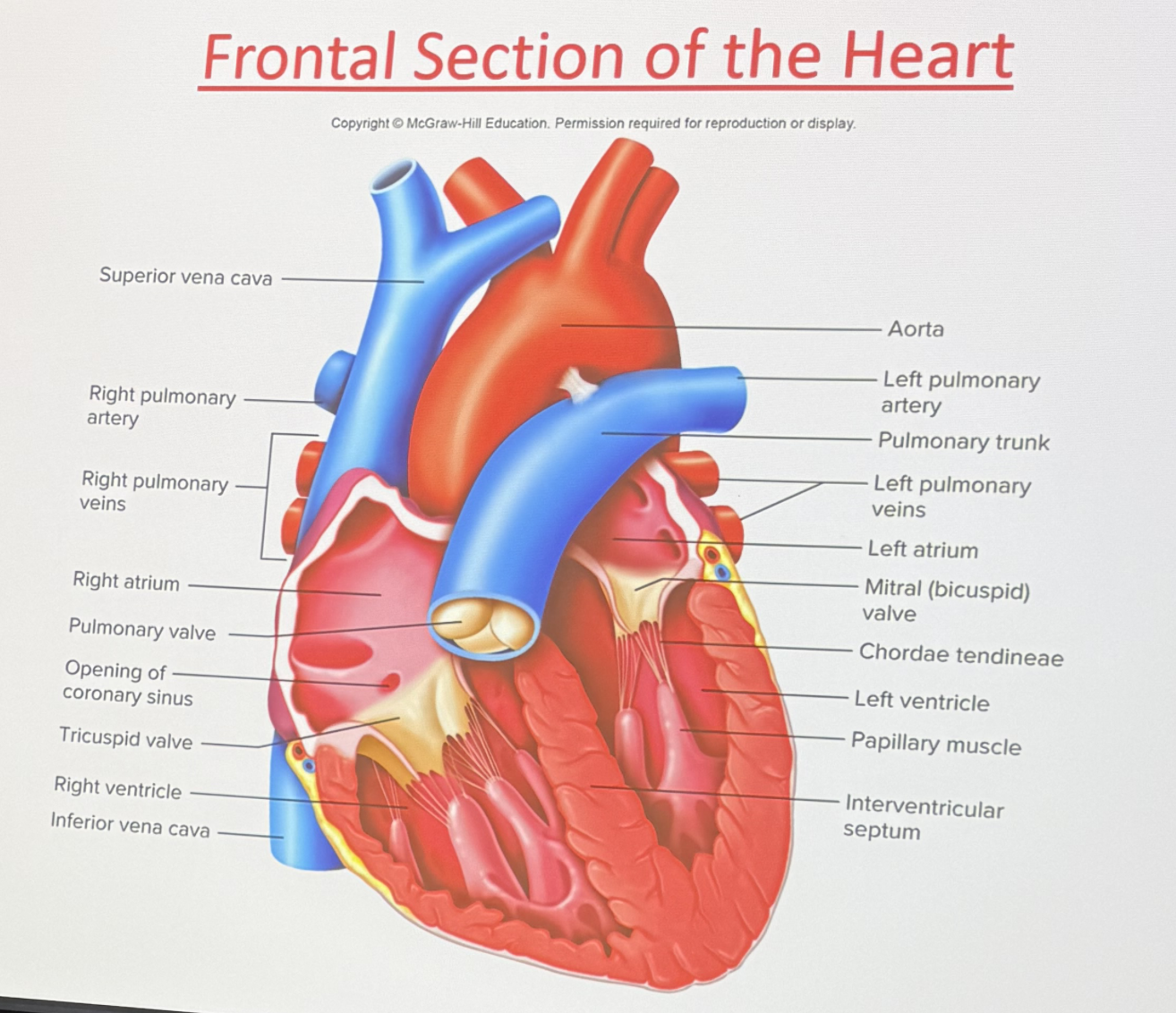
Papillary Muscles
Muscles located in the ventricles of the heart that attach to the chordae tendineae. They contract to help keep the atrioventricular valves closed during ventricular contraction.
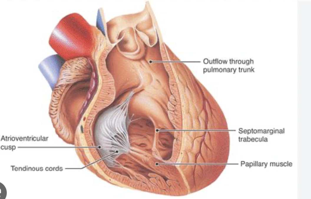

Right Atrioventricular ( Tricuspid) Valve
The right atrioventricular (tricuspid) valve is a valve located between the right atrium and right ventricle of the heart. It prevents blood from flowing back into the atrium during ventricular contraction.


Pulmonary Valve
The pulmonary valve is a heart valve located between the right ventricle and the pulmonary artery. It prevents the backflow of blood into the ventricles after it has been pumped to the lungs for oxygenation.


Left Atrioventricular ( Bicuspid/mitral) valve
The left atrioventricular (bicuspid or mitral) valve is a valve situated between the left atrium and left ventricle of the heart. It prevents blood from flowing back into the atrium during ventricular contraction.


aortic valve
The aortic valve is one of the four main heart valves. It sits between the left ventricle (the heart's primary pumping chamber) and the aorta (the body's main artery). [1]It prevents the backflow of blood into the ventricle after it has been ejected into the aorta.


ascending aorta
The ascending aorta is the initial section of the aorta that rises from the left ventricle of the heart. It carries oxygenated blood away from the heart to the upper parts of the body.

right and left coronary arteries
The right and left coronary arteries are blood vessels that arise from the base of the aorta and supply oxygenated blood to the heart muscle. The right coronary artery primarily nourishes the right side of the heart, while the left coronary artery supplies the left side.


aortic arch
The aortic arch is the curved section of the aorta that connects the ascending aorta to the descending aorta. It gives rise to major arteries that supply blood to the head, neck, and arms.


brachiocephalic artery
The brachiocephalic artery is a major blood vessel that branches from the aortic arch and divides into the right common carotid artery and right subclavian artery, supplying blood to the right arm and the head.

right subclavian artery
The right subclavian artery is a major blood vessel that branches off from the brachiocephalic artery and supplies blood to the right arm and portions of the thoracic cavity.


right common carotid artery
The right common carotid artery is a major blood vessel that branches off from the brachiocephalic artery, supplying blood to the right side of the head and neck.
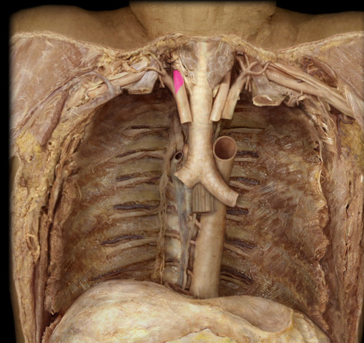

left common carotid artery
The left common carotid artery is a major blood vessel that branches directly from the aortic arch, supplying blood to the left side of the head and neck.

left subclavian artery
The left subclavian artery is a major blood vessel that branches directly from the aortic arch, supplying blood to the left arm and portions of the thoracic cavity.

pulmonary trunk
The pulmonary trunk is a large blood vessel that carries deoxygenated blood from the right ventricle of the heart to the lungs for oxygenation.
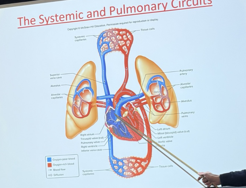
right and left pulmonary arteries
The right and left pulmonary arteries are blood vessels that branch from the pulmonary trunk, carrying deoxygenated blood from the heart to each lung for oxygenation.
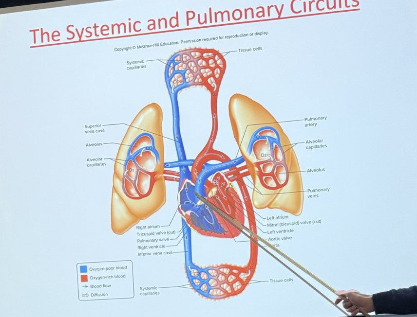
right and left pulmonary veins
The right and left pulmonary veins are blood vessels that transport oxygenated blood from the lungs back to the left atrium of the heart.

superior vena cava
The superior vena cava is a large vein that carries deoxygenated blood from the upper body to the right atrium of the heart.


inferior vena cava
A large vein that carries deoxygenated blood from the lower body to the right atrium of the heart.

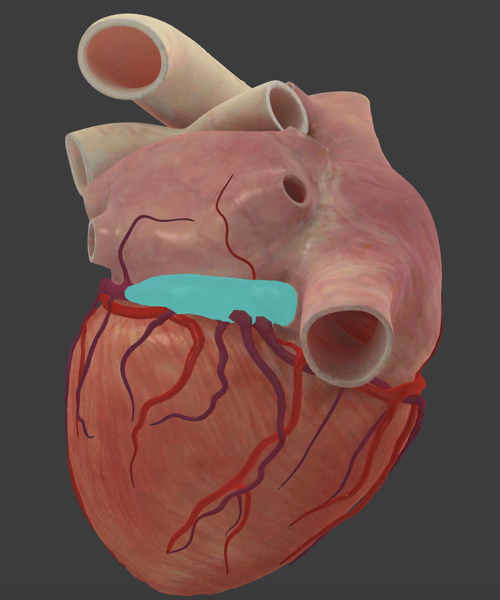
coronary sinus
The coronary sinus is a large vein that collects deoxygenated blood from the heart muscle and drains it into the right atrium.

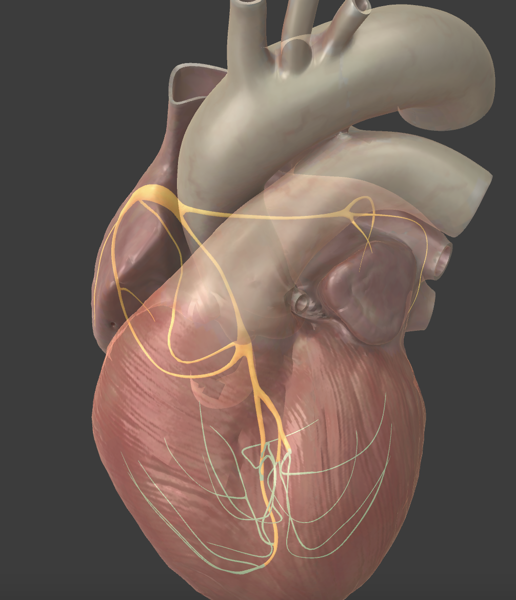
Intrinsic Conduction System
The intrinsic conduction system is a network of specialized cardiac muscle cells that generates and conducts electrical impulses to regulate the heartbeat and maintain the heart's rhythmic contractions. It consists of the sinoatrial node, atrioventricular node, bundle of His, and Purkinje fibers.

Atrioventricular bundle (Bundle of HIS)
A collection of heart muscle fibers that conducts electrical impulses from the atrioventricular node to the ventricles, facilitating coordinated contraction.

left and right bundle branches
The left and right bundle branches are pathways that conduct electrical impulses from the atrioventricular bundle (Bundle of His) to the respective ventricles, ensuring synchronized contraction of the heart.
The bundle branches are a part of the electrical system of the heart.

Purkinje fibers
The Purkinje fibers are specialized cardiac muscle fibers that spread throughout the ventricles and transmit electrical impulses from the bundle branches, promoting coordinated ventricular contraction.

Cardiac Muscle
The cardiac muscle is a type of striated muscle that is exclusively found in the heart. It is responsible for the contraction and pumping of blood throughout the circulatory system.