Fall DS3 Pathology Exam 1 Photos
 Call Kai
 Learn
Learn Practice Test
 Spaced Repetition
 Match
Match Flashcards
 Knowt Play
1/331

Earn XP
Description and Tags
Working on 9-13, 1-8 complete
Name | Mastery | Learn | Test | Matching | Spaced | Call with Kai | Chat |
|---|
No analytics yet
Send a link to your students to track their progress
332 Terms

Identify the finding shown.
Incomplete Cleft Lip (no nostril involvement)

Identify the finding shown.
Complete Cleft Lip (extends into nostril)
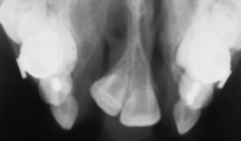
What do you see and what caused it?
Missing lateral incisors due to a complete cleft lip (complete cleft lip may involve alveolus)

What do you see and what caused it?
Supernumerary teeth due to complete cleft lip (complete cleft lip may involve alveolus)

Identify the finding shown.
Combined Cleft Palate and Cleft Lip

Identify the finding shown.
Cleft palate (no cleft lip)

Identify the finding shown.
Bifid Uvula (Cleft of uvula only)
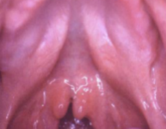
Identify the finding shown.
Sub-mucous palatal cleft
(Soft tissue/muscular defect, notching of palatal bone --> no true clefting)

What is this and what is it caused by?
Mandibular micrognathia associated w/ Pierre Robin sequence
(Pierre Robin sequence: mandibular micrognathia, cleft palate [U shaped], glossoptosis [airway obstruc- tion])

Pt presents with U shaped cleft palate, what do you think they might have?
Pierre Robin Syndrome

What is this and what is the syndrome associated?
Lateral Facial Cleft and the syndrome is Treacher Collins syndrome
(Cleft from commissure to ear is called macrostomia)
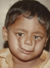
Identify the finding shown.
Oblique Facial Cleft
(Cleft from upper lip to eye)

Identify the finding shown.
Oblique Facial Cleft
(Cleft from upper lip to eye)

Identify the finding shown.
Oblique Facial Cleft
(Cleft from upper lip to eye)
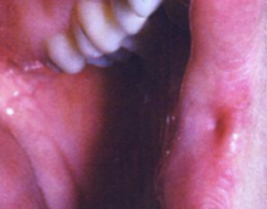
Identify the finding shown.
Commissural lip pits
(small invagination, 100% asymptomatic)
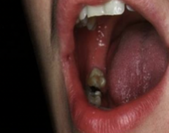
Identify the finding shown.
Commissural lip pits
(small invagination, 100% asymptomatic)

Identify the finding shown.
Paramedian lip pits
(symmetric, associated with van der woude syn- drome, congenital)

Identify the finding shown.
Paramedian lip pits
(symmetric, associated with van der woude syndrome, congenital)

Identify the finding shown.
Paramedian lip pits
(symmetric, associated with van der woude syndrome, congenital)
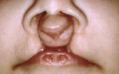
What syndrome is this likely associated with?
Van der woude syndrome
1. Paramedian lip pits
2. Cleft lip/palate
3. Hypodontia

What syndrome is this likely associated with?
Van der woude syndrome
1. Paramedian lip pits
2. Cleft lip/palate
3. Hypodontia

Identify the finding shown.
Double lip
(more common in upper lip, ascher syndrome, can cause double lip, can be associated with blepharochalasis and non-toxic thyroid enlargement in 50% of pa- tients)

Identify the finding shown.
Double lip
(more common in upper lip, ascher syndrome, can cause double lip, can be associated with blepharochalasis and non-toxic thyroid enlargement in 50% of pa- tients)

Identify the finding shown.
Blepharochalasis
(Associated with double lip which is more common in upper lip, ascher syndrome, can cause double lip, can be also associated with non-toxic thyroid enlargement in 50% of patients)

Identify the finding shown.
Blepharochalasis
(Associated with double lip which is more common in upper lip, ascher syndrome, can cause double lip, can be also associated with non-toxic thyroid enlargement in 50% of patients)
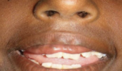
Identify the finding shown.
Double lip
(more common in upper lip, ascher syndrome, can cause double lip, can be associated with blepharochalasis and non-toxic thyroid enlargement in 50% of pa- tients)
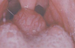
Identify the finding shown.
Lingual Thyroid
(Improper descent of thyroid bud --> ectopic thyroid tissue, more common in females, 90% in posterior dorsal tongue, reddish and associated with hypothyroidism. do not want to cut it out as for 70% of these patients it is the ONLY functioning thyroid tissue)

Identify the finding shown.
Lingual Thyroid
(Improper descent of thyroid bud --> ectopic thyroid tissue, more common in females, 90% in posterior dorsal tongue, reddish and associated with hypothyroidism. do not want to cut it out as for 70% of these patients it is the ONLY functioning thyroid tissue)

Identify the finding shown.
Lingual Thyroid
(Improper descent of thyroid bud --> ectopic thyroid tissue, more common in females, 90% in posterior dorsal tongue, reddish and associated with hypothyroidism. do not want to cut it out as for 70% of these patients it is the ONLY functioning thyroid tissue and risk of hemorrhage)

Identify the finding shown.
Fissured tongue

Identify the finding shown.
Hairy tongue
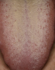
Identify the finding shown.
Coated tongue
(accumularion of bacteria and desquamated epithelial cells, no hair like projections)

Identify the finding shown.
Sublingual varices

Identify the finding shown.
Sublingual varices

Identify the finding shown.
Varices

Identify the finding shown.
Varices

Identify the finding shown.
Diascopy of varices??? idk maybe delete ?

Identify the finding shown.
Fordyce granules

Identify the finding shown.
Fordyce granules

Identify the finding shown.
Fordyce granules

Identify the finding shown.
Leukoedema

Identify the finding shown.
Leukoedema
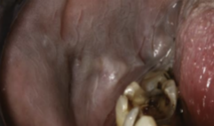
Identify the finding shown.
Leukoedema
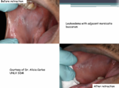
What is this showing?
Leukoedema can dissappear when the cheek is stretched --> do NOT rub it off

Identify the finding shown.
Exostoses

Identify the finding shown.
Exostoses

Identify the finding shown.
Exostoses

Identify the finding shown.
Exostoses (solitary exostoses)

Identify the finding shown.
Exostoses
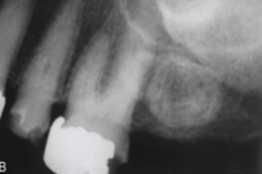
Identify the finding shown.
Exostoses

Identify the finding shown.
Exostoses

Identify the finding shown.
(Palatal) Exostoses

Identify the finding shown.
Reactice subpontine exostosis
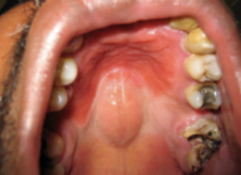
Identify the finding shown.
Torus palatinus (flat and denture stomatitis)

Identify the finding shown.
Torus palatinus (lobular)

Identify the finding shown.
Torus palatinus (nodular)

Identify the finding shown.
Torus palatinus AND palatal exostoses

Identify the finding shown.
Mandibular tori

Identify the finding shown.
Mandibular tori ("kissing")

Identify the finding shown.
Mandibular tori
(Radiopacity superimposed over roots of premolars)
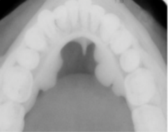
Identify the finding shown.
Mandibular tori
(Radiopacity superimposed over roots of premolars)

Identify the finding shown.
Stafne defect
(radiolucent, well defined, posterior mandible, below IA canal)

Identify the finding shown.
Stafne defect
(radiolucent, well defined, posterior mandible, below IA canal)

Identify the finding shown.
Salivary gland defect
(Anterior lingual)

Identify the finding shown.
Epstein pearls
(median palatal raphe)

Identify the finding shown.
Bohn nodules
(scattered over hard palate)

Identify the finding shown.
Epstein pearls
(median palatal raphe)

Identify the finding shown.
Bohn nodules
(scattered over hard palate)

Identify the finding shown.
Nasolabial cyst
(lateral upper lip, swelling of upper lip, elevation of ala of nose, obliteration of maxillary mucobuccal fold)
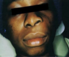
Identify the finding shown.
Nasolabial cyst
\(lateral upper lip, swelling of upper lip, elevation of ala of nose, obliteration of maxillary mucobuccal fold)

Identify the finding shown.
Nasolabial cyst
(lateral upper lip, swelling of upper lip, elevation of ala of nose, obliteration of maxillary mucobuccal fold)

Identify the finding shown.
Nasolabial cyst
(lateral upper lip, swelling of upper lip, elevation of ala of nose, obliteration of maxillary mucobuccal fold)

Identify the finding shown.
Nasolabial cyst
(lateral upper lip, swelling of upper lip, elevation of ala of nose, obliteration of maxillary mucobuccal fold)

Identify the finding shown.
Nasolabial cyst (lateral upper lip, swelling of upper lip, elevation of ala of nose, obliteration of maxillary mucobuccal fold)

Identify the finding shown.
Cyst of incisive papilla
(Specifically called this because it is entirely in soft tissue!)
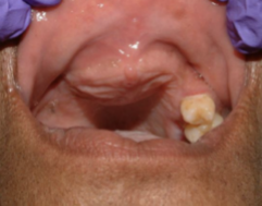
Identify the finding shown.
Nasopalatine duct cyst
(Tx: enucleation)

Identify the finding shown.
Nasopalatine duct cyst
(Tx: enucleation)

Identify the finding shown.
Nasopalatine duct cyst
(Tx: enucleation)
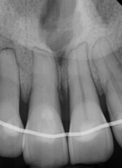
Identify the finding shown.
Nasopalatine duct cyst
(Look at location, it is on the anterior palate)
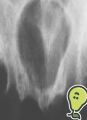
Identify the finding shown.
Nasopalatine duct cyst
(Tx: enucleation)

Identify the finding shown.
Nasopalatine duct cyst
(Tx: enucleation)
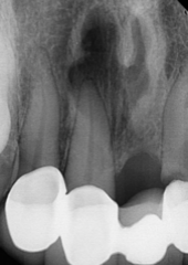
Identify the finding shown.
Nasopalatine duct cyst
(Tx: enucleation)

Identify the finding shown.
Nasopalatine duct cyst
(Tx: enucleation)

Identify the finding shown.
Nasopalatine duct cyst
(Tx: enucleation)

Identify the finding shown.
Median palatal cyst
(posterior midline hard palate)

Identify the finding shown.
Median palatal cyst
(posterior midline hard palate)

Identify the finding shown.
Dermoid cyst
(Occurs most in children and yound adults, midline floor of mouth is common location, swelling under tongue, "doughy")
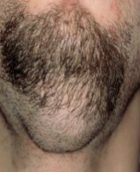
Identify the finding shown.
Dermoid cyst
(Occurs most in children and yound adults, midline floor of mouth is common location, swelling under tongue, "doughy")
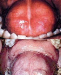
Identify the finding shown.
Dermoid cyst
(Occurs most in children and yound adults, midline floor of mouth is common location, swelling under tongue, "doughy")

Identify the finding shown.
Dermoid cyst
(Occurs most in children and yound adults, midline floor of mouth is common location, swelling under tongue, "doughy")

Identify the finding shown.
Thyroglossal duct cyst
(located at midline of neck, tx with sistrunk procedure, risk of malignant transformation --> papillary thyroid carcinoma)
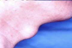
Identify the finding shown.
Thyroglossal duct cyst
(located at midline of neck, tx with sistrunk procedure, risk of malignant transformation --> papillary thyroid carcinoma)

Identify the finding shown.
Thyroglossal duct cyst
(located at midline of neck, tx with sistrunk procedure, risk of malignant transformation --> papillary thyroid carcinoma)

Identify the finding shown.
Thyroglossal duct cyst
(located at midline of neck, tx with sistrunk procedure, risk of malignant transformation --> papillary thyroid carcinoma)

Identify the finding shown.
Brachial cleft cyst caused by 2nd arch What is it and be specific (2nd arch most common --> clinically shows up in up- per lateral neck, anterior or deep to SCM)

Identify the finding shown.
Brachial cleft cyst caused by 2nd arch
(2nd arch most common --> clinically shows up in up- per lateral neck, anterior or deep to SCM)

Identify the finding shown.
Brachial cleft cyst
(idk if it would be the 2nd arch or 3/4 bc its lower neck (3/4) but also deep to SCM (2nd))

Identify the finding shown.
Brachial cleft cyst caused by 2nd arch
(2nd arch most common --> clinically shows up in up- per lateral neck, anterior or deep to SCM)

Identify the finding shown.
Brachial cleft cyst

Identify the finding shown.
Brachial cleft cyst