L30 - Molecular Basis of Cancer: Malignancy & Host Interactions
 Call Kai
 Learn
Learn Practice Test
 Spaced Repetition
 Match
Match Flashcards
 Knowt Play
1/48

There's no tags or description
Looks like no tags are added yet.
Name | Mastery | Learn | Test | Matching | Spaced | Call with Kai | Chat |
|---|
No analytics yet
Send a link to your students to track their progress
49 Terms
What are the 3 Major Tumor Categories?
Mesenchymal tumors
Epithelial tumors
Hematopoietic/round cell tumors
What is the main identifying feature of a Mesenchymal tumors?
Mesenchymal tumors arise from connective/supporting tissues and often make and secrete extracellular stroma/matrix
Examples of origin include:
Fibroblasts
Muscle
Adipose tissue
Bone/cartilage
Endothelial cells

What do Mesenchymal tumor cells typically look like histologically?
Elongated/spindle-shaped cells
Arranged in streams, fascicles, or bundles
Often accompanied by stroma/matrix
Usually have a higher nuclear:cytoplasm ratio

What question should you ask when trying to identify a Mesenchymal tumor?
“What are these cells making?”
The matrix/stroma produced helps identify the tumor's cell/tissue of origin
What types of matrix can Mesenchymal tumors produce?
Collagen
Osteoid (bone)
Cartilage
How could you distinguish a fibrosarcoma from an osteosarcoma?
Look at what the malignant mesenchymal cells are producing:
Fibrosarcoma → collagen/fibrous stroma
Osteosarcoma → osteoid (bone matrix)
How are Benign Mesenchymal tumors generally named?
Cell/tissue of origin + “-oma”
Examples:
Fibroblast → fibroma
Fat → lipoma
Bone → osteoma
Cartilage → chondroma
How are Malignant Mesenchymal tumor generally named?
Cell/tissue of origin + “-sarcoma”
Examples:
Fibrosarcoma
Liposarcoma
Hemangiosarcoma
Osteosarcoma
Chondrosarcoma
How does a fibroma differ from a fibrosarcoma?
Fibroma: benign, makes collagen, does not invade
Fibrosarcoma: malignant, neoplastic cells invade surrounding tissue

What is the key difference between Round Cell Tumors and Mesenchymal tumors?
Round cell tumors do NOT make stroma/matrix → they primarily consist of cell proliferation

What are the major Histologic features of Round Cell Tumors?
Individualized cells with clear spaces around them
Lack an orderly arrangement
Round to polygonal cells
Variable N:C ratio
Often infiltrate adjacent tissue

What are two Benign Examples of Round Cell Tumors from the lecture?
Histiocytoma — skin
Plasmacytoma — mucosal surfaces
What is the major identifying feature of Epithelial Tumors?
Epithelial cells like to form structures, including:
Glands
Tubules
Acini
Sheets

What do Epithelial Tumor cells typically look like histologically?
Large, polygonal cells
Defined cell borders
Cells remain grouped together
Arranged in sheets, tubules, acini, or glands

How are Benign Glandular Epithelial Tumors generally named?
Glandular: ______ adenoma
Lining epithelium: ______ papilloma
Mucosal: ______ polyp
How are Malignant Epithelial Tumors generally named?
Cell/tissue of origin + “-carcinoma”
If gland-forming: _____adenocarcinoma

What are 2 common gross features of Malignant Carcinomas?
Scirrhous response → reactive fibrous stroma → tumor feels firm
Necrosis → can cause ulceration and/or umbilication
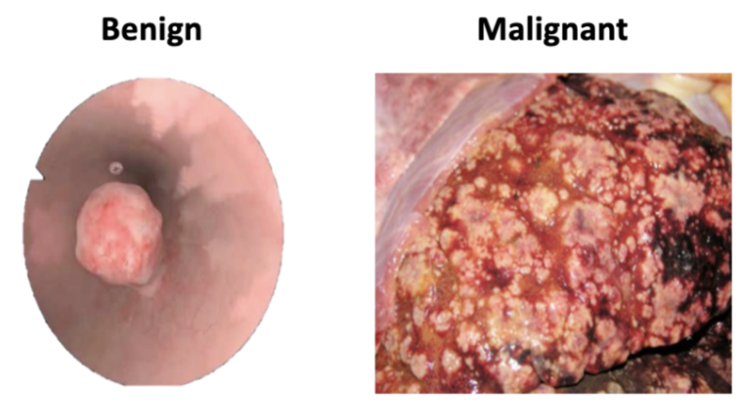
Why can Malignant Epithelial Tumors develop a necrotic center?
They may grow faster than their blood supply, causing central cells to become deprived of oxygen and undergo necrosis
What are 3 major general histologic hallmarks that help distinguish benign from malignant tumors?
Feature | Benign | Malignant |
Growth | Expansion/local | Invasion/infiltration ± metastasis |
Differentiation | Well differentiated | Poorly differentiated/anaplastic |
Mitotic activity | Low mitotic index | Increased mitotic index |
How does a benign tumor generally grow?
By local expansion, sometimes with pedunculation; it is usually well circumscribed and often encapsulated

How does a malignant tumor generally grow?
By expansion AND invasion/infiltration into surrounding tissues, with the potential for metastasis
How does cellular differentiation differ between benign and malignant tumors?
Benign: well differentiated → resembles tissue of origin
Malignant: poorly differentiated → may show anaplasia
How does mitotic activity differ between benign and malignant tumors?
Benign → low mitotic index
Malignant → increased mitotic index
What does normal glandular epithelium look like?
Orderly arrangement
Uniform cell morphology
No cellular atypia
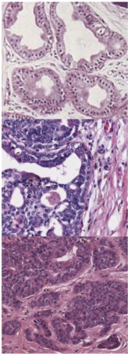
What does a Benign Epithelial Adenoma look like histologically?
Glandular arrangement still present
Less orderly; some cell jumbling/piling
Well circumscribed
Encapsulated
Mild cellular atypia

What does a Malignant Epithelial Carcinoma look like histologically?
Loss of glandular arrangement
Solid areas
Unencapsulated
Invades adjacent stroma
May invade lymphatic/blood vessels
Increased pleomorphism and atypia

Why is Vascularization critical for tumor growth?
Tumors require a blood supply to continue growing and spreading

Tumors require a blood supply to continue growing and spreading.
Tumors induce the formation of their own blood supply/vessels

How is cancer-associated vasculature different from normal vasculature?
Neoplastic vasculature has:
Abnormal structure
↑ Leakage
↑ Fragility
Abnormal paracrine and autocrine signaling
Continued vascular growth
This can contribute to edema and vascular fragility
What are the 4 Major Steps of Tissue Invasion?
Break cell-cell junctions
Bind extracellular matrix
Use type IV collagenase
Invade

Why is type IV collagenase important for tumor invasion?
It helps tumor cells degrade the extracellular/basement membrane barrier, allowing invasion

What are the 3 Major Routes of Metastasis?
Hematogenous → vascular spread
Lymphatic → lymphatic/vascular dissemination
Direct invasion or implantation → carcinomatosis
Which tumors commonly undergo Hematogenous metastasis?
Sarcomas
(Neuro)endocrine carcinomas
Venous spread is more common than arterial spread.
Which tumors are especially associated with Lymphatic metastasis?
Carcinomas > sarcomas
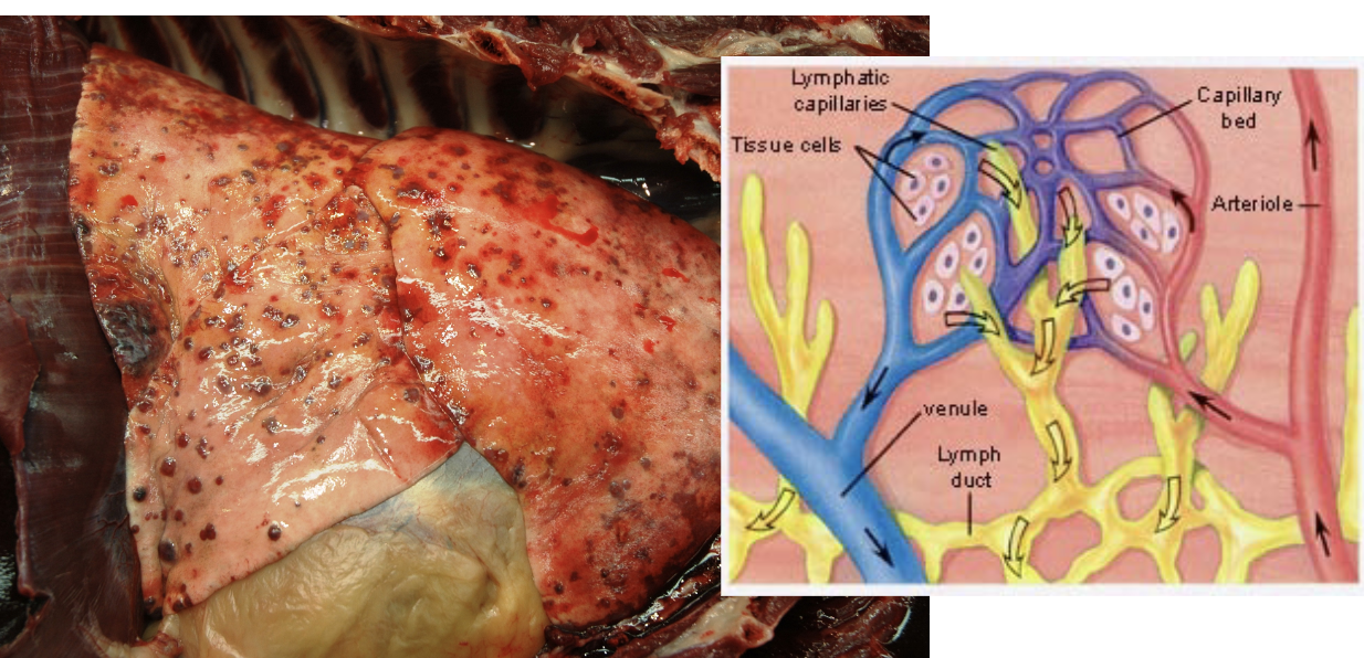
What is the general sequence for hematogenous metastasis to the lung?
Primary malignant tumor
Local tissue invasion
Tumor enters blood vessel (intravasation)
Travels through venous circulation
Reaches pulmonary capillary bed
Tumor cells lodge/adhere
Exit vessel (extravasation)
Establish metastatic focus
Angiogenesis + growth → pulmonary metastasis
Why is the lung a common site of hematogenous metastasis?
Tumor cells entering the venous circulation encounter the pulmonary capillary bed, making the lung an important site where circulating tumor cells can lodge
Venous blood → right heart → lungs → pulmonary capillaries → tumor cells lodge
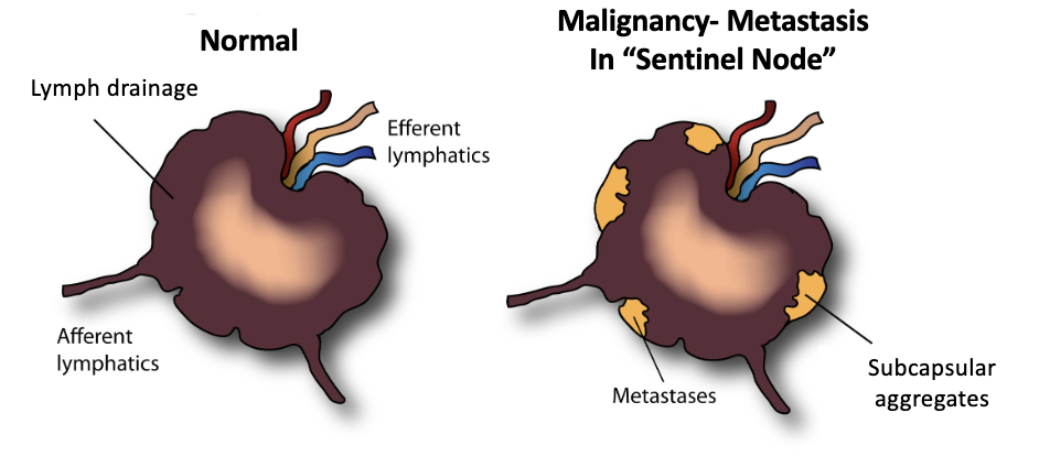
What lymph node is most likely to receive metastatic cells first?
The draining (sentinel) lymph node

How does a metastatic carcinoma enter and grow within a lymph node?
Primary carcinoma
Invades lymphatic vessel
Travels through afferent/draining lymphatics
Enters draining/sentinel lymph node
Reaches subcapsular sinus
Tumor cells establish metastatic aggregates
Metastatic tumor expands within lymph node
Where can metastatic tumor cells initially aggregate within a draining lymph node?
In the subcapsular region/sinus
What is carcinomatosis?
Direct spread/implantation of carcinoma throughout a body cavity
It does not require a blood vessel for spread
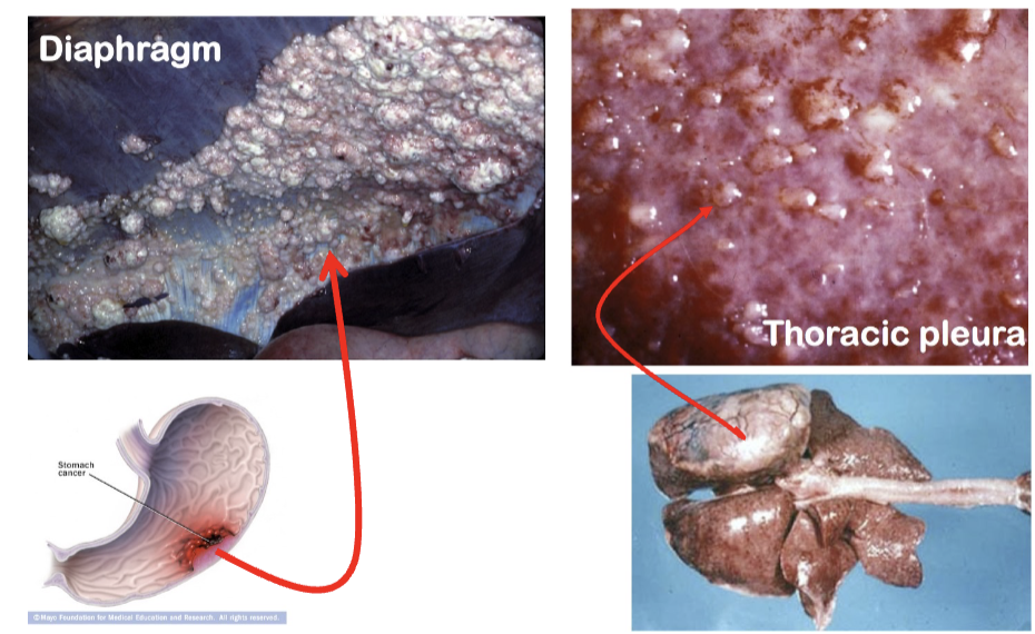
How is carcinomatosis different from hematogenous metastasis?
Hematogenous: tumor spreads through blood vessels
Carcinomatosis: tumor cells directly seed a body cavity
Tumor Classification SUMMARY
Mesenchymal = MATRIX | Epithelial = STRUCTURES | Round cell = INDIVIDUAL CELLS
Feature | Mesenchymal | Epithelial | Round Cell |
Main clue | Makes matrix/stroma | Makes structures | Cell proliferation; no matrix |
Cell shape | Elongate, spindle-shaped | Large, polygonal | Round → polygonal |
Arrangement | Streams, fascicles, bundles | Sheets, tubules, acini, glands | Individualized cells |
Matrix/stroma | Often present | Cells grouped into structures | Does not make stroma/matrix |
N:C ratio | Usually higher | — | Variable |
Malignant name | Sarcoma | Carcinoma | Nomenclature varies |
Recognition trick | What are they making? | What are they making? | Individual cells |
Identifying Mesenchymal Tumors SUMMARY
MESENCHYMAL TUMOR? → “What are the cells making?” → Identify the matrix → Identify tissue of origin
Tumor origin | What is produced? | Benign | Malignant |
Fibroblast clue | Collagen/fibrous matrix | Fibroma | Fibrosarcoma |
Fat | Adipose tissue | Lipoma | Liposarcoma |
Endothelium | Vascular tissue | Hemangioma | Hemangiosarcoma |
Bone | Osteoid | Osteoma | Osteosarcoma |
Cartilage | Cartilage | Chondroma | Chondrosarcoma |
Benign vs. Malignant SUMMARY
Feature | Benign | Malignant |
Spread | Local only | Metastasis possible |
Growth | Expansion/pedunculation | Expansion + invasion/infiltration |
Borders | Well circumscribed | Poorly circumscribed |
Capsule | Often encapsulated | Usually unencapsulated |
Differentiation | Well differentiated | Poorly differentiated / anaplastic |
Mitotic index | Low | Increased |
Angiogenesis | — | Often increased |
Necrosis | Less characteristic | Areas of necrosis |
Recurrence | Excision often curative | Local recurrence frequent |
Routes of Metastasis SUMMARY
Sarcoma → BLOOD → LUNG
Carcinoma → LYMPH → LYMPH NODE
Carcinomatosis → BODY CAVITY
Route | How? | Tumor association | Important destination/concept |
Hematogenous | Blood vessels | Sarcomas, (neuro)endocrine carcinomas | Lung |
Lymphatic | Lymphatics | Carcinomas > sarcomas | Draining/sentinel LN |
Direct implantation | Seeds body cavity | Carcinoma | Carcinomatosis |
Normal vs. Cancer Vasculature SUMMARY
Normal vessels | Neoplastic vasculature |
Organized structure | Abnormal structure |
Controlled permeability | ↑ Leakage |
Relatively stable | ↑ Fragility |
Normal signaling | Abnormal paracrine/autocrine signaling |
Regulated growth | Continued vascular growth |
Epithelial Malignancy Progression SUMMARY
Normal epithelium | Benign adenoma | Malignant carcinoma |
Orderly cells | Glandular pattern remains | Loss of glandular pattern |
Uniform morphology | Some jumbling/piling | Solid areas |
No atypia | Mild atypia | ↑ Pleomorphism + atypia |
— | Well circumscribed | Invasion |
— | Encapsulated | Unencapsulated |
Hematogenous spread → Lung SUMMARY
Primary tumor
Local invasion
Intravasation
Venous circulation
Right heart
Pulmonary capillary bed
Tumor cells lodge
Extravasation
Metastatic focus → angiogenesis → growth
Carcinoma → Lymph node SUMMARY
Primary carcinoma
Invades lymphatic
Afferent lymphatic
Draining/sentinel lymph node
Subcapsular sinus
Metastatic aggregates
Growth within LN