SDL: application of biomechanics (NMSK4)
 Call Kai
 Learn
 Practice Test
 Spaced Repetition
 Match
 Flashcards
 Knowt Play
1/39

There's no tags or description
Looks like no tags are added yet.
40 Terms
Review biomechanics terminology
Apply an understanding of biomechanics for the cause of fracture and management of fracture repair
Apply an understanding of biomechanics to assess conformation
biomechanics
applying mechanical laws to living things
especially forces and effects of these forces on and within the body
branch of physics
classical mechanics: how bodies (solids + fluids)/ systems of bodies respond to external forces
system of levers
musculo and skeletal systems
bones - form system of levers
most muscles act on tendons to change joint angles
lever = rigid object rotates around a fixed point
functional anatomy
principles of biomechanics used to work out how structures function
bone
look at the shape
wolff’s law: living bone adapts and remodels based in response to mechanical forces + physical loads
big prominence: suggests many muscles/ powerful muscles attach (eg/ the bumps that form based on force applied to bone - like at the epiphysis of femur)
muscles
ability to shorten to approx 50% of original length
determined by muscle length
parallel formation of fibres:
greater range of motion - less force developed (rectus abdominis can shorten by half)
amount of tendon + tendon:muscle ratio:
affects force and range of motion potential of a muscle
muscle power:
determined by cross sectional area of muscle
determined where fibres joint tendons at an angle (pennate muscles)
muscle architecture
variation in fibre arrangement in the muscle belly
strap muscle = infrahyoid muscles
fusiform muscles = spindle shaped
pennate
ACS
widest part of muscle belly
PCS
total area of muscle perpendicular to muscle fibres
pennate muscles
create large cross-section and force
BUT has lower range of motion for shortening
fusiform muscles
spindle shaped
pennate muscles
unipennate: diagonal fibres in one direction
bipennate: diagonal fibres in 2 directions (feather-like)
loading to failure
linear region = elastic region - reversible
yield region = plastic region - irreversible

how can loads be applied to muscles
tension (pull)
compression
shear (push either end in opposite direction)
torsion (twist either end in opposite direction)
wolff’s law - bone
bone acts like piezoelectric crystal
bending → microcurrents
osteocytes sense currents
compression - negative charge - lay down bown
tension - positive charge - remove bone
(use it (bone) or lose it)
important for:
fracture healing + rehab
slower than soft tissue adaptations
bone characteristics
not homogenous
mechanical behaviour influenced by direction of loading relative to orientation
bone = resistant to compression in all directions
what resists tensile (tension) force?
collagen fibres
what resists axial compression force?
mineral content
what resists torsion force?
not good at resistance
(torsion: mass of body changes direction when weight bearing)
fracture formation
external force → intermolecular resistance to prevent deformation: stress + strain → effected by force
most fractures = caused by tension
fracture type:
type of loading
rate that load is applied
fractures associated with type of loading

rate of loading - cortical bone
cortical bone:
slow = fracture starts at weak area + follows weakest path
rapid = energy stored in structure can cause multiple sites of disruption
not always along weakest path
high energy trauma = high comminution of bone (multiple parts)
rate of loading - cancellous bone (trabecular)
follows fracture patterns seen in cortical bone
compressive force: collapse + compaction
usually seen in vertebrae
stages of bone healing
inflammation → soft to hard callus → bone forms of original tissue
purpose of fracture fixation
minimise strain of fracture callus
allow healing to occur
spatial realignment
takes load until bone strong enough
weight bearing on limb
reconstruct the original structure
neutralise forces acting on fracture site
options of fracture fixation
Splints and casts
External fixator
Plate and screw
Intramedullary rod
Interlocking nails
uses of splints and casts
immobilise fracture
don’t directly contact bone
rigid material
what is external fixator + when is it used?
used for complex fractures
used when infection risk is high
increase rigidity via more pins
infinite variations of set up - suit range of needs

what is plate and screw and when is it used?
resist all three forces
screws:
apply friction between plate and bone
apply compression between fragments
plate:
internal splint - hold fractured parts in alignment
allows compression between fracture ends and load transfer
long bones loaded eccentrically:
results in tension side and compression side
position plate on tension side = helps counteract load and compressing on opposite side

intramedullary rod uses
little resistance to axial compression/ torsion
resist bending forces

interlocking nails uses
resist all three forces
central nail resists bending
screws/ bolts:
prevent collapse under compressive force
prevent rotation with torsional force
place away from fracture - avoid weakening it
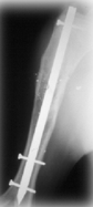
example of optimising surgical intervention
tibiotarsal bone plate:
Aim: improve surgical repairs
Validate surgical procedure
increase fatigue strength of surgically repaired canine tibiotarsal joints
Biomechanical study on canine hindlimb
strain gauges mounted to bone plates
test efficacy of added IM rod in this procedure
Tested by compressive loading noted:
increase in fatigue life via reduction in plate strains and increase in structural stability due to IM rod in tibia
implications of conformation (+ what is it?)
what is it?
alignment of bones + joints
critical to performance
implications:
determine shap§how the foot wears over time (e.g. horse)
the way the legs move during locomotion
some abnormalities simply reduce visual appeal, others can lead to lameness
knowing about proper conformation can help us to recognise potential problems before lameness occurs of foot
risks for dachshund
disc problem
risk for bulldogs
limb
respiratory problems
risks for pigs
conformation problems accentuated by farming
German Shepard risks
low hips, back problems
head and forelimb points to know
wing of atlas
proximal end of spine of scapula
posterior part of the greater tubercle of the humerus
elbow joint
lateral tuberosity of the distal end of radius
articulation of carpus and metacarpal bones
distal end of third metacarpal bone
articulation of first pastern joint and phalanx