Dental Radiology - Week 1
 Call Kai
 Learn
 Practice Test
 Spaced Repetition
 Match
 Flashcards
 Knowt Play
1/35

There's no tags or description
Looks like no tags are added yet.
36 Terms
Who discovered X-rays?
Wilhelm Conrad Roentgen
When were X-rays discovered?
November 8, 1895
What was the first X-ray taken?
An X-ray of Roentgen's wife's hand on December 22, 1895
What is digital imaging in dental radiography?
A method of taking dental X-rays using an electronic sensor instead of film

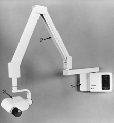
What are the three main parts of a dental X-ray machine?
Tubehead, Extension Arm, Control Panel
What does a periapical (PA) X-ray show?
The whole tooth, including the crown, root, and supporting bone

What does an interproximal (bite-wing/BW) X-ray show?
The crowns of both upper and lower teeth, useful for checking cavities

What does an occlusal X-ray show?
A large area of the maxilla or the mandible jaw in one picture.

What is direct digital imaging?
A method where the sensor in the mouth sends the image directly to the computer in real time
Components of a direct digital image are?
xray unit, sensor and computer
What is indirect digital imaging?
A method that converts an existing film into a digital image using a camera
Components of a indirect digital imaging are?
CCD camera and computer
What is storage phosphor imaging?
Wireless digital imaging system
it's a reusable, flexible plate that captures the image and is then scanned to make it digital

What is the function of a PID (Position-Indicating Device)?
To direct and position the X-ray beam toward the tooth and receptor

What is a complete mouth series (CMS)?
A full set of X-rays of all tooth-bearing areas, usually 14-20 images
PIMA - 18 ( 6 Anterior PAs, 8 Posterior PAs, 4 BW)

What are the advantages of digital imaging?
Better image detail, less radiation exposure, faster results, lower costs over time, and eco-friendly
What are the disadvantages of digital imaging?
High initial setup cost, variable image quality, sensors can be uncomfortable, and complicated infection control
What is a periapical image?
A traditional film X-ray showing the entire tooth and surrounding bone
*** 3mm of bone structure

What is a pixel in digital imaging?
A tiny square that makes up a digital image
What is the Rinn XCP system?
A holder with a color-coded ring, arm, and bite block that keeps the X-ray receptor and beam in the correct position

What does the blue ring in the Rinn XCP system indicate?
Anterior setup for front teeth X-rays

Anterior xrays

What does the yellow ring in the Rinn XCP system indicate?
Posterior setup for back teeth X-rays

Posterior teeth x-rays

What does the red ring in the Rinn XCP system indicate?
Bitewing setup for checking cavities between teeth

Bitewings
Left - Horizontal
Right- Vertical

What does the green ring in the Rinn XCP system indicate?
Endodontic setup for root canal procedures

What is the purpose of the bite block in the Rinn XCP system?
To hold the receptor in the correct position in the patient's mouth

What is the importance of aligning the PID with the center of the ring?
To ensure accurate and clear X-ray images
What are the key benefits of using the Rinn XCP system?
Keeps the receptor, tooth, and X-ray beam aligned, reduces retakes, and works with the paralleling technique
What should be checked after taking an X-ray?
The image for proper coverage and clarity
What are the types of Receptor Holders?
Uni-bite
Endoray
Molded plastic devices (snap-a-ray)
Disposable Styrofoam bite block

Disposable Styrofoam bite block (stabe)

Endoray
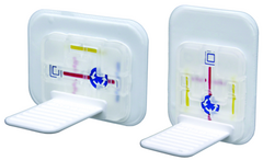
Uni-bite

Molded plastic devices (snap-a-ray)