Skeletal, Muscular, and Cardiovascular Systems
 Call Kai
 Learn
 Practice Test
 Spaced Repetition
 Match
 Flashcards
 Knowt Play
1/108

There's no tags or description
Looks like no tags are added yet.
109 Terms
What are the main functions of connective tissue?
Connecting and binding structures in the body
Supporting the weight of the body
Protecting our internal organs
Transporting medium through the body (via blood)
Insulation
What are the major components of all connective tissues?
Cells
Surrounding protein fibers — collagen, elastic, and reticular fibers
Ground substance — made up of water, glycosaminoglycans, proteoglycans, and glycoproteins
What are the four types of connective tissues and the cells that make them up?
Connective tissue proper (fibroblasts)
Loose/areolar, dense, reticular, and adipose/fat
Bone (osteoblasts)
Cartilage (chondroblasts)
Blood (blood cells)
What is the ECM? What cells secrete it and what is it composed of?
Extracellular matrix, secreted by fibroblasts
Composed of:
Fibrous proteins
Collagen — resistance to longitudinal tensile/pulling forces
Major organic portion of the bone tissue, large amounts in the cartilage
Elastic — stretching and relaxing
Tissue fluid (interstitial fluid)
Adhesion proteins
Proteoglycans
What are epithelial cells? What are its 3 membranes?
Makes up outer layer of organs, lines cavities, etc.
Cells bound closely together via adhesive junctions
Polarized:
Apical membrane — faces lumen/surface
Basal membrane — faces underlying cells
Lateral membrane — contacting other epithelial cells
Avascular but innervated
High regenerative capacity
What classifications exist for epithelia?
Ex 1: stratified squamous epithelium = epidermis of the skin
Ex 2: epithelium of the lung alveoli = simple squamous epithelium

What are the cells that secrete the cartilage matrix? What do they become when engulfed in lacunae cavities?
Chondroblasts → Chondroctyes
Why does cartilage recover slowly from injury?
Fewer blood vessels in the cartilage
Slower blood supply for healing
What are the three types of cartilage?

What are the three cell types in bone/osseous tissue? What functions do they have? What are some characteristics of mature bone cells?
Osteoblasts — bone deposition
Secrete the bone matrix (which is then calcified to harden)
Osteocytes — mature bone cells
Trapped inside the calcified bone matrix
Responsible for the maintenance of the bone matrix
Cannot differentiate
Have tight junctions
Act as sensors for degradation
Osteoclasts — bone resorption
What is the difference between bone tissue and the bone organ?
Bone tissue is a connective tissue
Bone tissue = one of many types of tissue that make up the bone organ
What are the major functions of our bone organ?
Protect and support our organs
Act as connection sites for muscles
Store calcium/phosphorus/magnesium salts
House our bone marrow
Store fat
What are the four types (shapes) of bones in the human body? What type are the phalanges?
Long bones (longer than wide)
Phalanges
Short bones (as long as wide)
Flat bones (thin and broad)
Irregular bones (etc.)
Where do we find the diaphysis and the epiphysis? What is located at the border of the epiphysis and diaphysis?
In long bones
The diaphysis is the shaft
The epiphysis is the rounded end
The epiphyseal/growth plate is located at their border in children and adolescents
What are the differences between spongy and compact bones?
Compact bone: hard, outer bone, which bears compression and twisting forces
Spongy bone: inner framework of the bone, where the bone marrow is housed
What is an osteon? What is it’s structure?
Osteon = fundamental functional and structural unit of compact bone
Osteocytes are on the ledges of each lamellae

What is the periosteum and what are its two layers? What is the endosteum? What cells line these layers and what cells are those differentiated from?
Periosteum:
Is the outer layer of the bone
Provides blood nourishment via blood vessels
Aids in bone growth and repair
Gives bones their sense of feeling
Two layers:
Outer fibrosis layer (collagen and elastin fibers)
Inner osteogenic layer
Endosteum: Lines the inner surfaces of the bone
Both layers are lined with osteoblasts (differentiated from osteoprogenitor cells)
Where is bone marrow found? What happens to bone marrow as you age?
Found in the epiphysis and diaphysis of the bone
Red bone marrow is found in the epiphysis (between the trabeculae at the proximal epiphyses) and during youth, the diaphysis
Red bone marrow in the diaphysis turns to yellow bone marrow with age → most fatty tissue

What is the bone matrix primarily composed of, and what would happen if each were removed?
Calcium
Allows for resistance to compression
Lack of calcium → flexible bone (demineralization)
Collagen
Allows for resilience to tension
Lack of collagen → brittle bones
How do bones grow in length (ie: ossification)? Why can’t osteocytes just multiply and grow the bone?
Chondrocytes at the epiphyseal plate grow and replicate upwards towards the epiphysis
Chondrocytes make and secrete an enzyme which calcifies the tissue
Crystallization and hardening halts diffusion of nutrients and oxygen (avascular, receive only via diffusion)
Chondrocytes die, creating a cavity
Osteoblasts come from the periosteum (diaphysis) via blood vessels and form new bone tissue
Epiphyseal plate “shifts” due to new osseous tissue
Osteocytes cannot divide, they’re fully differentiated
How does the bone know when to/how much to grow? What happens at puberty and then at the end of growth?
Growth hormone promotes bone and muscle growth during infancy and childhood
Insulin-like Growth Factor 1 secreted by liver in response to GH (IGF-1)
Puberty: bone growth increased dramatically by combined activities of growth hormone, thyroid hormone (anabolic hormone), and sex hormones
At the end of growth, bone formation wins over cartilage formation and growth ceases (entire plate is ossified)
What are the impacts of overproduction and deficits of growth hormone?
Overproduction: Gigantism
Deficits: Pituitary dwarfism
May be treated with growth hormone injectors
Everything is in proportion!
What is endochondral ossification and how does it work?
The process by which most long bones in the body develop in the growing embyro
Replacement of hyaline cartilage

What is intramembranous ossification?
Replacement of fibrous membrane (skull) in growing embryos
Fontanelles
What role do the osteoclasts play in endochrondral ossification? What the advantages and pitfalls of having a cavity in the long bone?
Protease activity
Necessary to stop/control building — balancing homeostasis
Creates the medullary/narrow cavity in the diaphysis
Advantage: lighter
Disadvantage: can’t hold as much weight
How is bone remodeling controlled by stress? What is Wolff’s Law in bone remodeling?
Mechanical stress/load
Increasing bone loading → bone remodeling to become stronger and to resist that loading
Osteocytes act as sensors
Forces on the bone determine how the bones are shaped
How is bone remodeling controlled by hormones?
Calcium levels are controlled by the parathyroid gland
The bones store majority of the body’s calcium (a little bit is in the blood)
The parathyroid glands secrete parathyroid hormone (PTH), which increases osteoclast activity and releases calcium into the blood
Contrarily, if the blood calcium levels rise, the thyroid gland produces calcitonin and osteoclast activity declines
What is osteoporosis? Who is at risk and how can we prevent it?
Osteoporosis occurs when there is a bone remodeling imbalance, and osteoclast activity overtakes osteoblast activity
People at risk:
Smokers
Females: estrogen promotes osteoblasts, in menopause, less estrogen → less osteoblasts
People with low calcium levels
Prevention: Lifting weights and resistance training promotes remodeling
What pathologic imbalances exist (other than osteoporosis) with the bones?
Osteomalacia: lack of mineralization (such as with Ca)
Rickets: analogous disease in children
Long bones become abnormally long
Defective mineralization prevents cartilage in the growth plates from hardening
This causes the growth plate to pile up and expand instead of turning into normal bone
What is the structure of a muscle? What are muscle cells also called? What are the epimysium, perimysium, and endomysium?
A single muscle contains approx. 1000 muscle cells (or fibers/myocytes)
These fibers run the length of the muscle
Epimysium: envelopes muscle tissue (outermost layer of connective tissue proper)
Perimysium: envelopes fascicles
Endomysium: envelopes each muscle cell
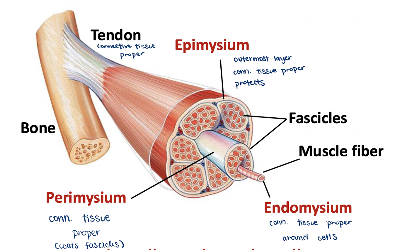
How are muscles connected to bones? Is skeletal muscle always connected to bones?
Via tendons (connective tissue proper)
No, skeletal muscle is not always connected to bone
Ex: the tongue
What are the three types of muscle? What other names can be used to refer to them?
Skeletal muscles – generally attached to skeleton, voluntary muscle contraction
Controlled by the motor cortex
AKA striated muscle due to alternating dark and light bands from myofibril overlap
Cardiac muscles – found in the heart, involuntary
Smooth muscles – found in the walls of hollow organs
AKA involuntary tissue or visceral muscle
What are some special characteristics of skeletal muscle cells?
Multinucleated (multiple nuclei due to increased size and need for constant protein synthesis)
Also has many mitochondia
Special organelles
Sarcoplasmic reticulum: endoplasmic reticulum of the muscle cell
Sarcolemma: membrane of the muscle cell
Sarcoplasm: cytoplasm of the muscle cell
T-tubules
What are myofibrils?
Bundles of specialized proteins involved in muscle contraction
Arranged specifically in different types of muscle cells
Run along the length of the muscle cell
Characterized by circumferential bands along the length of the fiber
Contains many repetitive, continuous units of myofilament units
What are myofilaments? What filaments make these up? What is a cross bridge?
Proteins composing bands of a myofibril
Light bands are thin filaments (Actin)
Act as the contractile apparatus of the cells
Drive cell dynamics (changes in shape, migration, etc.)
Has binding sites for Myosin
Dark bands are thick or thick/thin filaments (Myosin)
Acts as the motor driving contraction
Cross bridges connect the thin and thick filaments
What is a sarcomere? What are the landmarks of each sarcomere?
The basic unit of muscle contraction
Formed by myofilaments

How does is muscle mass correlated with exercise over the lifespan?
As one ages, muscle deterioration occurs and the appendages are made up of increasing fat tissue
With regular exercise, more muscle tissue is retained proportionally
How do actin and myosin interactions lead to muscle contractions?
Sliding filaments theory
Actin has Myosin binding sites
Once bound, a change in the angle of the myosin head will pull the actin filaments
Thin filaments slide past the thick ones
Myosin won’t move since bound to M-line
Z-lines come closer to one another
Sarcomere shortens
When a sarcomere contracts, what happens to the myofibrils and the muscle fibers? What happens to the muscle tissue? Do all muscle cells need to contract for the muscle organ to contract?
Contracted sarcomeres — all the sarcomeres in a specific filament contract simultaneously (will pull if not)
All sarcomeres shorten = myofibril shortens
All myofibrils shorten = muscle cell shorterns
Muscle cell contracting =/= whole muscle tissue
In order for a muscle organ to contract, all aspect must contract at the muscle cell level, but not all cells need to contract
How does degree of Action/Myosin overlap impact muscle contraction?
In relaxed state, thin and thick filaments overlap slightly
Contraction → overlap greater
If the thin and think filaments did not overlap, no tension would be exerted because myosin heads can’t bind to actin
AKA overextending muscles
The greater the overlap of actin and myosin before contraction, the weaker the contraction
Actin closer to the M-line → greater force applied
What are tropomyosin and troponin? What role does calcium play?
At resting state, tropomyosin physically blocks myosin from binding to actin
Calcium binds to troponin, leading to the release of tropomyosin from the myosin-binding sites
Ca2+ binding changes the configuration and tropomyosin slides from binding sites
What is the cross bridge cycle? Explain the steps, the molecules involved, and the following concepts: high/low energy configuration, hydrolysis, and the power stroke.
Myosin head attaches to the actin myofilament, forming a cross bridge (Myosin head in high energy configuration)
Inorganic phosphate generated in previous contraction cycle is released, initiating the power stroke.
The myosin head pivots and bends as it pulls on the actin filament, sliding it towards the M-line
ADP is released
As new ATP attaches to the myosin head, the link between myosin and actin weakens, and the cross bridge detaches
Myosin head in low-energy configuration
As ATP is split into ADP and P (hydrolysis by ATPase), the myosin head is energized — cocked into the high-energy conformation

What is rigor mortis and why does it occur? What is muscle tone?
Body muscle stiffening a few hours after a person dies
Ca2+ causes progressive binding, not immediately on or off
Our muscles are always contracting (muscle tone)
After death: anything binded will stay binded, so the muscles are stuck
How are skeletal muscles excited for contraction?
Skeletal muscles do not contract spontaneously — must be excited @ neuromuscular junction = excitation contraction coupling
A nerve ending excites the muscle cell through a neural impulse (action potential)
The muscle cell reacts to the neuronal excitation and physically contracts
How is the neuromuscular junction specialized? What neurotransmitter is released and how is it different?
The motor-end plate is a part of the sarcolemma where neurotransmitter is received
The neurotransmitter is acetylcholine
Only EPSPs and depolarization
How are action potentials generated at the neuromuscular junction? What effect does this action potential have?
Action potential propagates along the sarcolemma (in both directions) and its invaginations, the t-tubules
The action potential stimulates release of calcium from the SR to the sarcoplasm
Mediated by calcium voltage-gated channels that are physically coupled to mechanically-gated channels on the SR
How can we make sure that calcium is not constitutively released?
Stop secreting Ca from the SR: removing acetylcholine from the synaptic cleft via ACh esterase
Do something about the Ca that was already released: throw Ca back in the sarcoplasmic reticulum/terminale cisternae (requires a pump, goes against the concentration gradient
What is Myasthenia gravis? How would you help a patient, and how would this treatment impact a healthy person?
An auto-immune response attacking the ACh receptor
Less depolarization = compromised muscle contraction
Treatment: inhibit ACh esterase → ACh in the synapse will last longer
If a healthy person were exposed to an inhibitor of acetylcholine-esterase, they may die due to uncontrolled muscle contractions
More ACh in the synapse
Uncontrolled contraction (ex: over contraction of the diaphragm will cause death)
Sarin gas is an inhibitor of ACh esterase → continued contraction in the absence of additional neuronal stimulation
What is muscle tension? What is load?
Muscle tension: the force exerted by a contracting muscle on an object
Load: the opposing force, exerted by the weight of the object on the muscle
Does muscle contraction always lead to muscle shortening, and thus movement? What is isometric contraction and why does it occur?
No, muscle contraction doesn’t always mean movement
Isometric contraction = muscle contraction without movement or changing muscle length
Load is greater than muscle tension
Ex: lifting weights too heavy
Isometric may provide more tension, which is why we try to lift to failure
Sarcomeres shorten, but the muscle does not — the tendon stretches instead, and all force remains in the elastic components
What is isotonic contraction, and what types exist? Do muscles always shorten?
Muscle contraction with movement and a change in length
Tension is greater than load
Concentric = muscle shortening
When standing up after a squat, the quadriceps contract
Eccentric = muscle lengthening
While holding a squat, the quadriceps oppose gravity and elongate
What is a motor unit? How do motor units respond to stimuli? Can the response be different across regions of the body?
Motor unit: a somatic neuron and all the skeletal muscle fibers it innervates
Generated AP in one unit + all fibers that respond
Each muscle cell is innervated by one neuron, but one neuron can innervate many muscle cells
Response of a motor unit to a single action potential = a muscle twitch
Not all motor units look the same — the soleus muscle in the trunk has a longer response than an extraocular muscle

How do muscle contractions vary in strength in response to different demands? What is fused vs. unfused tetanus and why does it occur (in reference to Ca2+ ions)? What is muscle tone?
Increasing stimuli frequency increases tension
Increased response of motor unit
Ca2+ ions don’t leave in wave summation (not pumped back to SR) → more myosin heads bind and the response is stronger
When all binding sites have been bound = fused tetanus
Increased strength of the stimulus
Increased number of motor units = increased contraction
Muscle tone: continuous contraction of the muscles, helps to maintain posture, etc.

How do we have ATP for muscle contraction? (Steps, don’t explain)
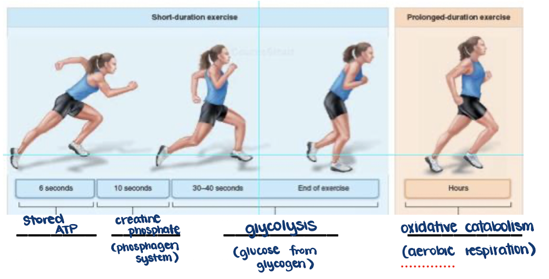
Why is stored ATP only an acceptable source of energy for muscles for the first few seconds of exercise? What are the critical enzymes of the next reaction?
ATP is regenerated by a reaction with creatine phosphate, but muscles only contain so much of it
Myokinase and creatine kinase are critical in the phosphogen system
Kinases phosphorylate (add a PO4 group) to substrates
Where do muscles cells obtain glucose for glycolysis? How does glycolysis compare to aerobic respiration in terms of products? What are the sources of oxygen for aerobic respiration?
Glucose may enter from the bloodstream, but mostly from stored glycogen in the muscle fibers/liver
Aerobic respiration/oxidative catabolism produces more ATP then glycolysis over a longer period of time
Myoglobin (in the muscles) and the cardiovascular/respiratory systems via hemoglobin and perfusion
How do endurance/resistance training impact muscle performance? Do muscles that use more oxygen have more mitochondria?
Endurance:
Increases amounts of oxidative enzymes, mitochondria and mitochondrial proteins, and blood vessels
Enhances the oxidative capabilities of the muscle
Increased need for oxygen (more use) = more mitochondria
Resistance:
Hypertrophy (increase in cell size)
Increase in number of myofibrils
Deceased proportion of mitochondrial proteins and blood supply
What is the difference between red and white muscle fibers? Can a muscle fiber change types?
Red fibers = slow oxidative (1)
White fibers = fast glycolytic (2 subtypes — 2x and 2a)
Muscles contain mixed types, but one type will predominate a specific muscle
With increased exercise comes better blood supply and more mitochondria, and a transformation of fiber types
2x can become 2a, and 2a can go to 1 with increased preparation for endurance and long contraction
What are the three layers of the heart wall? Which is the thickest?
Epicardium: most superficial layer
Myocardium: second and thickest layer
Endocardium: third/deepest layer
What are the main functions of the fibrous skeleton in regards to the heart?
Offers structural support to cardiac muscle cells
Acts as an insulator for the heart’s electrical activity
What are the two major sets of arteries and veins?
Arteries: pulmonary trunk (right ventricle) and aorta (left ventricle)
Veins: superior/inferior venae cavae and pulmonary veins
What are the chambers of the heart and how do they function? What is the interventricular septum? What is the difference between the walls of the two inferior chambers?
Superior chambers = atria, receive blood returning via the veins
Inferior chambers = ventricles, pump blood into the arteries
Interventricular septum: depression located between the right and left ventricles
Walls: the left ventricular wall is thicker than the right wall since it must overcome greater resistance and thus pumps harder
What is the purpose of the heart values? What role do the papillary muscles play?
Ensure one-way blood flow and thus normal heart functioning
Papillary muscles prevent valves from blowing open or prolapsing backward during contraction
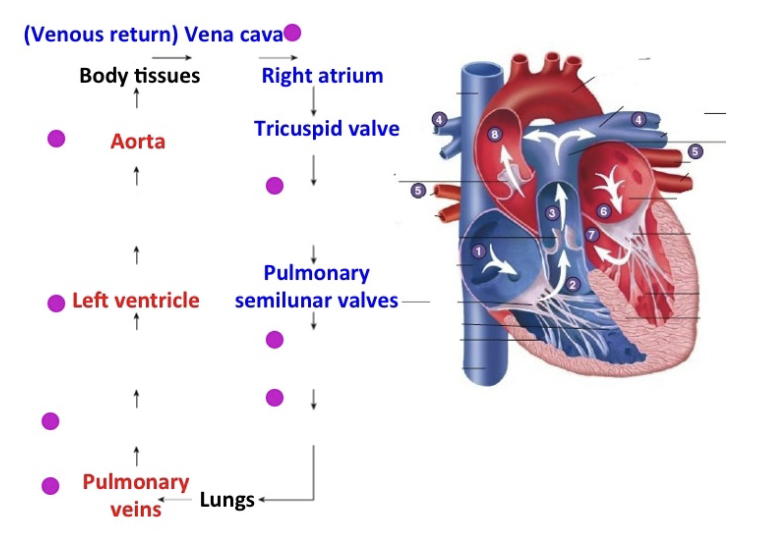
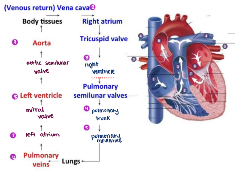
What is missing from this diagram?
What is the purpose of blood? What is it composed of?
Carries nutrients, oxygen, CO2, amino acids, etc. through the body
Composition 55% plasma, 45% formed elements
Plasma = fluids, proteins, nutrients
Formed elements = red blood cells (erythrocytes), white blood cells (leukocytes), and platelets (thrombocytes)
What is the plasma of the blood composed of? What do these elements do?
Water
Proteins
Carriers, like albumin, shuttle molecules (ex: non-soluble hormones) through circulation
Keep water in the bloodstream, contributing to osmotic pressure
Role in clotting (fibrinogen, clotting factors)
Ions and nutrients
Involved in osmotic pressure
Hormones, amino acids, glucose, etc.
Why are erythrocytes important in the blood? How are they distinguished from normal cells?
Make up 45% of whole blood
Transport of gases (oxygen and CO2)
Oxygen to the tissues, CO2 to the lungs
Based on concentration gradient
RBCs have a biconcave shape and no nucleus/certain other organelles
Carry more oxygen
What is the major component of RBCs which allows for oxygen binding? How does this work?
Hemoglobin protein (pigmented)
Composed of four globin polypeptides, each bound to a heme group
Each heme group bears an atom of iron, which can bind to one oxygen molecule
Each hemoglobin molecule can thus transport four molecules of oxygen
Oxygenated heme - red color
Hemoglobin reversibly binds with oxygen
Carries 20% of the CO2 molecules back to the heart
Why are leukocytes important in the blood? How are they different from erythrocytes?
Involved in defense and immune systems
Less than 1% of whole blood
Use the blood for transport
Can leave the blood and migrate through connective tissues
Larger and fewer than erythrocytes, have a nucleus and normal organelles
What is hematopoiesis? How does it work?
Blood cell formation
Red and white blood cells and platelets are synthesized in the bone marrow
RBCs live for 120 days
The kidney’s erythropoietin hormone promotes RBC synthesis
Triggers include decreased oxygen levels and increased demand for oxygen
Differentiated from stem cells
How does the blood clot?
Platelets form a temporary plug that helps seal breaks in blood vessels
Through a complex chain of activations, clotting factors from the plasma convert the plasma protein fibrinogen into fibrin
A fibrin net traps blood cells, platelets, and fluid
What are cardiomyocytes? How are they organized and why is this important?
Cells of the myocardium
Short cells attached by desmosomes and electrically/chemically coupled by gap junctions (electrical synapses)
The heart muscle acts as a single functional unit
Cells contract in unison (to an extent)
How does the contraction of a cardiomyocyte differ from skeletal muscle contraction and why? How is this beneficial for the cardiovascular system?
Takes about 200 ms → very slow = efficient ejection of blood from the chambers
The action potential is much longer
Depolarizes similarly
Upon repolarization, slow calcium channels in the membrane of the cardiac muscle compete with potassium channels, resulting in a plateau phase (Ca and K in equilibrium)
Repolarization continues when Ca2+ channels close
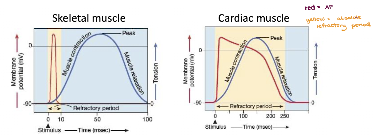
How is the absolute refractory period of a cardiomyocyte different? Why is this necessary?
The absolute refractory period lasts throughout the plateau period (Na activation gates still closed
No new contraction until the muscle completes contraction and relaxation
This allows time for the ventricles to refill and prevents tetanus
How are action potentials triggered in the heart muscle?
We have an intrinsic conduction system with autorhythmic cells
Spontaneous APs propagate to cardiomyocytes and cause contraction
Repolarization stimulates the opening of funny Na+ channels, followed by Ca2+ channels
No real resting potentials
What is the SA node?
Sinoatrial (SA) node
Collection of autorhythmic cells that can generate APs faster than any other part of the heart
More voltage-gated channels
Sends ions through gap junctions
Pacemaker of the heart — determines the heart rate
How do electrical signals travel through the heart based on the SA node? How do we control where contraction starts?
SA node in the right atrial wall generates an AP that travels via gap junctions to the AV node and the left atrial wall
At the AV node, the impulse is delayed for 0.1 s, allowing for the atria to start and complete their contraction before ventricles contract (promotes blood flow in the heart)
From the AV node, the impulse spreads to the bundle and its branches ( = only electrical connection between the atria and the ventricles)
No gap junctions exist between the cells in the septum and their neighboring myocytes so we can control where contraction begins — contract at the bottom so the blood pushes upwards
The impulse sweeps to the ventricular wall via the Purkinje fibers and ventricular muscle cells contract
What occurs is there is homeostatic imbalance in the heart? How can we fix a faulty SA node?
Arrhythmias = defects in the intrinsic conductive system
Ex: SA stops functioning, AV node takes over pacemaker role (lowering HR)
An artificial pacemaker is an instrument placed beneath the skin, equipped with electrodes that extend to the heart
Provides an electrical stimulus to the heart at a set frequency
What is Ventricular Fibrillation? How do we treat this?
Different autorhythmic cells fire at their own pace, not following the SA node — many pacemakers
Ventricles fail to function as pumps → circulation stops and brain death occurs
Defibrillation = electric shock to the heart
Simultaneous depolarization to all muscle cells
If works, SA takes over again, being the fastest autorhythmic cell
What is the cardiac cycle? What drives it?
All events associated with blood flow through the heart during a single heart beat
Blood flow is driven by pressure changes that are produced as a result of muscle contraction (or addition of fluid)
Systole = contraction of the heart muscle
Diastole = relaxation of heart muscle
Both atria and ventricles have their own systole and diastole
What are the stages of the cardiac cycle?
Late atrial and ventricular diastole (all chambers relaxed)
AV valves open
Semilunar valves closed
Ventricular filling from the atria
Atrial systole (ventricles are still relaxed)
AV vales open
Semilunar valves closed
Ventricular filling from the atria, more blood is now flowing into the ventricles
Ventricular systole (atrial diastole)
AV valves closed — pushed by backflow of blood from ventricles
Semilunar valves closed
Internal pressure is generated in the ventricles
Isometric contraction
Ventricular systole — ejection phase (atria in diastole)
AV valves closed
Semilunar valves open (ventricular pressure rises to 120 mm Hg)
Ventricular pressure exceeds the pressure in the aorta and blood is expelled into the aorta
Isotonic contraction
Early diastole
AV valves closed
Semilunar valves closed (ventricular pressure drops and backflow from aorta closes valves)
Most blood ejected to aorta, some leftover in ventricles
The atria, still in diastole is filled with blood from the veins
→ step 1, passive blood flow into ventricles

What are End Diastolic Volume (EDV) and End Systolic Volume (ESV)?
End Diastolic Volume (EDV): amount of blood in a fully relaxed ventricle (~120 mL)
At the end of diastole
End Systolic Volume (ESV): amount of blood left in the ventricle after contraction (~ 50 mL)
Blood REMAINING at the end of systole
What is Stroke Volume? How is it related to Cardiac Output?
Stroke Volume: amount of blood pumped out by the ventricles during each heartbeat
= EDV - ESV
~ 70 mL
Cardiac Output: amount of blood ejected from the left ventricle into the aorta per minute
= Stroke Volume * HR
About 5.25 L/min in a healthy adult
How can we change cardiac output? How do we increase SV (in terms of EDV and ESV)? What are the three ways we can regulate SV?
CO = stroke volume * HR
We can increase SV by either increasing EDV (blood entering the ventricle) or decreasing ESV (blood left in the ventricle)
Regulating SV:
Preload — the amount of tension (degree of stretching) of the ventricular muscle prior to its contraction
Contractility — amount of force produced during a contraction at a given preload
Afterload — pressure that the ventricle has to overcome to force open the semilunar valve
What is preload? How does it impact stroke volume and cardiac output?
Preload: the amount of tension of the ventricular muscle before it contracts
More overlap = greater contraction
Increases blood volume (EDV) → Increase in preload
Frank-Starling’s Law of the Heart: The more stretched the resting fibers, the more forceful the contraction
Increased venous return → increased ventricular filling → increased preload → increased force of contraction → increased stroke volume → increased cardiac output
What is contractility and how does it affect stroke volume and cardiac output? What systems control contractility?
Contractility: amount of force produced during a contraction at a given preload
More Ca2+ to bind to troponin = more contraction
Controlled by the nervous system and endocrine system
Sympathetic neurons secrete norepinephrine
Adrenal medulla secretes norepinephrine and epinephrine
→ opening of Ca2+ channels in the cardiac muscle (increase of intracellular Ca2+, more myosin-actin interactions, stronger contraction)
What is afterload and how does it affect stroke volume and cardiac output?
Afterload: the pressure that the ventricle has to overcome to force open the semilunar valves
Resulting from the blood pressure in the aorta (how strong the backflow pushes the valves)
Increased arterial pressure → increased afterload → decreased blood volume ejected into the artery → increased ESV → decreased stroke volume
How does the autonomic nervous system and endocrine system regulate the heart rate?
Parasympathetic innervation:
Releases acetylcholine (ACh) — ACh has inhibitory effect in cardiac muscles, and excitatory effect in skeletal muscles
Hyperpolarizes membranes of SA nodal cells
Lowers heart rate
Sympathetic innervation:
Releases norepinephrine
Depolarizes membranes of SA nodal cell
Raises heart rate
Adrenal medulla hormones:
Epinephrine and norepinephrine released
Same effects as sympathetic nerves (raises HR)
What are the main types of blood vessels? Which are rich in oxygen?
Arteries: carry blood away from the heart
Systemic (rich in oxygen) and pulmonary
Veins: carry blood toward the heart
Systemic and pulmonary (rich in oxygen)
Capillaries: contact tissue cells and directly serve cellular needs
Gas/nutrient exchange
Waste collection
Circulation
What is the driving force for blood flow through the body?
Blood pressure gradient → highest to lowest
Blood pressure (∆P): force per unit area exerted on the wall of a blood vessel by its contained blood
Highest BP in the aorta (closest to the pump), lowest in the venae cavae — falls gradually due to friction
Is 0 mm Hg in the right atrium
How does arterial pressure fluctuate?
With heart contractions
Ventricular contraction:
Ventricle contracts
Semilunar valve opens
Aorta and arteries expand and store pressure in elastic walls
Ventricular relaxation:
Isovolumetric ventricular relaxation
Semilunar valve shuts, preventing back flow into the ventricles
Elastic recoil of arteries sends blood forward into the rest of the circulatory system

What is systolic and diastolic pressure? What is MAP?
Systolic pressure — pressure exerted on arterial walls during ventricular contraction (120 mm Hg)
Diastolic pressure — lowest level of arterial pressure during a ventricular cycle (80 mm Hg)
Creates afterload
Mean Arterial Pressure (MAP): mean between systolic and diastolic pressures
Pressure that propels the blood to the tissues
94 mm Hg — diastole is longer than systole
Systemic BP = Arterial BP
Why does the local blood pressure decline in the arterioles?
The diameter of the arterioles is smaller than one of the arteries (more resistance)
The length of the arterioles is greater than one of the arteries (more mileage)
How does blood return to the heart given that veins encounter weak BP? How does exercise influence this?
BP declines to <15 mm Hg when it reaches the veins
The contraction of skeletal muscles “milk” blood toward the heart
Valves prevent back flow during venous return
Exercise causes more squeezing, increasing venous return and therefore EDV
What are blood flow, blood pressure, and resistance? How are they related at the systematic level?
Blood Flow (F): volume of blood flowing through a vessel, an organ, or the entire circulation at a given period
Blood Pressure (∆P): force per unit area exerted on the wall of a blood vessel by its contained blood
Resistance (R): opposition to flow (measure of the amount of friction blood encounters
Cardiac Output = MAP / Peripheral Resistance

Why do we need to keep systematic blood pressure relatively constant? How do we do this?
If MAP is:
Too low → not enough blood flow
Increase cardiac output and/or resistance
Too high → damage to walls of the blood vessels
In normal conditions, cardiac volume is relatively constant, so we change resistance by changing vessel diameter
How does changing BV diameter impact systemic BP?
Resistance varies inversely to the fourth power with vessel radius
We constrict/dilate the arterioles because there is more muscle tissue around them, which impacts MAP more generally
BP too low → vasoconstriction via muscle contraction
BP too high → vasodilation via decreasing signaling
Original state is partially constricted (vasomotor tone)
How do we control systematic BP in the long-term as opposed to the short term?
Short-term: Counteracting moment to moment fluctuations by altering peripheral resistance and heart rate
Long-term: Regulating blood volume (e.g. reducing cardiac output by increasing urine production)
How is blood flow regulated locally?
Smooth muscle cells (sphincters) encircle capillaries as they branch from the arterioles
Regulation of blood flow into the capillary is achieved through contraction and relaxation of the sphincters
Closing sphincters will reduce local blood flow, but increase MAP
What are some impacts of chronic high blood pressure? What are some treatments?
Hypertension can cause local ruptures, hemorrhage, and damage to wall elasticity (which may result in a stroke)
Increased resistance to ventricular contraction = increased afterload
Causes the left ventricle to need to push more in response
May result in a heart attack
Treatment: drugs that promote vasodilation and drugs that increase urine production (reducing blood volume)