Biology Lab practical Exam I: Studyguide
 Call Kai
 Learn
Learn Practice Test
 Spaced Repetition
 Match
Match Flashcards
 Knowt Play
1/38

There's no tags or description
Looks like no tags are added yet.
Name | Mastery | Learn | Test | Matching | Spaced | Call with Kai | Chat |
|---|
No analytics yet
Send a link to your students to track their progress
39 Terms
Lab 1: Microscope Handling
Proper handling:
One hand on the arm, support the bottom
Lab 1: Microscope Usage
When using:
Focus light on specimen, then close iris diaphragm, then look
Adjust light if needed
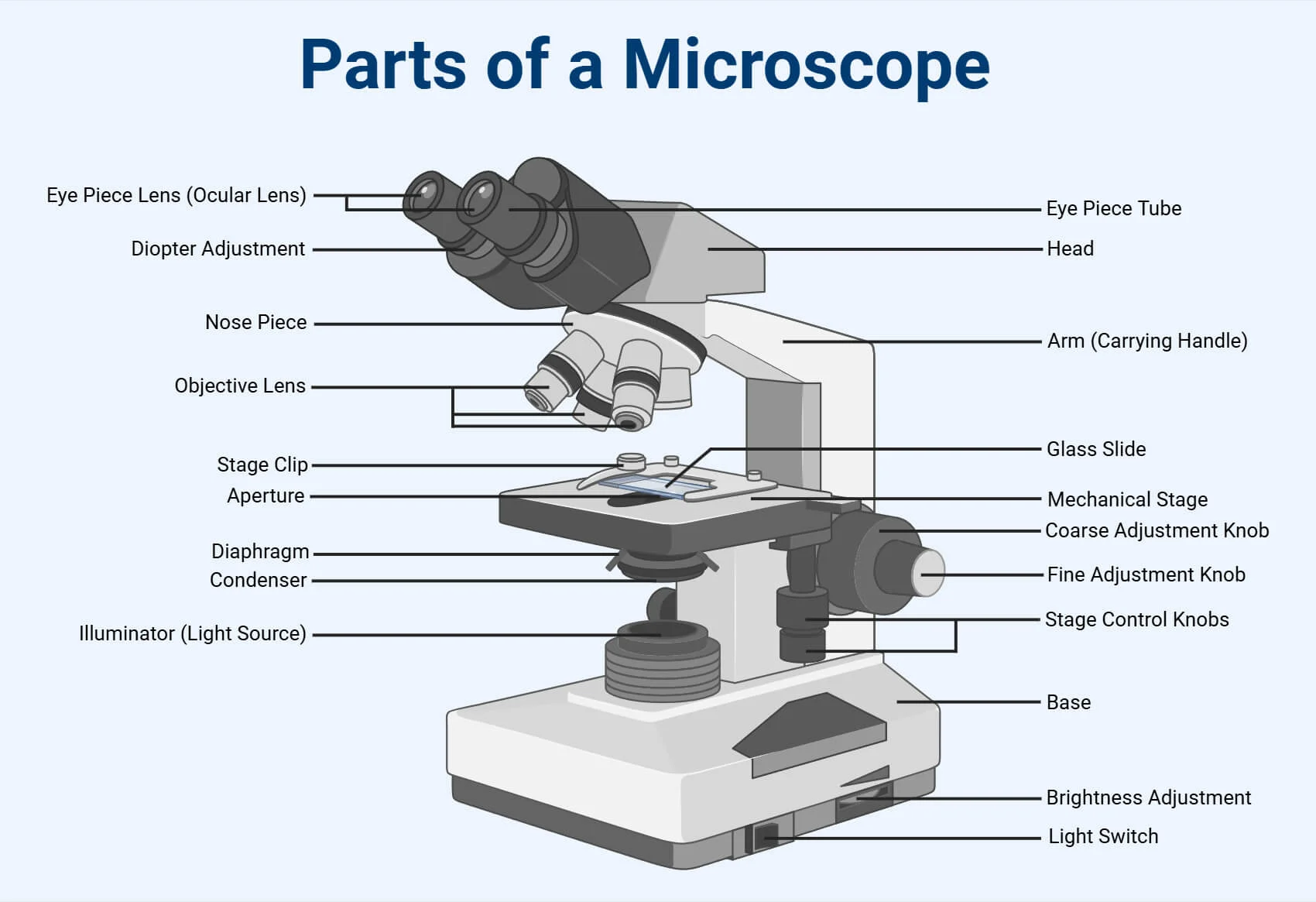
Lab 1: Microscope Anatomy
Lab 1: Letter e Slide
Letter e slide:
Naked eye = normal
Microscope = upside down and flipped
Lab 1: Total magnification
Ocular lens (usually 10) x (Objective lens) = total magnification
Example 10 × 4 (objective lens) = 40x (total magnification)
Lab 1: Diameter
Increasing diameter = field of view decreases
Lab 1: Wet Mount
Drop size of a penny
45 degree angle drop
Lab 2: Dialysis Tubing
In bag = 1% albumin, 3% NaCl
In solution = 0.25% starch, 1% Na2SO4
Thesis: The larger particles will not move. Smaller ones will move. water moves from high to low concentration.
Starch stays outside the bag, albumin stays inside the bag. Na2SO4 stays outside the bag then moves inside. NaCl will stay inside the bag then move outside the bag.
Lab 2: Methods
Sample 4 1mL samples for the inside and outside then place into tubes
Fill dialysis bag then weigh
Cover bag with solution
Rise off bag
At 15m intervals, swirl the beaker
Remove bag and dry, reweigh
Take samples from inside (tubes, 1mL)
Put liquid into beaker
Take samples
Lab 2: Results
Na2SO4
Start: outside yes
End: outside yes
Start: inside no
End: inside yes
NaCl
Start: outside no
End: outside yes
Start: inside yes
End: inside yes
Protein
Start: outside none
End: outside none
Start: inside none
End: inside yes
Starch
Start: outside yes
End: outside none
Start: inside no
End: inside none
Lab 2: Tests
Add stripes to tubes. When green, protein is present
Add 3 drops of 2% BaCl = tests for suflate
Add 3 drops of LUGOL to tubes = tests for starch
Add 3 drops of 5% of NaCl to tubes = test for cl ions
Add 3 drops of silver nitrate = test for iodide ions
Lab 2.5: Spectrophotometer
Plug in spectrophotometer, let it warm up and initialize
Use modern interface and live display
Blank the spectrophotometer, set to 0 A (absorbance)
Bromophenol demonstration
Measure absorbance, highest absorbance at wavelength it rh optimal wavelength
580
Lab 2.5 Spectrophotometer test - Concentration and Absorbance for 8 Dye Dilutions at 580nm
Set wavelength to 580nm
Label tubes
Fill according to chart then chart absorbance
Lab 2.5: Charting
Find total mg Dye = mL of dye added x dye concentration (0.02mg/ml)
Divide this 4 by = concentration
Chart the concentration (x) and absorbance (y)
Using the absorbance of the unknown, dot over to the line of best fit, go down and now you have the concentration
Lab 3: Enzymes
Enzyme substrate complex and induced fit models are more accurate
Puts strain on bonds to lower activation energy and form product
Lab 3: Enzyme Methods
Fill tubes according to chart
Adjust spectrophotometer to 0A at 470nm
Mix tubes in pairs (start timer), place in spectrophotometer and record absorbance in 20s intervals
Lab 3: Enzyme tests
Concentration
pH
Lab 3: Graphs
Graph absorbance (y) over time (s)
Find the slope
Graph slopes (pHs)
Lab 3: Results
pH 5 was optimal (highest slope/increase)
Lab 4: Plants Intro
Gymnosperm: non-flowering plants with exposed or "naked" seeds usually found on cones
Angiosperm: flowering plants with seeds enclosed in a fruit
Monocotyledon: one seed leaf
Dicotyledon: two seed leaves
Lab 4: Organs, tissue, cells
Organs
Roots
Stem
Leaves
Tissues
Dermal
Vascular
Ground
Cells
Parenchyma
Collenchyma
Sclerachyma
Lab 4: Growth
Apical meristems: grow up
Lateral meristem: grow in girth
Primary: tips
Secondary: girth
Lab 4: Plant Terms
Xylems: regulate movement of water, growth in rings
Phloem: outer bark, transports photosynthetic product
Lab 4: Dicot and Monocot
Roots:
Monocot: Vascular bundles neatly in ring
Dicot: Vascular bundles scattered, vascular bundle in the center
Leaf:
Monocot: small skull big skull small skull
Dicot: Vascular line, more open space, larvae
Stem:
Monocot: Vascular bundles scattered
Dicot: Vascular bundles on outer ring
Lab 4: Diagram
Xylem vessels are for bulk water transport
Casparian strip forces water to flow through cells
Sieve tube has no nucleus, transports sugar/water
Pith: center
Lab 4: Stem Tip
Apical meristem
Lateral buds
Lab 4: Leaflet growth
Simple grows single from lateral bud
Compound grows leaflets extending from one compound
Lab 4: Phyllotaxy
Alternate with buds
Opposite with two on each side of the buds
Whole is multiple on one bud
Lab 5: Muscle Types Smooth
Smooth
lines digestive system
walls of blood vessel
spindle shaped

Lab 5: Muscle Type Cardiac
Cardiac
striations, intercalated discs, single nucleus

Lab 5: Muscle Type Skeletal
Skeletal
striations, multiple nuclei, cylindrical fibers

Lab 5: Muscle fiber and anatomy

Lab 5: inside the Myofibril

Lab 5: Bone
Canaliculi - pathway
Lacunae - cavities
Osteocyte - bone cell in mineralized bone matrix
Haversian canal - transmits blood vessels and fibers
Lamellae - structural support

Lab 5: Skeleton
Exoskeleton - spiders, crabs, beetles
Protection, locomotion
Endoskeleton
Lab 5: Axial and Appendicular
Axial: skull, vertebral column, ribs
Cervical (neck)
Thoracic (torso)
Lumbar (lower back)
Sacral (tiny lower triangle)
Cocyx (tail)
Appendicular: pectoral girdle (clavicle, scapula, sternum), forelimb (humerus, radius, ulna), pelvic girdle (coxa, sacrum), hindlimb bones (femur, tibia, fibula)
Lab 5: Joint
Ball and socket
Humerus and pectoral girdle
Femur and pelvic girdle
360 degree motion
Hinge joint
Limits motion to plane
Femur and tibia
Fingers
Fibula
Humerus and ulna
Pivot joint
Radius and humerus
Suture joint
Immovable parts of skull
Fused bomes of the coxa/skull
Lab 5: Labeling the Skeleton
