ANAT 100- Module 3 muscles 낱말 카드
 Call Kai
 Learn
Learn Practice Test
 Spaced Repetition
 Match
Match Flashcards
 Knowt Play
1/50

There's no tags or description
Looks like no tags are added yet.
Name | Mastery | Learn | Test | Matching | Spaced | Call with Kai | Chat |
|---|
No analytics yet
Send a link to your students to track their progress
51 Terms
Functions of Muscles
- movement (moving the skeleton)
- beating heart
- digestion
- constriction of blood vessels
Types of Muscle
- skeletal
- cardiac
- smooth
Skeletal Muscles
- helps the human body move
- most skeletal muscle is attached to the bones via tending
- striated
- multi nucleated
- Voluntary Movement
- makes up about 1/2 of a persons weight
- each muscle is composed of skeletal muscles, epithelial, connective, and nervous tissue

Cardiac Muscles
- found in the heart
- contracts rhythmically from signals from neurons and hormones
- striated
- uni/bi nucleated
- involuntary movement
Smooth Muscle
- controlled by the nervous system or hormones
- may be generally inactive and then response to stimulation, or it may be rhythmic
- NOt striated
- uni nucleated
- involuntary movement
Characterization of Muscle
Morphological characterization
Functional Characterization
Morphological characterization
Striated Muscle
Smooth Muscle
Morphological characterization: Striated Muscle
- cardiac and skeletal muscle
- marked by light and dark band and long muscle fibres
- nuclei are located PERIPHERALLY for skeletal muscles
- nuclei are located centrally for cardiac

Morphological characterization: Smooth Muscle
- found in blood vessels, the digestive system, and other viscera
- each smooth muscle fibre contains a single centrally located nucleus
- fusiform-shape (spindle like) cells without striations

Functional Characterization
Voluntary muscles
- muscles that are consciously controlled
- skeletal muscles
Involuntary Muscles
- muscles that are no consciously controlled
- cardiac and smooth muscle
How to Muscles Function
Excitability:
- the muscle can respond to electrical signals from nerves or stimulation from hormones
Contractility
- this then causes the muscle to shorten, causing a contraction
Skeletal Muscle: Functions
- contracts to move parts of the body
- most muscles are attached to 2 bones across a joint, so the contracted muscles bring those 2 bones closer together
- maintain posture and stabilize joints
- control excretion (pee and poo)
- produce heat
- support and protect internal organs
Organization of Skeletal Muscle
- connective tissue surrounds the muscle tissue and attaches the end of the muscles to the bone
Connective Tissue in Muscles
1. Epimysium
2. perimysium
3. Endomysium
1. Epimysium
- layer of connective tissue on the outside of the entire muscle that becomes a tendon

2. Perimysium
- layer of connective tissue that surround bindles of muscle fibres (fascicles) inside the muscle

3. Endomysium
- layer of connective tissue that surround an individual muscle cell (muscle fibre/myofibre) in each bundle (fascicle)

Muscle cell is called
- myofibre
Microscopic anatomy of muscle tissue
- inside a muscle cell (myofibres), there are MYOFIBRILS which contain MYOFILAMENTS (the units of contraction from the muscle cell)

Microscopic anatomy of muscle tissue: Sarcolemma
- cell membrane surrounding the muscle cell (myofibre)
- different than the endomysium
- under the sarcolemma are nuclei, myofibrils, and sarcoplasm (cellular fluid)

Microscopic anatomy of muscle tissue: Nuclei
- skeletal cells are multinucleated
- nuclei are located towards the outside if the myofibre (muscle cell)
Microscopic anatomy of muscle tissue: Myofibril
- structure unit of muscle cells (myofibres)
- contain contractile myofilaments

Microscopic anatomy of muscle tissue: Myofilaments
- contractile unit of the muscle cell

Microscopic anatomy of muscle tissue: Sarcoplasmic reticulum
- surround each myofibril and is where the muscle cell stores calcium (important for muscle function)

Microscopic anatomy of muscle tissue: T-tubules
- extensions of the sarcoplasmic that surround the myofibrils and transmit nerve stimulation to the sarcoplasmic reticulum within the cell
Myofilaments: Sarcomeres
- myofilaments are organized into repeating structure called sacromeres
- when shortened, sarcomeres cause contraction of the muscle (each unit consists of actin or myosin)
- 1 sacromere spans from 1 Z line to the next
Sarcomere
- on diagram in book, myosin is the thick brown centre lines, actin is the little red ones attaching to it

Sarcomere Banding
A band
- made up of thick and thin filaments
- appears darker
Z-line
- protein that makes a zig zag line marking the beginning and end of each sarcomere
- in the muddle of the I-band and its where the thick filaments attach
M-line
- Protein down the centre of the sacromere
- thick filaments attach
I-Band
- made up of thin filaments
- appears lighter

Muscle Contraction
- actin and myosin filaments on sarcomeres slide over each other, which shortens the sarcomere
- increases muscle tension

The lighter and darker parts of sarcomeres
I-Band
- light because it is all light filaments
Z-line
- dark because it has lots of protein
A-Band
- dark because it is all thick filaments, even darker where is overlaps with actin (on the side)
M-line
- dark because it has lots of protein

Axial Muscles: Facial Expression Muscles
- muscles of facial expression insert into the skin
- contracting muscles move the skin
zygomaticus
orbicularis oris
orbicularis oculi
frontalis
Facial Expression Muscles: Zygomaticus
- extends from the zygomatic arch to the corners of the mouth
- draws the angle of the mouth superiorly and posteriorly, creating a smile

Facial Expression Muscles: Orbicularus oris
- surrounds the mouth and puckers the lips
- originates on the maxillary bone
- kissing muscle

Facial Expression Muscles: Orbicularis oculi
- muscle surrounding the eye that forcefully closes it
- originates in the frontal and maxillary bones

Facial Expression Muscles: Frontalis
- muscle that covers the frontal bones and lifts the eyebrows, causing the forehead to wrinkle

muscles of mastification (chewing)
Temporalis
massester
Temporalis
- muscle that extends from the temporal fossa of the parietal bone to the cornalid process of the mandible
- pulls the mandible back

Masseter
- powerful muscle that extends from the zygomatic arch to the angle of the mandible (lower jaw)
- elevates and protracts (move jaw forward) the jaw

3 muscles that move the head
- sternocleidomastoid muscle (anterior)
- semispinalais capitis (posterior)
- splenius capitis (posterior)
Sternoclediomastoid
- flexes the neck, and rotates the head to the opposite side
Semispinalis capitis
- turns the face slightly to the opposite side

Splenious Capitis
- extends the neck, causing flexion and lateral rotation of the neck
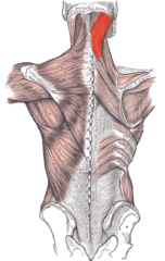
Muscles of the Thorax (muscles that help with the process of breathing)
External Intercostal
Internal Intercostal
external intercostals
- most superficial
- help in breathing in
- fibres run: imagine putting hands in to pockEt

internal intercostals
- deeper than external intercostals
- help in breathing out
- fibres run: imagine putting hands on tIts
Muscles of the Back
Erector Spinae muscles
- run down both sides of the spinal column

Muscles of the abdomen wall (help with breathing)
External Oblique
Internal Oblique
Transversus Abdominus
Rectus Abdominus
External Oblique
- most superficial
- functions:
bilateral- flexes the vertebral column and compresses the abdominal wall, used in forced expiration
unilateral- lateral flexion and rotation of the spine
FIBRES: ANTERIORLY AND INFERIORLY

Internal Oblique
- deeper than the external oblique
functions:
- bilateral- flexes the vertebral column and compresses abdominal wall
- unilateral- lateral flexion of the vertebral column
FIBRES: ANTERIORLY AND SUPERIORLY

Transversus Abondminus
- runs horizontally, deep to the internal oblique
functions:
- bilateral: flexes the vertebral column and compresses the abdominal wall. Used in forced expiration
- unilateral: lateral flexion of the vertebral column

Rectus Abdominus
- lies on either side of the line alba (line of connective tissue down the middle of the abdomen)
- separated why tendinous intersections
function:
- flexion of the trunk
- forced expiration
