Intro to Diagnostic Imaging, Intro to Gross Anatomy 1&2
 Call Kai
 Learn
Learn Practice Test
 Spaced Repetition
 Match
Match Flashcards
 Knowt Play
1/75

There's no tags or description
Looks like no tags are added yet.
Name | Mastery | Learn | Test | Matching | Spaced | Call with Kai | Chat |
|---|
No analytics yet
Send a link to your students to track their progress
76 Terms
Conventional X-ray
Use a form of ionizing radiation to create images of the body for clinical evaluation
Best for evaluating skeletal structures
Air in Xray
Due to simple molecular structure, most of the xray beams will pass through and reach the detector leading to it appearing as dark regions on xray images
Fat in Xray
It is slightly denser than air but less than soft tissue. Appears darker gray shade on radiographs
Soft tissue in Xray
Soft tissues such as muscles and organs have higher density than fat, allowing them to absorb more x-rays. They appear as lighter grays
Bone in Xray
Denser and contains calcium absorbs a greater amount of xrays compared to soft tissue and fat
Metal in Xray
A very dense structure and contains atoms with heavy atomic weights, which absorb most of the incident x-rays
CAT scan/CT Scan
uses x-ray to produce using different technique but provide more information than conventional x-ray
Good for tissue evaluation
CAT scan/CT Scan
Housed in a rotating gantry that spins around the patient on a horizontal table
A helical CT, both rotates and the table moves continuously, creating a spiral data path for faster and smoother images
Magnetic Resonance Imaging(MRI)
Produces images similar to CT but shows greater structural detail
Excellent for soft tissue evaluation but not for bone
Generation of MRI Images
Protons of the body is aligned with the caudal.cranial axis by the magnetic field of the MRI
Protons snap back to align with the magnetic field when the
radio wave ceases. They snap back into alignment with the
magnetic field, as protons liberate energy.
Liberated energy is detected by sensor coils surrounding
specific regions of the patient’s anatomy (e.g. knee coils,
head coils).
signal strength
Different tissue appearance is dependent on the ________ when protons snap back to align with the magnetic
field.
Ultrasound
Relies upon high frequency, inaudible and sound arising from a piezoelectric crystal, in a hand held transducer, for the creation of images
acoustic gel
Ultrasound transmission is facilitated by —— which helps to minimize the air-gap
The returning sound is altered in wavelength and intensity
and arrives back to the probe after being reflected from
tissues of different depths beneath the skin.
○ The ultrasound machine assigns a pixel brightness to a
particular tissue
gray scale
complex, computer driven, algorithm determines the
characteristics of the returning echoes and displays the
sound on a
Echoic
Sound is reflected back from the tissue to be
measured by the ultrasound machine.
○ Anatomy appears white when sound waves are
reflected back to the ultrasound transducer.
Anechoic
Sound is not reflected back to the ultrasound
probe from the tissue.
○ Anatomy appears black when sound waves are not
reflected back to the ultrasound transducer.
Hypoechoic
The sound reflected back to the ultrasound
probe is decreased in comparison to some other tissue in
the anatomy being assessed
○ The bile in the gallbladder
Hyperechoic
The sound reflected back is increased in
comparison to some other tissue in the anatomy being
assessed
○ The liver is hyperechoic compared to the bile in the
gallbladder.
Doppler principle
Principle which is utilized by the ultrasound to analyze the flowing fluids and determine if there is a frequency shift over the length of a structure
Color can also be applied
BART
Blue away, Red towards
Convex/Curved transducer
ultrasound transducer that is Optimal for abdominal and obstetric imaging,
Linear probes
Ultrasound transducer that is Best adapted to vascular and small superficial structures (i.e. thyroid)
Long, wand-shaped ultrasound transducer
Can reach the gland via the rectum (endo-cavitary
probe).
Can be used for intra-vaginal image acquisition for
the assessment of female pelvic anatomy (i.e.
uterus, endometrium, and ovaries).
Higher frequency
Characteristic for transducer: Limited depth of field but provide better image
resolution of the near field anatomy
Lower Frequency
Best for deep tissue imaging
■ Do not provide imaging details of the subcutaneous
tissues
X-Axis (Horizontal / Transverse)
Paired axis with sagittal plane
Y-Axis (Vertical)
Paired axis with Horizontal plane
Z-axis(Antero-posterior)
Paired axis with coronal plane
Hyperextension:
Extension beyond the neutral position
Can occur in the shoulder, hip wrist and neck
Dorsiflexion
Foot and toes are off the ground
Plantar flexion
Bends the foot or toe towards the ground
■ Similar to standing on tiptoes
Abduction and adduction
Movement that generally occur in the coronal and z axis
Trunk and neck
Lateral flexion occurs in which parts
Ulnar deviation
deviation: hand and fingers move to the ulnar
side (pinky side)
Radial deviation
hand and fingers move to the radial
side (thumb side)
Inversion
sole of the foot faces towards the median
plane
Eversion
sole of the foot faces away from the median
plane
Circumduction
movement that involves sequential flexion,
abduction, extension, and adduction of the extremity
Internal rotation

External rotation
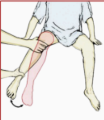
Protraction and retraction
Movements in the temporomandibular joint and scapula on the thoracic wall
Opposition
Movement of the pad of the 1st digit of the thumb to the pad of other digits
Coronal plane
Plane of the thumb when the Movement is flexion/extension
Sagittal plane
lane of the thumb when the Movement is Abduction/Adduction
Plane: Sagittal
Axis:X-axis
Movements: Flexion/Extension
Elbow/Knee
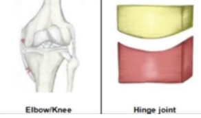
Plane: Horizontal
Axis:Y-axis
Movements: Rotation of one bone around another
Top of the neck

Plane: Multiple PLanes
Axis: Multiple Axes
Movements: Flexion/Extension, Abduction/Adduction, Internal/External Rotation
Shoulder/Hip

Plane: Coronal and SAgittal planes
Axis: Z and X-axis
Movements: Flexion/Extension, Abduction/Adduction, Circumduction
Saddle Joint

Plane: Coronal and SAgittal planes
Axis: Z and X-axis
Movements: Flexion/Extension, Abduction/Adduction, Circumduction
Condyloid Joint

Ipsilateral
Laterality meaning same side
Contralateral
Laterality meaning opposite side
Bilateral
Laterality meaning paired; both sides
Unilateral
Laterality meaning one side
size, shape, border, consistency, mobility and laterality/location
Creating a correct anatomical description
Consistency
Soft, firm, hard
Border
Smooth or irregular
size
cm
shape
if its round or oval
Medical record
Immortalized version of what happened
on the day of physical examination.
SEE
Observe the symptom or sign. Identify what is
actually present before interpreting it.
SAY
Describe it with standard anatomical and
examination terminology.
DECIDE
Use location and distribution to narrow the
differential diagnosis.
DO
Choose the next examination, investigation,
procedure, or management step safely.
Site & Side, Region & Depth, Landmark relation, Distribution and radiation
A clinically useful description should answer:
History
— A symptom without a precise location remains
broad. Localization converts a complaint into an anatomical
problem.
Physical examination
A sign without a landmark is
difficult for another examiner to reproduce or compare.
Procedure/management
An intervention without
a map risks the wrong site, wrong layer, wrong structure, or
injury to adjacent anatomy.
Localization error in the history
An error which propagated downstream which led to attention directed to the wrong organ system
Imprecise examination description
An error which propagated downstream which led to the next clinician who cannot reproduce the finding
Incorrect laterality
An error which propagated downstream to the wrong side investigation, procedure and treatment
Failure to appreciate anatomical variation
An error which propagated downstream which led to the increased procedural or operative injury
Poor contemporaneous documentation
An error which propagated downstream which led to weak continuity of care and weak medico-legal defensibility
Patient’s perspective
The laterality(R & L) always refer to the —— not the examiners
Supine
Face upward
Lateral decubitus
Face downward
Trendelenburg
Head-down tilt/ semi prone
Lithotomy
Hips/knees flexed’ thighs abducted