Nucleic Acids Part 2
 Call Kai
 Learn
Learn Practice Test
 Spaced Repetition
 Match
Match Flashcards
 Knowt Play
1/19

Earn XP
Description and Tags
Lecture
Name | Mastery | Learn | Test | Matching | Spaced | Call with Kai | Chat |
|---|
No analytics yet
Send a link to your students to track their progress
20 Terms
Nuclei acids are not just carries of genetic information, but also capable of forming……
Key message: Nucleic acids are versatile molecules, not just carriers of genetic information but also capable of forming complex shapes for catalysis, regulation, and binding.
This lecture blends history, chemistry, and structural biology, giving you a clear understanding of how discoveries in nucleic acids have shaped molecular biology and biotechnology.
Info card on what this lecture is about
This lecture sets the stage for a deeper exploration of DNA, RNA, and functional nucleic acid structures beyond basic definitions, focusing on their structural diversity and biological roles.
Lecture Summary
This lecture provides a comprehensive overview of nucleic acids, starting with their historical discovery and moving through structural features and functional diversity:
Key historical milestones: Summarizes contributions of pioneers like Friedrich Miescher, Phoebus Levene, Frederick Griffith, Oswald Avery, Erwin Chargaff, Rosalind Franklin, and the Nobel Prize-winning work of Watson, Crick, and Wilkins.
Chemical and structural insights: Discusses Chargaff’s rules, Franklin’s X-ray data, and the evidence that led to the double-helix model, including variations such as A-DNA, B-DNA, and Z-DNA.
Functional molecules and secondary/tertiary structures:
Examines usual structural motifs such as G-quadruplexes.
Introduces synthetic molecules (DNA aptamers and DNAzymes) and their applications.
Explores natural RNA molecules—tRNA cloverleaf to L-shaped 3D form, riboswitch aptamers, ribozymes, and rRNA secondary and tertiary structures.
Key message: Nucleic acids are versatile molecules, not just carriers of genetic information but also capable of forming complex shapes for catalysis, regulation, and binding.
This lecture blends history, chemistry, and structural biology, giving you a clear understanding of how discoveries in nucleic acids have shaped molecular biology and biotechnology.
Briefly mention what these scientists contribution to genetic material and DNA structure
Friedrich Miescher
Phoebus Levene
Frederick Griffith
Oswald Avery
Erwin Chargaff
Maurice Wilkins and Rosalind Franklin
James Watson, Francis Crick, Linus Pauling,
This slide covers the genetic material and DNA structure, emphasizing the historical context and major contributors to the discovery.
Top section: Features photographs of three scientists (with names and Nobel Prize share noted) who were awarded the 1962 Nobel Prize in Physiology or Medicine for their work on DNA: Francis Crick, James Watson, Maurice Wilkins. The Nobel citation recognized their discoveries concerning the molecular structure of nucleic acids (specifically DNA) and its significance for information transfer in living material.
Middle section: Notes that the Nature publication by Watson and Crick’s DNA double helix model (discussed in Lecture 1) marked the culmination of nearly a century of research into genetic material and nucleic acids.
Bottom section: Lists other key scientists whose work paved the way for this breakthrough, including:
Friedrich Miescher (discovered nuclein)
Phoebus Levene (identified sugar and phosphate components)
Frederick Griffith (transformation experiments)
Oswald Avery (DNA as genetic material)
Erwin Chargaff (base pairing rules)
Maurice Wilkins and Rosalind Franklin (X-ray diffraction images)
James Watson, Francis Crick, Linus Pauling, and many others
The slide highlights that DNA’s structure and function emerged from decades of cumulative research, culminating in the double helix model, which provided the framework for modern molecular biology.

What are the key pieces of evidence used to elucidate the DNA structure, which together enabled Watson and crick to build their model?
This slide explains the key pieces of evidence used to elucidate the DNA structure, which together enabled Watson and Crick to build their model.
Main points:
Chemical and physical dimensions – Knowledge of the components of DNA, including deoxyribose sugars, nitrogenous bases, and phosphate groups, provided the basic building blocks and size constraints for the model.
Chargaff’s rules – Experimental findings showed consistent 1:1 ratios of adenine to thymine and cytosine to guanine across species, suggesting base-pairing rules.
X-ray diffraction data by Rosalind Franklin – High-quality X-ray patterns revealed critical details about DNA’s helical structure and its repeating dimensions.
Wilkins’ measurements – Additional X-ray diffraction studies by Maurice Wilkins provided estimates for DNA’s diameter and pitch (helix turn length).
Linus Pauling’s protein helix work – Demonstrated that biological polymers could form helical conformations, influencing the thinking about DNA’s shape.
gave Watson and Crick the idea of 10 base pairs between helical turn
The slide emphasizes that understanding DNA’s structure required combining chemical insights, quantitative rules, and structural imaging techniques, leading to the iconic double helix model.

What did Fredrick Miescher do?
Discovery of nuclei acids
This slide introduces Friedrich Miescher and his pioneering work on the discovery of nucleic acids.
Content:
Johannes Friedrich Miescher (1844–1895) was a Swiss physician and biologist, recognized as the first scientist to isolate nucleic acids.
In 1869, working in Felix Hoppe-Seyler’s laboratory at the University of Tübingen, Germany, Miescher extracted phosphate-rich compounds from the nuclei of white blood cells.
He named these substances nuclein, now known as nucleic acids. This discovery laid the foundation for understanding DNA and RNA as key biological molecules.
The slide underscores the historical significance of Miescher’s work, showing that the identification of nucleic acids began with chemical isolation long before their genetic role was understood.

What does Phoebus Levine do?
Chemical structure of nucleotides
This slide highlights Phoebus Levene and his contributions to understanding the chemical structure of nucleotides.
Content:
Phoebus Aaron Theodore Levene (1869–1940) was a Russia-born American biochemist known for his work on the structure and function of nucleic acids.
Levene identified that DNA contains adenine, guanine, thymine, cytosine, deoxyribose, and a phosphate group, and clarified that these components are linked in the order phosphate–sugar–base to form a unit known as a nucleotide.
He further showed that DNA consists of a string of nucleotides joined together by phosphate groups, laying the groundwork for understanding DNA’s chemical composition and its polymeric nature.
The slide underscores Levene’s key role in defining the building blocks of DNA, even though the overall double-helix structure was still unknown at the time.
determine DNA contain ATGC and the Sugar is deoxribose and contains a phosphate group
He thought nuclei acid was cyclical
He also said nucleotide or nucleic acid is a polymer but got it wrong by saying nucleic acid molecules are cyclic
Not able to explain polymeric structure of nucleic acid but determine the chemical composition of nucleic acid is super important towards the discovery of double helical structure

Who is Frederic Griffith and what did he discover?
Transforming principle
This slide focuses on Frederick Griffith and his discovery of the transforming principle, a landmark finding in genetics.
Content:
Frederick Griffith (1877–1941) was a British bacteriologist whose work provided the first widely accepted demonstration of bacterial transformation.
In January 1928, Griffith conducted experiments showing that Streptococcus pneumoniae could change from one strain to another. A harmless strain could be transformed into a virulent one when exposed to material from a heat-killed virulent strain.
He proposed the existence of an unidentified “transforming principle” or factor responsible for this change. This was a key insight suggesting that genetic information could be transferred between organisms.
The transforming factor was later identified as DNA by Oswald Avery and colleagues, confirming its role as the genetic material.
The slide emphasizes that Griffith’s work was pivotal in revealing that genes could be transferred, paving the way for the molecular era of genetics.

Who is Oswald Avery and what has he done?
DNA as genetic material
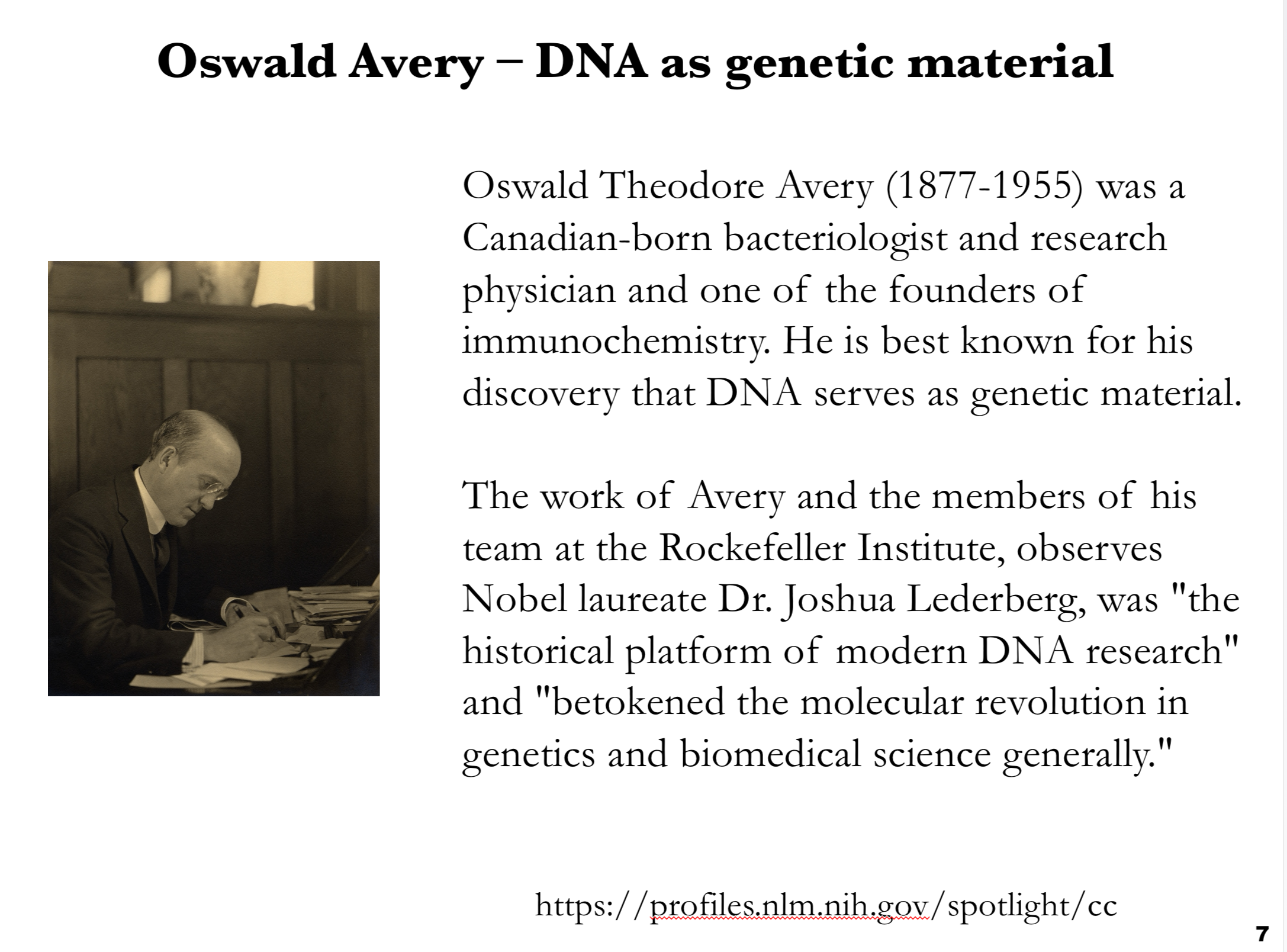
This slide highlights Oswald Avery and his pivotal role in identifying DNA as the genetic material.
Content:
Oswald Theodore Avery (1877–1955) was a Canadian-born bacteriologist and research physician, widely regarded as one of the founders of immunochemistry.
Avery is best known for his landmark discovery that DNA, not protein, is the substance responsible for heredity.
Working with colleagues at the Rockefeller Institute, Avery’s studies built on Frederick Griffith’s findings, identifying DNA as the “transforming principle.”
Nobel laureate Joshua Lederberg praised Avery’s work, calling it “the historical platform of modern DNA research” and noting that it marked the molecular revolution in genetics and biomedical science.
The slide emphasizes Avery’s critical contribution to molecular biology, establishing DNA as the chemical basis of genes and setting the stage for all subsequent genetic and genomic research.
Erwin Chargaff
G triple bond C and A=T
This slide introduces Erwin Chargaff and his discovery of the base composition rules that became key to understanding DNA structure.
Content:
Erwin Chargaff (1905–2002) was an Austro-Hungarian American biochemist and professor at Columbia University.
He is best known for establishing Chargaff’s rules, which state that in DNA:
The number of guanine (G) units equals the number of cytosine (C) units.
The number of adenine (A) units equals the number of thymine (T) units.
These relationships hinted at base pairing in DNA, suggesting complementary structures that later explained how DNA stores genetic information.
The slide emphasizes that Chargaff’s quantitative analyses were critical clues leading to the double helix model proposed by Watson and Crick.

Rosalind Franklin
Crystallographic data of DNA
This slide focuses on Rosalind Franklin and her crucial contributions to understanding DNA structure.
Content:
Rosalind Elsie Franklin (1920–1958) was an English chemist and X-ray crystallographer whose research was fundamental to revealing the molecular structures of DNA, RNA, viruses, coal, and graphite.
Her X-ray diffraction studies produced some of the most important structural data for DNA, including “Photo 51,” which provided clear evidence of the molecule’s helical nature.
While Franklin’s work on coal and viruses was recognized during her lifetime, her contributions to DNA’s discovery were mostly celebrated after her death, highlighting the delayed acknowledgment of her role in this scientific milestone.
The slide emphasizes Franklin’s central but historically underappreciated role in solving the structure of DNA, and how her data became essential for Watson and Crick’s double helix model.

What are the types of duplex DNA structures? When are they formed and what are their properties?
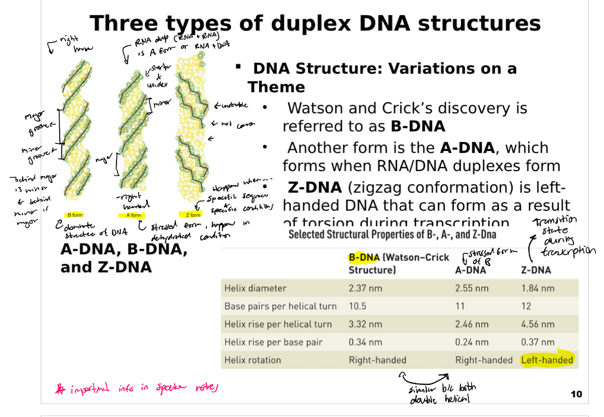
This slide introduces the three types of duplex DNA structures and explains their key differences.
Content:
B-DNA: The familiar right-handed double helix discovered by Watson and Crick. It is the predominant form of DNA under physiological conditions.
A-DNA: Another right-handed helix that is shorter and more compact. It often appears in dehydrated DNA samples or in DNA/RNA hybrid duplexes. RNA/RNA duplexes are also A-type.
Z-DNA: A left-handed helix with a zigzag backbone. It can form transiently during transcription or under torsional stress, and is thought to have regulatory roles.
Illustrations:
The slide includes side-by-side visualizations of the three forms, showing their helical handedness and shape differences.
Table of properties:
B-DNA: Diameter ~2.37 nm, 10.5 base pairs per turn, helix rise 3.32 nm per turn (0.34 nm per base), right-handed.
A-DNA: Diameter ~2.55 nm, 11 base pairs per turn, helix rise 2.46 nm per turn (0.24 nm per base), right-handed.
Z-DNA: Diameter ~1.84 nm, 12 base pairs per turn, helix rise 4.56 nm per turn (0.37 nm per base), left-handed.
The slide emphasizes that DNA is structurally dynamic, capable of adopting different conformations based on environment and function.
What are Hoogsteen Hydrogen bond and what can they form?
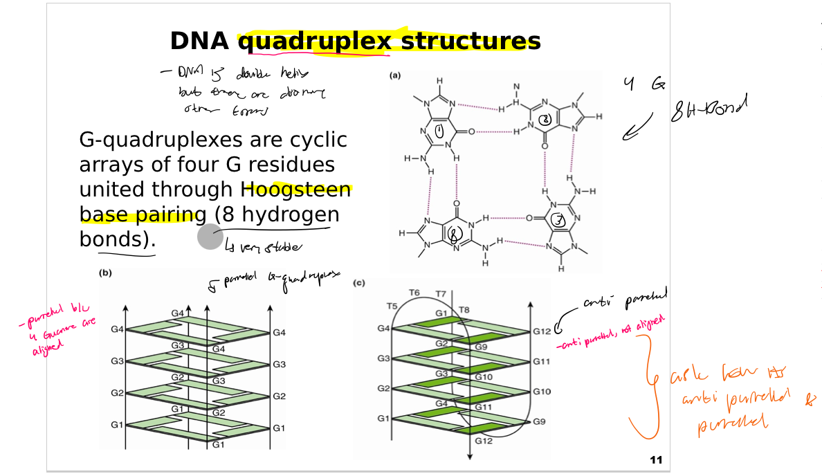
This slide introduces the DNA G-quadruplex structure, a special form of DNA folding based on guanine-rich sequences. It emphasizes how guanine (G) bases can organize into unique arrangements stabilized by Hoogsteen hydrogen bonds.
Key concept: G-quadruplexes are cyclic arrays of four guanine residues (G-tetrads) that stack on top of each other. Each tetrad is stabilized by Hoogsteen base pairing, forming eight hydrogen bonds among the four guanines.
Panel (a): Shows the chemical detail of a single G-tetrad, highlighting the cyclic arrangement and the network of hydrogen bonds (dashed lines) connecting the bases.
Panel (b): Illustrates a simple stacked G-quadruplex, where several G-tetrads stack vertically to create a stable structure. Each layer consists of four G residues, and the arrows indicate the orientation of strands. This is a parallel G-quadruplex.
Panel (c): Shows another G-quadruplex formed between two DNA molecules with connecting loops (labeled T5–T8) and additional guanines (G9–G12) forming an extended structure. This is an anti-parallel G-quadruplex.
G-quadruplexes can vary in complexity, accommodating different loop lengths and orientations.
G-quadruplexes are widely studied because they can form in telomeric DNA, regulatory regions of genes, and synthetic nucleic acid designs. Their compact and stable structures are useful for molecular recognition, aptamer design, and nanotechnology.\
(The bolder passage in easier terms) This passage means that G-quadruplexes are special, stable DNA/RNA structures that play a massive role in human biology, medicine, and advanced technology.
Here is a breakdown of what the text is saying in plain, simple terms:
1. Where they are found in biology
The text states that these structures form in specific, crucial parts of our genetic code:
Telomeric DNA: Telomeres are the protective caps at the ends of our chromosomes (like the plastic tips on shoelaces). They control cell aging and prevent DNA damage.
Regulatory regions of genes: These are the "control switches" of your DNA that turn genes on or off. G-quadruplexes found here often act as speed bumps or volume knobs for controlling how proteins are made.
2. Why their shape matters
Compact and stable structures: Instead of the classic, long double-helix ladder we usually picture for DNA, G-quadruplexes fold into tightly packed, sturdy four-stranded squares. Because they are so stable, they don't easily fall apart.
3. How scientists use them
Because of their unique shape and stability, scientists use them as building blocks and tools for three main things:
Molecular recognition: Because they have a distinct 3D shape, they can act like a precise puzzle piece to find and lock onto specific molecules in the body.
Aptamer design: Aptamers are synthetic DNA or RNA strands engineered to bind to specific targets (like a customized biological velcro). They are used to create targeted medicines or diagnostic tests.
Nanotechnology: Because they are so tiny, tough, and predictable, scientists use them as structural building blocks to build microscopic machines, sensors, or drug-delivery systems.
In short: G-quadruplexes are exciting because they help us understand how our bodies control genes, and they give scientists a powerful, sturdy tool to design next-generation medicines and nanotech.
What is Hoogsteen base pairing?
A Hoogsteen base pair involving 8 hydrogen bonds describes a Guanine quartet (G-quartet or G-tetrad), which is a square planar cyclic structure formed by four guanine bases held together by Hoogsteen hydrogen bonding. [1, 2]
What is a Hoogsteen Base Pair?
Alternative pairing: Discovered by Karst Hoogsteen, it is a non-Watson-Crick way that nucleic acid bases can bind.
Syn conformation: The purine base (like guanine or adenine) rotates 180° around its glycosidic bond into a syn conformation.
Major groove interaction: Bonding happens using the "Hoogsteen edge" of the purine rather than the standard Watson–Crick face. [1, 2, 3, 4, 5]
The 8 Hydrogen Bond Structure (G-Quartet)
Four-way association: Four guanine bases arrange in a flat, square ring. [1]
Dual edge sharing: Each individual guanine base uses its Watson–Crick face to hydrogen-bond with one neighbor and its Hoogsteen face to hydrogen-bond with the other neighbor. [1]
Hydrogen bond count: This cyclic arrangement creates a network of 8 total hydrogen bonds (two per guanine neighbor interface). [1]
G-Quadruplexes: These 8-hydrogen-bond quartets stack on top of each other to form stable secondary structures called G-quadruplexes, which play roles in telomeres and gene regulation. [1, 2]
Further Exploration
Learn more about the structural formation of Guanine Quadruplex DNA.
Read about the general geometry of Hoogsteen Base Pairs on Wikipedia. [1, 2]
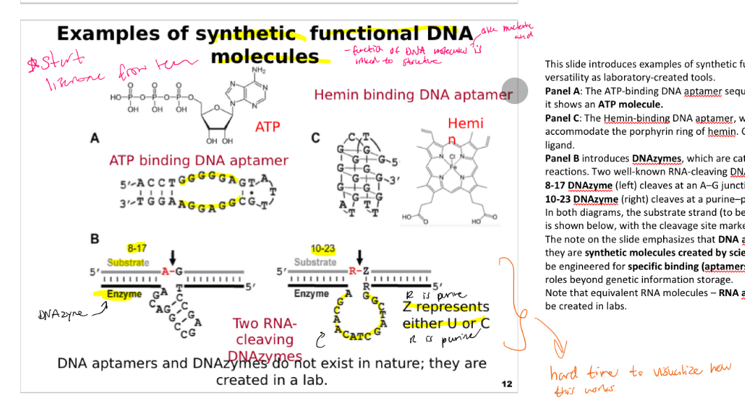
What are some examples of synthetic functional DNA molecules?
This slide introduces examples of synthetic functional DNA molecules, highlighting their versatility as laboratory-created tools.
Panel A: The ATP-binding DNA aptamer sequence is shown in a stem-loop structure. At the top, it shows an ATP molecule.
Panel C: The Hemin-binding DNA aptamer, which folds into a G-quadruplex structure to accommodate the porphyrin ring of hemin. On the right is a Hemin molecule (right) as target ligand.
Panel B introduces DNAzymes, which are catalytic DNA molecules engineered to carry out reactions. Two well-known RNA-cleaving DNAzymes are illustrated:
8-17 DNAzyme (left) cleaves at an A–G junction.
10-23 DNAzyme (right) cleaves at a purine–pyrimidine junction (R–Z, where Z is U or C).
In both diagrams, the substrate strand (to be cut) is shown on top, and the catalytic DNA strand is shown below, with the cleavage site marked by a red line under an arrow.
The note on the slide emphasizes that DNA aptamers and DNAzymes do not exist in nature; they are synthetic molecules created by scientists. These examples demonstrate how DNA can be engineered for specific binding (aptamers) and catalytic activity (DNAzymes), expanding its roles beyond genetic information storage.
Note that equivalent RNA molecules – RNA aptamers and RNA enzymes (ribozymes) can also be created in labs.
How Do Secondary and Tertiary Structures Enable DNA Function?
This slide highlights the structural basis of function for synthetic DNA molecules, focusing on an ATP-binding aptamer and the 8-17 DNAzyme. The key message is that their activities depend on their ability to adopt defined secondary and tertiary structures.
Top left shows the secondary structure of an ATP-binding DNA aptamer: a short DNA sequence folded into a stem-loop, with complementary base-pairing creating the stem and a loop region forming the binding pocket for ATP.
Top right illustrates the tertiary structure of the same aptamer: a 3D rendering of the folded DNA (green) with two ATP molecules (multi-colored spheres) bound within its pocket, demonstrating how aptamers create a precise shape for target recognition.
Bottom left depicts the secondary structure of the 8-17 DNAzyme, showing its substrate strand (Sub) aligned with the catalytic strand (Dz36). Key paired regions (P1–P4) are marked, and the scissile phosphate site is indicated where cleavage occurs.
Bottom right presents the three-dimensional structure of the 8-17 DNAzyme, with distinct helical segments (P1, P2) and the catalytic core in green. This structure demonstrates how DNAzymes fold to create an active site that can cleave RNA-containing substrates.
The accompanying note at the top reinforces that the unique functions of DNA aptamers and DNAzymes come from their ability to fold into specific structures, allowing them to bind targets or catalyze reactions like proteins or ribozymes.

How Does tRNA's Structure Enable Its Function?
Transfer RNA (tRNA) folds into specific secondary and tertiary structures that directly enable it to carry amino acids and decode messenger RNA (mRNA) during protein synthesis.
This slide explains the secondary and tertiary structures of transfer RNA (tRNA), emphasizing how structure underlies its function.
Left panel: Secondary structure
Shows the cloverleaf diagram of tRNA with four key arms:
Acceptor stem (top) ending with 3’-CCA, where amino acids attach.
D loop (left) containing modified nucleotides.
Anticodon loop (bottom) containing the anticodon triplet for pairing with mRNA.
TψC loop (right) important for ribosome interaction.
A variable loop is also present.
Colored circles indicate constant nucleotides (green) and constant purines/pyrimidines (orange).
Right panel: Tertiary structure
Depicts the three-dimensional conformation of tRNA.
The molecule folds into an L-shaped structure, stabilized by base-pairing interactions between loops.
This arrangement places the anticodon arm and amino acid acceptor stem at opposite ends, crucial for translation.
The slide highlights that the functions of tRNA—decoding mRNA and carrying amino acids—depend on its ability to form stable and precise secondary and tertiary structures.
DNA is a very robust molecule
Without nucleolus DNA can survive for a long time
But RNA on the other hand can only survive four day
Doesn’t have to be strictly in the freezer like RNA
You can make RNA make DNA
Watson and Crick made long artificial nucleic acids and you can use that to make enzymes, aptamers and other stuff
Additionally you can create enzymes that will recognize stuff and to other stuff after recognition
RNA can also be made synthetically

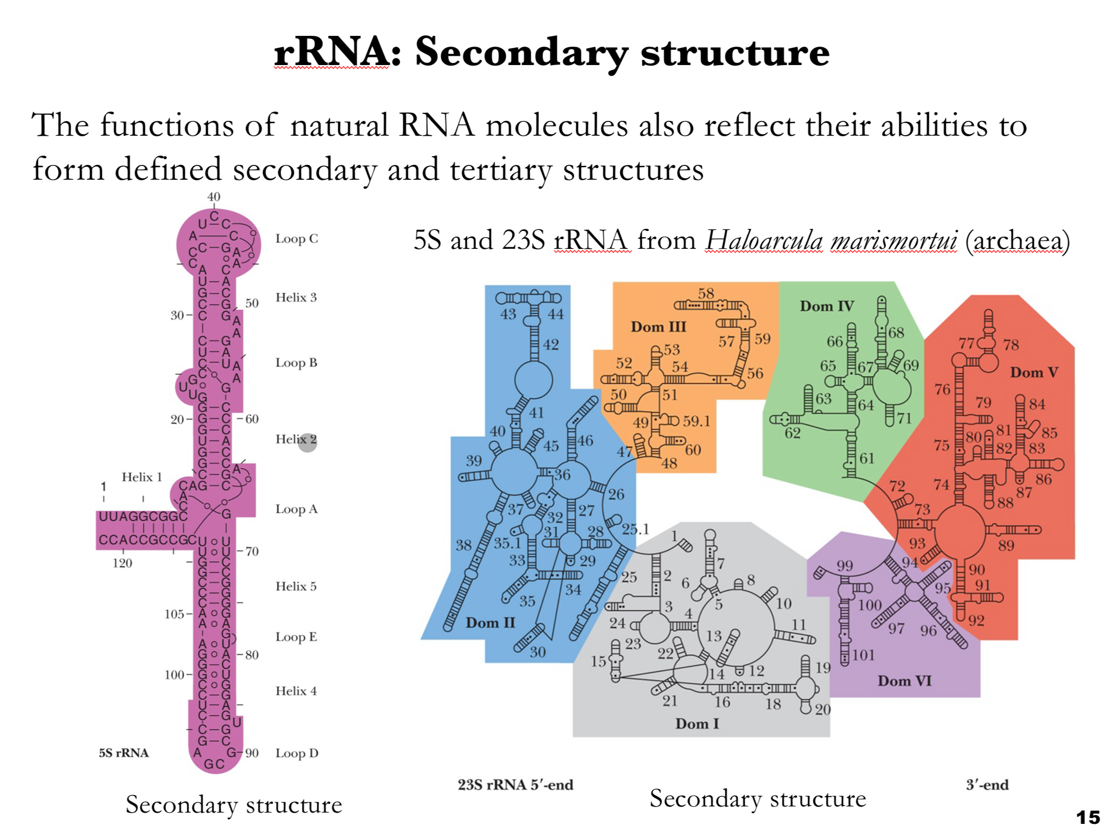
How Does rRNA Secondary Structure Support Ribosome Function?
This slide focuses on the secondary structures of ribosomal RNA (rRNA), using 5S and 23S rRNA from the archaeon Haloarcula marismortui as examples.
Left panel: 5S rRNA
Shows the secondary structure of 5S rRNA, approximately 120 nucleotides long.
It folds into a compact structure with multiple helices (Helix 1–5) and loops (A–E), including the long Loop C at the top.
The arrangement highlights the highly structured and conserved nature of rRNA, critical for its role in ribosome assembly and function.
Right panel: 23S rRNA
Displays the secondary structure of the much larger 23S rRNA, broken into colored domains (I–VI) for clarity:
Domain I (gray)
Domain II (blue)
Domain III (orange)
Domain IV (green)
Domain V (red)
Domain VI (purple)
Each domain contains multiple helices and loops, forming a complex scaffold essential for protein synthesis.
Key point:
rRNA molecules are highly structured RNAs whose extensive base-pairing and conserved motifs are essential for their catalytic and structural roles in the ribosome.
What is the tertiary structure of rRNA, and why is it important?
A: The tertiary structure is the 3D folding of rRNA, where helices and loops from the secondary structure fold and interact with each other. Base-pairing and interactions between unpaired regions, especially loops, stabilize the structure. This 3D structure is essential for ribosome assembly, protein synthesis, and catalytic function.
This slide shows the tertiary structure of ribosomal RNA (rRNA), building on the secondary structures introduced in slide 15.
Content:
Depicts the three-dimensional structure of the 5S and 23S rRNAs from the archaeon Haloarcula marismortui.
The complex structure integrates the helices and loops from the secondary structures into a tightly packed, functional form.
Multiple colors highlight the different domains, illustrating how they fold and interact.
The text explains that the structure contains extensive base-pairing and interactions between unpaired regions, particularly in the loop areas, which help stabilize the molecule.
Key message:
The tertiary structure is essential for the rRNA’s role in ribosome assembly and function, forming a scaffold for proteins and providing catalytic sites for translation.

What is the TPP aptamer, and how does its structure allow it to bind TPP?
This slide highlights natural RNA aptamers found in riboswitches, using the thiamine pyrophosphate (TPP) aptamer as an example. The key point is that many natural RNA molecules regulate genes by forming precise secondary and tertiary structures that bind small molecules.
Riboswitches are regulatory RNA elements in bacteria that sense metabolites. The TPP riboswitch binds thiamine pyrophosphate and controls gene expression.
Left panel (A): Secondary structure – The schematic shows the RNA sequence folded into stems (paired regions) and loops (unpaired regions). It contains five paired elements (P1–P5) and six unpaired regions (L3, L5, J2/3, J2/4, J4/5, and J1/4). These form the scaffold that holds the binding pocket for TPP.
Right panel (B): Tertiary structure – This 3D rendering shows how the RNA folds to create a binding site. The helices are colored (blue, pink, and purple), and the TPP ligand is shown in yellow nestled in the center. Additional structural elements, such as the pyrophosphate sensor helix and pyrimidine sensor helix, help secure the ligand and stabilize the switch.
Bottom: The chemical structure of TPP is shown for reference.
The accompanying text emphasizes that the tertiary structure is stabilized by extensive base pairing and interactions, including those involving unpaired regions. This structural organization enables the RNA to sense and bind TPP tightly and undergo conformational changes that affect gene expression.

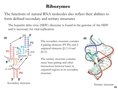
What is the HDV ribozyme, and why does it need to form a specific 3D structure?
The HDV ribozyme is an RNA molecule that acts like an enzyme during hepatitis delta virus replication. Its secondary structure folds into a specific 3D tertiary structure, creating a catalytic site that brings certain nucleotides together and allows the RNA to cut itself at a specific location. This self-cleavage helps process the long viral RNA into the correct pieces needed for replication, showing that RNA structure determines function and that RNA can catalyze chemical reactions.
This slide focuses on ribozymes, using the hepatitis delta virus (HDV) ribozyme as an example. It emphasizes that some natural (and synthetic) RNA molecules can act as enzymes, and that their catalytic functions depend on forming defined secondary and tertiary structures.
The HDV ribozyme is embedded in the HDV genome and is essential for viral replication, catalyzing self-cleavage of the viral RNA.
Left panel (A): Secondary structure – The diagram shows the RNA sequence folded into helices and loops. There are four paired elements (P1–P4), shown in different colors, and two unpaired regions (J1.1/4 and J4/2). These structural domains create the framework for catalysis.
Right panel (B): Tertiary structure – The 3D rendering depicts how the HDV ribozyme folds into a compact catalytic core. The colored helices (red, blue, gray) are connected by loops and junctions, while the surrounding mesh indicates the detailed atomic model. This structure brings distant nucleotides into close proximity to form an active site.
The accompanying note highlights that the tertiary structure is stabilized by extensive base pairing and other interactions, particularly in unpaired regions, enabling catalytic activity.