Biotech Exam 2
 Call Kai
 Learn
 Practice Test
 Spaced Repetition
 Match
 Flashcards
 Knowt Play
1/180

There's no tags or description
Looks like no tags are added yet.
181 Terms
L10
proteins functions
most of cell’s dry mass
versatile functions
storage
transport
movement - motor
structure
signal transduction - receptors
growth/development - hormones
immune protection - defense
catalysis - enzyme
structure determines function
categories of amino acid side chains
acidic (negative charge) polar
basic (positive charge) polar
uncharged polar
nonpolar
polypeptide chain directionality
orientation of a protein chain, running from an N-terminus (amino end) to a C-terminus (carboxyl end)
protein domains
in large proteins
compact modular units that fold more or less independently
40-350 continuous amino acids
diff domains associated w diff functions
ex: cytochrome b562, NAD binding domain of lactate dehydrogenase, SH2 domain, Src protein formed from 3 domains
complex assemblies
dimer/homodimer → a protein complex containing of 2 identical subunits
tetramer → hemoblobin; 4 heme groups
sacromere → myosin and actin
protein denaturation
when a protein is converted into a randomly coiled peptide without its normal activity
primary structure is not active
due to heating, chemical denaturants, oxidizing or reducing agents
may lose biological function
native protein
a protein in its folded and active form
protein biosynthesis
translation of nucleotide sequences into amino acid sequences
same basics across all kingdoms of life
ribosomes are responsible for this process
codon
set of 3-letter combinations of nucleotides, which corresponds to a specific amino acid or stop signal
transfer RNA (tRNA)
adaptor molecule in protein synthesis; contains an amino-acid recognition site as well as a template-recognition site (anticodon)
each tRNA sequence can be arranged in a cloverleaf pattern
activated amino acid is attached to a hydroyl group of the A residue in amino-acid-attachment site of CCA sequence at 3’ end
anticodon loop - makes 3 bases that constitute the anticodon accessible for interaction w an mRNA codon
activation of amino acid
formation of an adenylated amino acid
attachment of amino acid to corresponding tRNA
aminoacyl-tRNA synthetase
enzyme that catalyzes the aa activation reaction and linkage of aa to tRNA
activates tRNA by coupling it w its respective tRNA molecule

L10 NOT DONE YET
L13
irreversible inhibition
covalent binding of inhibitor to the enzyme
cannot be broken
forms stable complex
reversible inhiibition
inhibitor forms noncovalent bonds w enzyme
can dissociate
inhibitor can be unbound
suicide inhibition
an irreversible form of enzyme inhibition
inhibitor binds to active site of enzyme and forms an irreversible complex thru a covalent bond during normal catalysis reaction
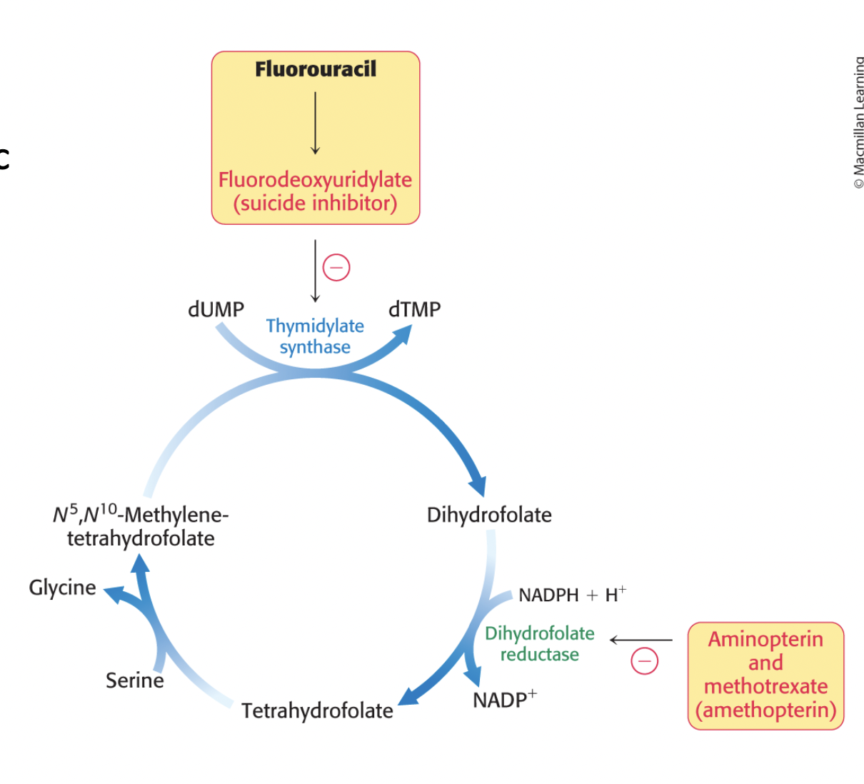
ex: aspirin, penicillin, fluorouracil
fluorouracil (5-FU)
anticancer drug that acts as suicide inhibitor of thymidylate synthase
converted in vivo into flouorodeoxyuridylate (F-dUMP)
F-dUMP irreversibly inhibits thymidylate synthase after acting as a normal substrate through part of the catalytic cycle
used to treat breast cancer, colon or rectal cancer, pancreatic cancer, and stomach cancer
types of reversible inhibition
competitive inhibition
uncompetitive inhibition
non-competitive inhibition
competitive inhibition
the inhibitor competes w the substrate for binding to the enzyme; binds to:
active site
anywhere else on the enzyme
ex: statins, ACEi

allosteric competitive inhibitor
binds to allosteric site, changes the conformation of the active site and the substrate can no longer bind there
statins as competitive inhibitors
competitively inhibit HMG-CoA reductase enzyme
cholesterol-lowering drugs

uncompetitive inhibition
inhibitor binds only to the enzyme-substrate complex
can prevent catalysis rather than substrate binding
ex: camptothecin (topoisomerase inhibitor that binds to topoisomerase I and DNA complex - anti-cancer drug)

non-competitive inhibition
the inhibitor binds either the enzyme or enzyme-substrate complex
can prevent catalysis rather than substrate binding
ex: doxycycline - collagenase inhibitor → preserves collagen in tissues; used in periodontal disease, osteoarthritis, RA

types of non-competitive inhibitors
pure non-competitive inhibitors
mixed non-competitive inhibitors
pure non-competitive inhibitors
bind equally well to the enzyme w or without substrate bond
mixed non-competitive inhibitors
bind preferentially to either the free enzyme or the enzyme-substrate complex
enzyme kinetics
V = rate of enzyme reaction
Vmax = max rate of a reaction
Km = substrate conc at which the reaction velocity is half of the max

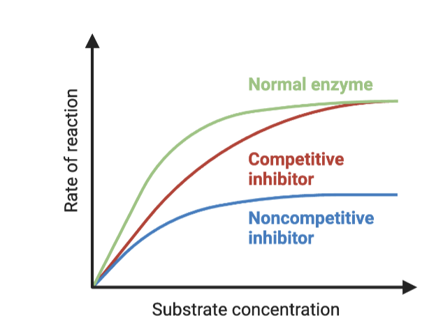
competitive inhibitor enzyme kinetics
inc in apparent value of Km
Vmax remains unchanged
can be overcome by a sufficiently high [S]

non-competitive inhibitor enzyme kinetics
lowers conc of functional enzyme
Vmax is dec
Km is unchanged
cannot be overcome by inc substrate conc
uncompetitive inhibitor enzyme kinetics
inhibitor binds to ES (enzyme substrate) to form ESI (enzyme substrate inhibition complex), depleting ES
Vmax is dec
Km is dec
dec in free enzyme correspond to an enzyme w greater affinity for its substrate

competitive inhibitor as drugs
a higher conc of the inhibitor is req for competitive inhibition when substrate conc is high
equally potent
equally selective
noncompetitive inhibitor as drugs
unaffected by high substrate conc
equally potent
equally selective
majority of molecular targets of FDA approved drugs
human and pathogen proteins
pic - drug targets span a variety of protein classes

antibiotics
compounds produced by bacteria and fungi which are capable of killing or inhibiting competing microbial species
penicillin
one of the most widely used antibiotics in the world
penicillium mold naturally produces the antibiotic penicillin
against many bacterial infections caused by staphylococci and streptococci
treatment of diphtheria, gonorrhea, pneumonia, syphilis, and others
estimated to have saved > 200 million lives
penicillin mechanism of action
interferes w synthesis of cell walls of reproducing bacteria
defective walls cause bacterial cells to burst
human cells are not affected bc they do not have cell walls
irreversibly inhibits transpeptidase
transpeptidase → enzyme that catalyzes the last step in bacterial cell-wall biosynthesis
suicide inhibitor

aspirin
extracts form the bark and leaves of the willow tree
active compound → salicylic acid
acetylsalicylic acid (aspirin) made by neutralizing salicylic acid w a base and acetyl chloride
aspirin mechanism of action
irreversible inhibitor of COX (cyclooxygenase) enzymatic activity of prostaglandin H2 synthase (PGH2)
prostaglandins promote the inflammatory response (swelling, pain, fever)
COX inhibitors blunt this response

protease inhibitors
block protease activity
ex: HIV-1 protease, thrombin, ACE
ACEi (angiotensin converting enzyme inhibitor)
competitively block the ACE enzyme, which prevents to production of angiotensin II
relax veins and arteries to lower bp
protect kidney function in patients w diabetes or mild kidney disease
angiotensin II
hormone that circulates in blood
main role is to constrict blood vessels
can cause high bp
present in abnormally high quantities in many cardiovascular diseases
ACE
zinc metalloproteases
hydrolyze peptides
convert inactive angiotensin I to angiotensin II by removing a dipeptide
thrombin
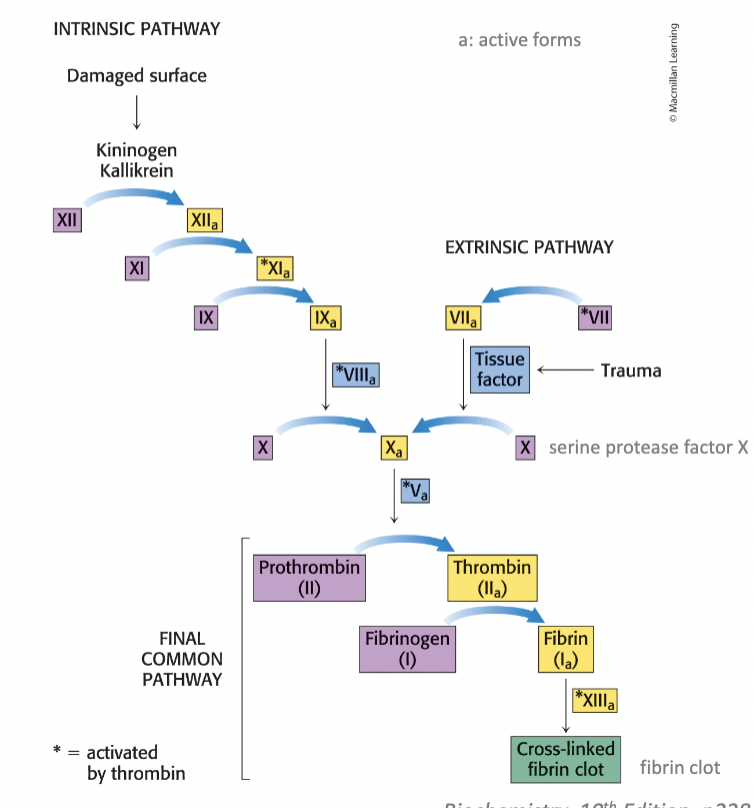
converts fibrinogen to fibrin to create a blood clot
serine protease, final enzyme of blood coagulation cascade
blood clotting cascade
enzymatic cascade - series of zymogen activations → rapid response
activated form of one clotting factor catalyzes the activation (enzyme) of the next precursor
signal is amplified at each step
a very small amt of initial factors suffice to trigger the cascade → rapid response to trauma
anticoagulant drugs
meds that reduce blood clot formation; prevent stroke, myocardial infarction, and pulmonary embolism in patients w inc clotting risk
DTIs (direct thrombin inhibitors)
anticoagulant drugs; serine protease inhibitor
univalent → bind only to active site
ex: argatroban, melagatran
bivalent → bind to both active site and regulatory sites
ex: hirudin, bivalirudin

warfarin
anticoagulant drug; vitamin K antagonist
inhibits enzymes req to regenerate the active form of vit K
vitamin K: essential for the synthesis of prothrombin and several other clotting factors

HIV protease
cleaves multidomain viral proteins into their active forms
aspartyl protease
HIV protease inhibitors
inhibit HIV protease
ex: indinavir, retrovir
blocking this process completely prevents this virus from being infectious
in combo w inhibitors of other key enzymes have dramatically reduced deaths to AIDs

indinavir
one of the 1st HIV protease inhibitor
designed to resemble a peptide substrate of the enzyme
OH group interacts w 2 aspartate residues of the active site of HIV proteases but generally absent in cellular aspartyl proteases → specificity
paxlovid
SARS-CoV-2 protease inhibitor; antiviral drug combo that treats COVID-19
bioavailable orally
prevents transmission
reduce hospitalization rate and mortality
ex: nirmatrelvir and ritonavir
nirmatrelvir
3C-like (3CL) protease inhibitor
3CL → protease that SARS-CoV-2, the virus that causes COVID-19, uses to make more copies of itself
ritonavir
strong cytochrome p450 (CYP3A4) inhibitor and pharmacokinetic boosting agent
kinases
transfer a gamma-phosphate group from ATP to serine, threonine, or tyrosine residues of a protein
> 518 protein kinases on the human genome (1.7% of human genes)
kinases phosphorylate more than 30% of the cellular proteins
classified by amino acid substrate preference
serine-threonine kinases
tyrosine kinases
dual kinases (phosphorylate serine, threonine, or tyrosine residues)

phosphatatases
transfer the phosphate from a phosphoprotein to a water molecule

major kinase classes and immunoreceptor signaling

timeline of approval of key protein kinases inhibitors
some kinase inhibitors are used to treat cancers

imatinib (gleevec)
breakthrough in cancer treatment
highly specific inhibitor of bcl:abl kinase
forms due to chromosomal translocation and overexpression of bcr:abl fusion genes
used to treat CML
occupies the ATP-binding site
targets tumor cells specifically, minimizing side effects
83.3% rate of 10-year overall survival
inhibitor of several tyrosine kinases
c-kit, a protein tyrosine kinase in GI stromal tumors
PI3K in solid tumors
serine/threonine kinase BRAF in melanoma
the receptor tyrosine kinase EGFR in lung cancer
serine/threonine kinase mTOR in renal tumors

FLT3 inhibitors for the treatment of AML
[2017] midostaurin → first approved FLT3 inhibitor in US
[2018] gilteritinib → received FDA approval in US
[2019] quizartinib → recieved approval in Jaban
[2023] quizartinib → received approval in US

L14
maintaining homeostasis
req metabolic regulation that coordinates the use of nutrient pools
diet
proteins
amino acids
body protein
amino acid pool
gluconeogenesis
urine
carbohydrates
glucose
glycogenesis
lipogenesis
glucose pool
brain metabolism
lipids
free fatty acids + glycerol
fat stores
free fatty acid pool
metabolism in most tissues

overall protein digestion
dietary proteins → degraded to amino acids which are absorbed by the intestine and transported in blood
cellular proteins are degraded to amino acids bc of damage, misfolding, or changing metabolic demands
excess amino acids → cannot be stored or directly excreted, so they must be used for protein synthesis or degraded to ammonia
protein homeostasis
rate of synthesis = rate of degradation
required to maintain a physiological concentration req for life
essential amino acids
in humans must be obtained through diet; cannot be synthesized by body; 9 amino acids

protein digestion
begins in stomach where acidic environment denatures proteins into random coils by pepsin
partly digested proteins move from stomach to beginning of small intestine (duodenum) → stimulating secretion of NaHCO3 and proteolytic enzymes from pancreas
aminopeptidases in plasma membrane of intestinal cells enhance digestion
free amino acids, dipeptides, and tripeptides are transported into intestinal cells
at least 7 diff transporters specific to a diff group of amino acids
absorbed amino acids are released into blood by a number of Na+ - amino acid antiporters

pepsin
the primary proteolytic enzyme of the stomach; max active at pH 2
protein turnover
degradation and resynthesis of proteins
takes place constantly in cells
essential for removing short-lived or damaged proteins
half-lives of proteins range over several orders of magnitude
ornithine decarboxylase (catalyzes synthesis of polyamines) - 11 minutes
hemoglobin - life of RBC
lens protein crystallin - life of organism
ubiquitin
a small (76 aa) protein that tags proteins for destruction
present in all eukaryotic cells
highly conserved
has extended carboxyl terminus, which is activated and linked to proteins targeted for destruction

regulation of protein turnover
ubiquitin attaches by its carboxyl-terminal Gly residue to the epsilon-amino groups of 1+ Lys residues on the target protein
req ATP hydrolysis
forms an isopeptide bond bc epsilon- rather than alpha-amino groups are targeted

attachment of ubiquitin to a protein
ubiquitin-activating enzyme (E1) → adenylates ubiquitin and transfers it to a sulfhydryl group of a Cys residue of E1
req ATP
ubiquitin-conjugating enzyme (E2) → transfers ubiquitin to one of its own sulfhydryl groups
ubiquitin-protein ligase (E3) - transfers ubiquitin from E2 to an epsilon-amino group on the target protein
brings E2 and the target protein together
ubiquitin be transferred directly or be passed to a Cys residue of E3 first

multiple ubiquitin molecules are typically added to a single protein substrate
E3 can remain bound to target protein and generate a chain of ubiquitin molecules
E3 can dissociate after the first ubiquitin addition, and a chain can be extended by another E2/E3 pair
ubiquitin can be added onto any of the 7 Lys or the N-terminus of the previous ubiquitin
a chain of 4+ ubiquitin molecules linked via Lys 48 is an especially effective signal for protein degradation

E3 ubiquitin ligases provide protein target specificity
in humans
there are 2 E1 ligase genes
there are 30-50 E2 ligase genes
there are over 600 E3 ligase genes
in tetraubiquitin chain, 4 ubiquitin molecules are linked
linked by isopeptide bonds
the epsilon-amino group of a Lys residue of one ubiquitin is linked to the terminal carboxylate of another
this unit is the primary signal for degradation when linked to a target protein

importance of E3 proteins to cell function
proteins not degraded bc of a defective E3 may accumulate, causing a disease of protein aggregation
angelman syndrome → severe neurological disorder of an unusually happy disposition, cognitive disability, absence of speech, uncoordinated movement, and hyperactvitity
caused by defect in a member of E3 family
overexpression of E3 ligase (UBE3A) is strongly linked to certain forms of autism
inappropriate protein turnover can lead to cancer
many tumor suppressors, including BRCA1, are E3 ligases
additional roles of ubiquitination
regulates proteins involved in
DNA repair
chromatin remodeling
innate immunity
membrane trafficking
autophagy
proteasome
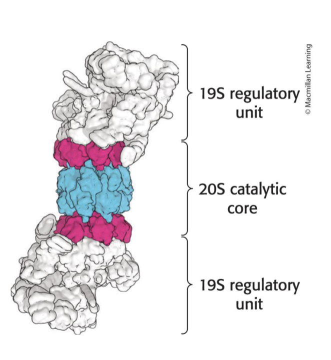
26S proteasome - large, ATP-driven protease complex that digests ubiquitinated proteins
complex of 2 components
one 20S catalytic unit arranged as a barrel
two 19S regulatory units that control access to interior of the 20S catalytic subunit
function of 19S regulatory unit
contain ubiquitin receptors that bind specifically to polyubiquitin chains
use ATP to unfold polyubiquitinated chains and direct them into the catalytic core
contain an isopeptidase that cleaves off intact ubiquitin molecules so that they can be reused
6 ATPases of the AAA+ class
AAA+ class → class of chaperone-like ATPases associated w assembly, operation, and disassembly of protein complexes
20S catalytic unit
barrel-shaped
made up of 28 homologous subunits
alpha-type and beta-type are arranged in 4 rings of 7 subunits each
some of beta-type subunits include protease active sites at the amino termini
active sites in the beta subunits
chymotrypsin-like → cleaves after large hydrophobic amino acids
trypsin-like → cleaves after basic amino acids
caspase-like → cleaves after acidic amino acids
all active sites employ an N-terminal Thr residue
the OH group of Thr residue attacks the carbonyl groups of peptide bonds, forming acyl-enzyme intermediates
substrates are degraded in a processive manner without intermediate release
substrates are reduced to peptides ranging from 7-9 residues before release

proteasome and other proteases generate free amino acids
ubiquitinated proteins are processed to peptide fragments
ubiquitin is removed and recycled prior to protein degradation
peptide fragments are further digested to yield free amino acids, which can be used for biosynthetic reactions, most notably protein synthesis
alternatively, the amino group can be removed and processed to urea and the C skeleton can be used to synthesize carbohydrate or fats or used directly as a fuel for cellular respiration

processes regulated by protein degradation
many biological processes are controlled, at least in part, by protein degradation via the ubiquitin-proteasome pathway

bortezomib (velcade)
dipeptidyl boronic acid inhibitor of the proteasome; used as a therapy for multiple myeloma

HT1171
suicide inhibitor of the proteasome of M. tuberculosis; has no effect on human proteasomes

amino acid degradation
amino acids not needed as building blocks are degraded to compounds able to enter the metabolic mainstream
major site of degradation → liver
muscles also readily degrade the branched-chain amino acids (Leu, Ile, Val)
amino acid degradation steps
amino group is removed & remaining carbon skeleton is metabolized to a glycolytic intermediate or to acetyl CoA
alpha-amino groups transferred to alpha-ketoglutarate, yielding glutamate
glutamate is oxidatively deaminated in the liver to yield ammonium ion
ammonium ion is converted to urea, which is excreted

aminotransferases
catalyze the transfer of an alpha-amino group from an alpha-amino acid to an alpha-ketoacid; req coenzyme pyridoxal phosphate (PLP) - derivative of pyridoxine (vit B6)
reactions are reversible and can be used to synthesize amino acids from alpha-ketoacids
presence of alanine and aspartate aminotransferases in the blood is an indication of liver damage
liver damage due to:
viral hepatitis
long-term excessive alcohol consumption
reaction to drugs
liver cell membranes are damaged and aminotransferases leak into blood

aspartate aminotransferases (AST)
catalyzes the transfer of amino group of aspartate to alpha-ketoglutarate

alanine aminotransferase (ALT)
catalyzes the transfer of amino group of alanine to alpha-ketoglutarate

pyridoxal phosphate (PLP) enzymes
at the alpha-carbon of amino acids, PLP-dependent enzymes catalyze
decarboxylations
deaminations
racemizations
aldol cleavages
at the beta-carbon and gamma-carbon of amino acids, PLP-dependent enzymes catalyze
elimination
replacement reaction
glutamate dehydrogenase
mitochondrial enzyme that converts N atom in glutamate to a free ammonia ion by oxidative deamination
liver-specific enzyme
can use NAD+ or NADP+
proceeds by dehydrogenation of the C-N bond, followed by hydrolysis of the ketimine
allosterically inhibited by GTP and stimulated by ADP in mammals

serine and threonine can be directly deaminated
serine dehydratase and threonine dehyrdatase directly deaminate their respective amino acids
PLP is the prosthetic group
no transfer of the alpha-amino group to alpha-ketoglutarate is req
dehydration precedes deamination
serine → pyruvate + NH4+
threonine → alpha-ketobutyrate + NH4+

N from amino acids is cleared through the urea cycle in the liver

peripheral tissues transport N to the liver as glutamine
N is transported to liver as glutamine thru glutamine synthetase to avoid high levels of circulating ammonia
N of glutamine are eliminated by incorporation into urea in the liver

glutamine synthetase
catalyzes the synthesis of glutamine from glutamate and NH4+
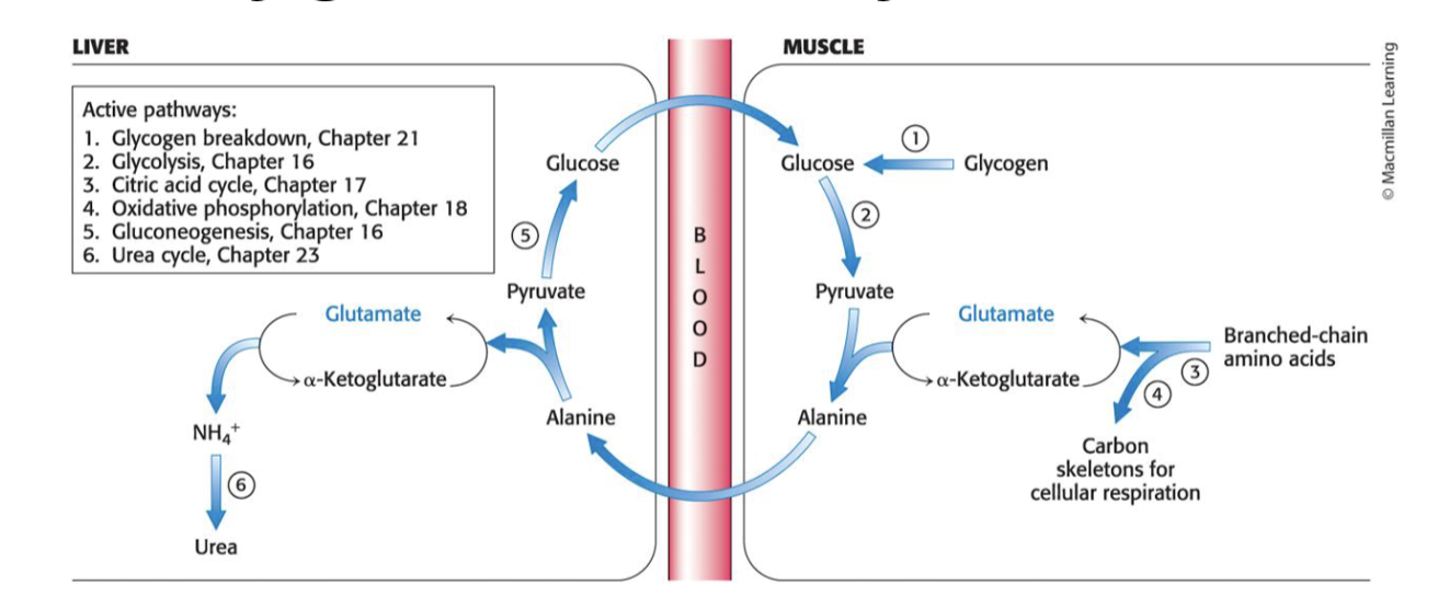
muscle cells transport N to liver as alanine by glucose-alanine cycle
during prolonged exercise and fasting, muscle used branched-chain amino acids as fuel
the N removed is transferred (through glutamate) to alanine, which is released into the blood stream
in liver, alanine is taken up and converted into pyruvate for the subsequent synthesis of glucose
urea cycle overview
2 N atoms enter the cycle and leave as urea
CO2 is simultaneously eliminated as it is hydrated to bicarbonate, which then enters the cycle
urea cycle, gluconeogenesis, and transamination of oxaloacetate are linked by fumarate and aspartate

urea cycle steps
ammonia + bicarbonate → carbamoyl phosphate via carbamoyl phosphate synthetase I in the mitochondria
carbamoyl phosphate synthetase I req allosteric regulator N-acetylglutamate for activity
carbamoyl phosphate + ornithine → citrulline via ornithine transcarbamoylase in the mitochondria
citrulline is transported into the cytoplasm
citrulline + aspartate (donor of second N in urea) → argininosuccinate via argininosuccinate synthetase in the cytoplasm
argininosuccinate → arginine + fumarate via argninosuccinase
fumarate is hydrated to malate, which is oxidized to oxaloacetate
oxaloacetate converted to glucose by gluconeogenesis OR transaminated to aspartate for another round of urea synthesis
arginine is hydrolyzed → ornithine + urea
ornithine is transported back into the mitochondria
urea is excreted

carbamyl phosphate synthetase I
catalyzes the coupling of ammonia with bicarbonate to form carbamoyl phosphate
mammals have 2 isozymes
req 2 molecules of ATP, making the rxn essentially irreversible
req the allosteric regulator N-acetylglutamate for activity
inhibited by acetylation and stimulated by deacetylation
N-acetylglutamate synthase
catalyzes the synthesis of N-acetylglutamate
activated when amino acids are readily available

ornithine transcarbamoylase
catalyzes the transfer of the carbamoyl group of carbamoyl phosphate to ornithine, forming citrulline
occurs in the mitochondria
