lecture 7 regulation of translation
 Call Kai
 Learn
 Practice Test
 Spaced Repetition
 Match
 Flashcards
 Knowt Play
1/32

There's no tags or description
Looks like no tags are added yet.
33 Terms
cells can regulate protein production by controlling how long an mRNA lasts and how often translation starts. how does each affect protein output?
mRNA degradation: a longer mRNA half-life gives ribosomes more time to translate it.
translation initiation: more frequent initiation produces more copies of the protein from the same mRNA.
the 5′ cap protects mRNA from degradation. what happens when the cap is removed?
the exposed 5′ end can be degraded by a 5′ → 3′ exonuclease.
how do the poly(A) tail and AU-rich sequences in the 3′ untranslated region (3′ UTR) affect mRNA half-life?
as the poly(A) tail shortens, the mRNA generally becomes less stable. AU-rich sequences in the 3′ UTR can recruit proteins that promote degradation. many growth factor mRNAs have these sequences and therefore have short half-lives.
cells need iron. what carries iron in the blood, and how do cells take it up?
transferrin carries iron in the blood. iron-loaded transferrin, called ferrotransferrin, binds the transferrin receptor and enters the cell by receptor-mediated endocytosis.
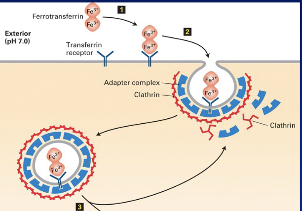
after ferrotransferrin enters the endosome, what causes it to release iron, and where does the iron go?
the endosome’s low pH weakens transferrin’s grip on iron. the released iron moves into the cytosol, where it can be stored in ferritin.

once transferrin releases its iron, what happens to the iron-free transferrin and its receptor?
apotransferrin (iron-free transferrin) stays bound to the receptor at low pH. both recycle to the plasma membrane, where the higher extracellular pH causes apotransferrin to detach.

where is the iron response element (IRE) in transferrin receptor mRNA, and what binds it when iron is low?
the IRE forms a stem-loop in the 3′ untranslated region (3′ UTR). when iron is low, the IRE-binding protein binds and stabilizes the stem-loop, protecting the mRNA from degradation.

once transferrin receptor mRNA is protected from degradation, how does that increase iron uptake?
the mRNA lasts longer and can be translated more times, so the cell makes more transferrin receptors. those receptors bring more iron-loaded transferrin (ferrotransferrin) into the cell.

when iron levels rise, what happens to the protein protecting transferrin receptor mRNA?
high iron causes the iron response element (IRE)-binding protein to detach from the IRE in the 3′ UTR. this leaves the mRNA exposed to degradation by ribonucleases.
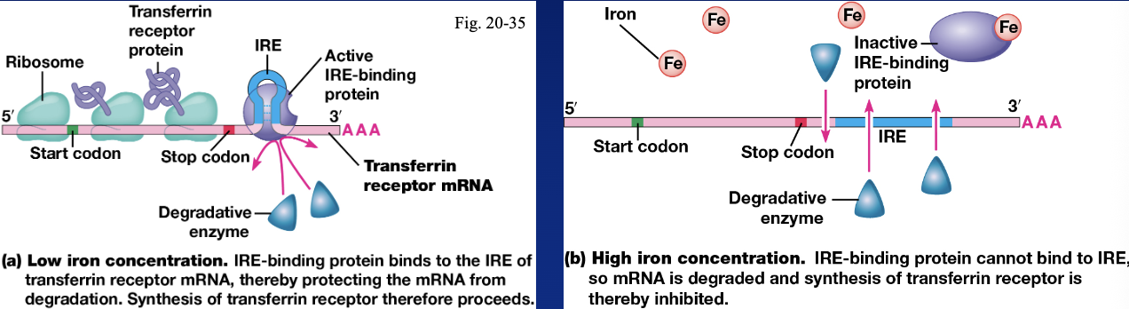
once transferrin receptor mRNA is degraded, how does that affect iron uptake?
less mRNA is available for translation, so the cell makes fewer transferrin receptors and takes up less iron-loaded transferrin.

where is the iron response element (IRE) in ferritin mRNA, and what happens to it when iron is low?
the IRE forms a stem-loop in the 5′ untranslated region (5′ UTR). when iron is low, the IRE-binding protein binds and stabilizes the stem-loop.

once the IRE-binding protein binds the ferritin mRNA stem-loop, how does it affect translation initiation?
the bound protein blocks the small ribosomal subunit from starting its 5′ → 3′ scan for the start codon in the Kozak sequence. translation initiation is repressed, so the cell makes less ferritin.

when iron levels rise, how does ferritin mRNA become available for translation?
high iron causes the IRE-binding protein to change shape and detach from the IRE in the 5′ UTR. the small ribosomal subunit can then scan 5′ → 3′ to find the start codon in the Kozak sequence. translation begins, producing more ferritin to bind and store cytosolic iron.

a reticulocyte is an immature red blood cell with ribosomes still making globin. what does globin combine with, and how much of the mature red blood cell’s dry weight does the product make up?
globin combines with heme to form hemoglobin, which makes up about 98% of a mature red blood cell’s dry weight.
what happens to globin synthesis when heme is scarce, and what controls this response?
the heme-controlled inhibitor (HRI) becomes active and inhibits translation initiation, reducing globin synthesis until heme is available.
when heme is scarce, how does the heme-controlled inhibitor (HCI) suppress translation in reticulocytes?
HCI acts as a kinase and phosphorylates eIF2. this reduces the supply of eIF2–GTP available to bind initiator Met-tRNA, so fewer translation initiation complexes form. translation decreases broadly, including globin synthesis.

when heme is available again, how does it allow globin synthesis to resume?
heme binds and inactivates HCI. eIF2–GTP can then bind initiator Met-tRNA and form the translation initiation complex, allowing globin synthesis to resume.

RNA interference begins with double-stranded RNA in the cytosol. how do dicer and RISC turn it into a guide strand?
dicer cuts double-stranded RNA into 21–22-base-pair fragments called small interfering RNAs (siRNAs). the RNA-induced silencing complex (RISC) loads an siRNA, discards one strand, and keeps the other as the guide strand.
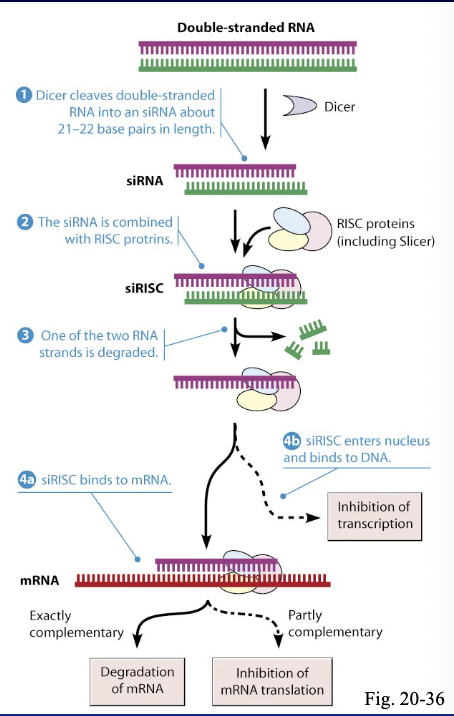
once RISC has a guide strand, how does the match between that strand and its target mRNA affect the outcome?
high complementarity → RISC cleaves the target mRNA, leading to its degradation.
partial complementarity → RISC inhibits translation and can also promote mRNA decay.
what are microRNAs (miRNAs), and why are they important during embryonic development?
about 1,000 genes produce miRNAs, short RNA molecules that guide RISC to target mRNAs. this reduces gene expression by inhibiting translation or promoting mRNA degradation, helping control which proteins are made and when during embryonic development.

how can miRNAs leave one cell and reach distant cells?
cells can package miRNAs into extracellular vesicles, such as exosomes, and release them. these vesicles can enter the bloodstream and be taken up by distant cells.
once a distant cell takes up the miRNAs, what can they do, and why is this signaling endocrine-like?
the delivered miRNAs can regulate gene expression by targeting mRNAs in the receiving cell. this is endocrine-like because the signal travels through the blood and acts on distant cells.
after translation, what six processes can modify, process, or degrade a protein?
proteolysis — cuts the polypeptide at specific sites.
phosphorylation — adds a phosphate group.
lipid conjugation — attaches a lipid, which can help anchor the protein to a membrane.
glycosylation — attaches sugars to the protein.
disulfide bond formation — links two cysteine residues, helping stabilize the folded protein.
proteasomal degradation — breaks down the protein into short peptides.
what is a proteasome, and where are its proteases located?
a proteasome is a hollow, cylindrical complex made of multiple proteins. proteasomes are abundant in the cytosol, and their proteases line the inner chamber.
how is a misfolded protein tagged for proteasomal degradation?
a ubiquitin ligase helps attach ubiquitin, a 76-amino-acid protein, to a lysine residue on the target protein. multiple ubiquitins can form a chain, and the target can be tagged at more than one lysine.
once a protein has a ubiquitin chain that marks it for degradation, what happens next?
the proteasome recognizes the tag, unfolds the target protein, and feeds it into its inner chamber. the proteases cut it into short peptide fragments.
what symptoms characterize Creutzfeldt–Jakob disease (CJD), and what are two related diseases?
CJD causes progressive memory loss and difficulty performing routine tasks. related diseases include kuru, historically documented in Papua New Guinea, and scrapie, which affects sheep.
why did researchers originally suspect a “slow virus” caused diseases like scrapie?
scrapie could be transmitted in animal studies, and symptoms took a long time to appear. researchers originally suspected a slow virus. the infectious agents were later identified as prions, misfolded proteins that cause other copies of the same protein to misfold.
what did Stanley Prusiner propose in 1982 after studying scrapie, and why was the idea controversial?
Prusiner found that the infectious material contained protein, with no detectable viral DNA or RNA. he proposed that the infectious agent was itself a protein and called it a prion. the idea challenged the prevailing assumption that infectious agents needed nucleic acid.
CJD, scrapie, and kuru are prion diseases. what’s the difference between PrPᶜ and PrPˢᶜ, and how can misfolding begin?
PrPᶜ is the normally folded prion protein. PrPˢᶜ is the misfolded form. a genetic mutation can make PrP more likely to misfold, and existing PrPˢᶜ can cause normally folded PrPᶜ to adopt the misfolded shape.
why do prion diseases cause sponge-like changes in the brain, and what does TSE stand for?
PrPˢᶜ resists protein breakdown and accumulates in aggregates. as neurons are damaged and die, small spaces called vacuoles give the brain tissue a sponge-like appearance. TSE stands for transmissible spongiform encephalopathy.
what is bovine spongiform encephalopathy (BSE), and how did it historically spread among cattle?
BSE, also called mad cow disease, is a prion disease in cattle. it historically spread through feed containing meat-and-bone meal contaminated with prions.
who won the 1997 Nobel Prize in Physiology or Medicine for discovering prions, and why was the discovery significant?
Stanley Prusiner. his discovery showed that a misfolded protein can act as an infectious agent without carrying DNA or RNA.