A&P
 Call Kai
 Learn
Learn Practice Test
 Spaced Repetition
 Match
Match Flashcards
 Knowt Play
1/110

There's no tags or description
Looks like no tags are added yet.
Name | Mastery | Learn | Test | Matching | Spaced | Call with Kai | Chat |
|---|
No analytics yet
Send a link to your students to track their progress
111 Terms
Digastirc
Origin: Mastoid process (posterior belly)
Insertion: Mandible near midline (anterior belly), hyoid bone
Nerve: Posterior belly—facial nerve; anterior belly—mandibular division of trigeminal nerve
Action: Depresses and retracts mandible; elevates hyoid bone

Geniohyoid
Origin: Mental protuberance of mandible
Insertion: Body of hyoid bone
Nerve: Fibers of C1 and C2 via hypoglossal nerve
Action: Protracts hyoid bone; depresses mandible

Mylohyoid
Origin: Body of mandible
Insertion: Hyoid bone
Nerve: Mandibular division of trigeminal nerve
Action: Elevates floor of mouth and tongue; depresses mandible when hyoid bone is fixed

Stylohyoid
Origin: Styloid process
Insertion: Hyoid bone
Nerve: Facial nerve
Action: Elevates hyoid bone

Omohyoid
Origin: Superior border of scapula
Insertion: Hyoid bone
Nerve: Upper cervical nerves through ansa cervicalis
Action: Depresses hyoid bone; stabilizes hyoid bone during mouth opening

Sternohyoid
Origin: Manubrium and first costal cartilage
Insertion: Hyoid bone
Nerve: Upper cervical nerves through ansa cervicalis
Action: Depresses hyoid bone; stabilizes hyoid bone during mouth opening

Thyrohyoid
Origin: Thyroid cartilage
Insertion: Hyoid bone
Nerve: Upper cervical nerves, traveling with hypoglossal nerve
Action: Depresses hyoid bone and elevates thyroid cartilage of larynx; stabilizes hyoid bone during mouth opening

Sternothyroid
Origin: Manubrium and first or second costal cartilage
Insertion: Thyroid cartilage
Nerve: Upper cervical nerves through ansa cervicalis
Action: Depresses larynx; stabilizes hyoid bone during mouth opening

Sternocleidomastoid
Origin: Manubrium and medial clavicle
Insertion: Mastoid process and superior nuchal line
Nerve: Accessory nerve (cranial nerve XI)
Action: Single contraction: laterally flexes head/neck to same side and rotates head/neck to opposite side. Bilateral contraction: flexes neck.

Scalene Muscles
Origin: C2–C6 vertebrae
Insertion: First and second ribs
Nerve: Cervical and brachial plexuses
Action: Flexes, laterally flexes, and rotates neck
Anterior: elevation of rib 1; lateral flexion and rotation of neck
Middle: elevation of rib 1; lateral flexion of neck
Posterior: elevation of rib 2; lateral flexion of neck

Longissimus Capitis
Origin: Upper thoracic and lower cervical vertebrae
Insertion: Mastoid process
Nerve: Dorsal rami of cervical nerves
Action: Extends, rotates, and laterally flexes neck
Oblique Capitis Superior
Origin: Atlas
Insertion: Occipital bone (inferior nuchal line)
Nerve: Dorsal ramus of C1
Action: Extends and laterally flexes neck; turns head
Rectus Capitis Posterior
Origin: Axis, atlas
Insertion: Occipital bone
Nerve: Dorsal ramus of C1
Action: Extends and rotates neck; turns head
Semispinalis Capitis
Origin: C4–T6 vertebrae
Insertion: Occipital bone
Nerve: Dorsal rami of cervical nerves
Action: Extends and rotates neck
Splenius Capitis
Origin: C4–T6 vertebrae
Insertion: Superior nuchal line and mastoid process
Nerve: Dorsal rami of cervical nerves
Action: Extends, rotates, and laterally flexes neck
Trapezius
Origin: Occipital protuberance, nuchal ligament, spinous processes of C7–T12
Insertion: Clavicle, acromion process, and scapular spine
Nerve: Accessory nerve (cranial nerve XI)
Action: Extends and laterally flexes neck
Superior fibers: elevation of scapula; superior rotation of scapula (glenoid cavity directed superiorly)
Middle fibers: retraction (adduction) of scapula
Inferior fibers: depression of scapula
Origin:
Occipital bone (superior nuchal line)
Nuchal ligament
Vertebra prominens (spinous process of C7 vertebra)
Spinous processes of T1-12 vertebrae
Insertion:
Clavicle (lateral)
Scapula (spine and acromion)
Innervation:
Accessory nerve (CN XI)

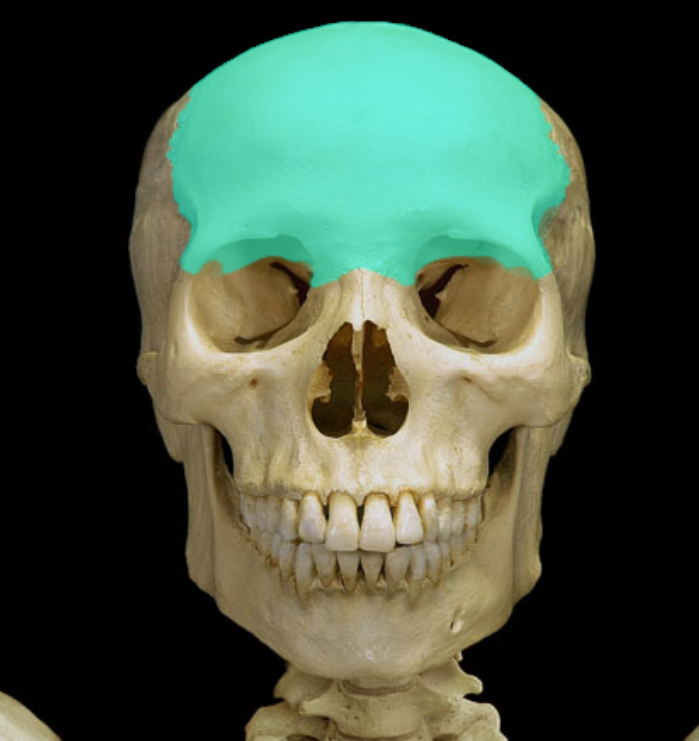
Frontal Bone
Greater wing of sphenoid bone

Inferior or middle nasal conchae

Infraorbital foramen

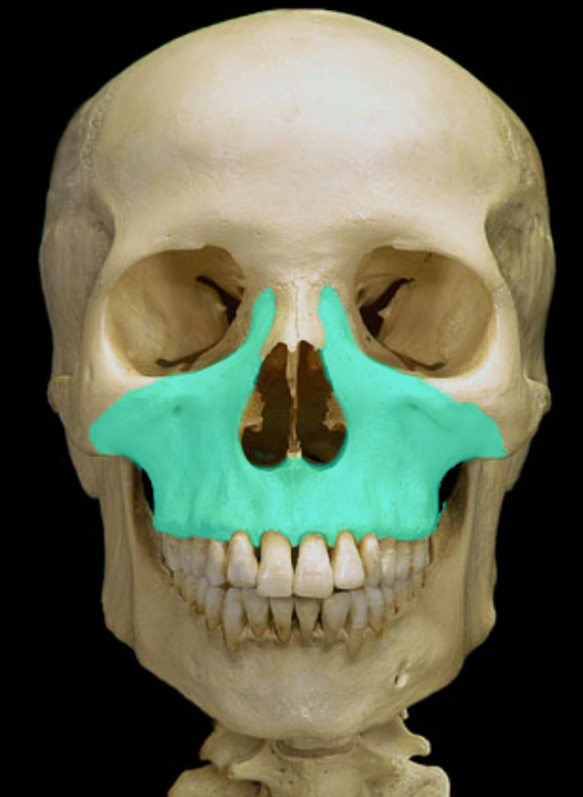
Maxilla
Mental foraemen

Nasal bones

Perpendicular plate of ethmoid

Sphenoid bon

Zygomatic bone

Vomer

Condylar process of mandible

Coronid process of mandible

External acoustic meatus

Mastoid process

Parietal bone

Pterion

Ramus of mandible

Squamous part of temporal bone

Styloid process of temporal bone

Temporal bones

Temporal process of zygomatic bone
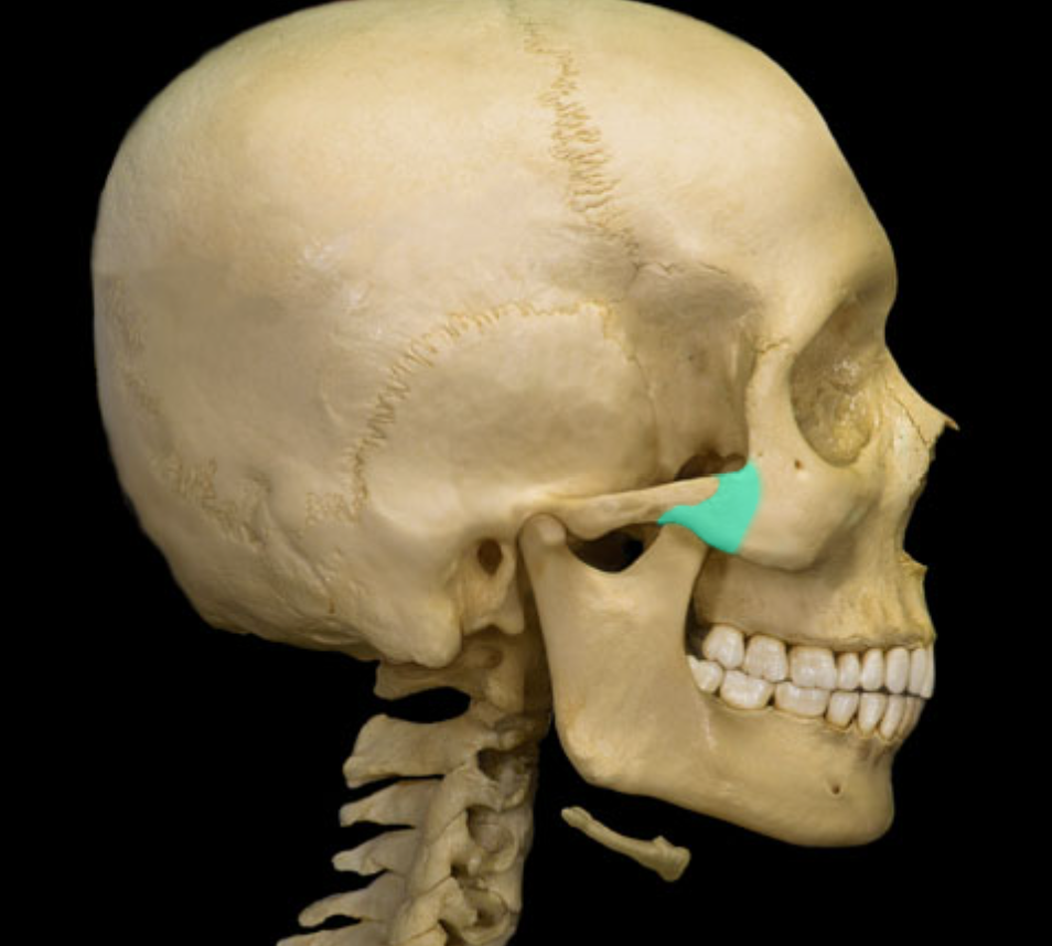
Temporal process of zygomatic bone

Zygomatic arch

Zygomatic process of temporal bone

Buccinator m.
Action:
Compression of cheek (e.g., inflating a balloon; playing a wind musical instrument)
Origin:
Pterygomandibular raphe
Maxilla (lateral aspect)
Mandible (lateral body)

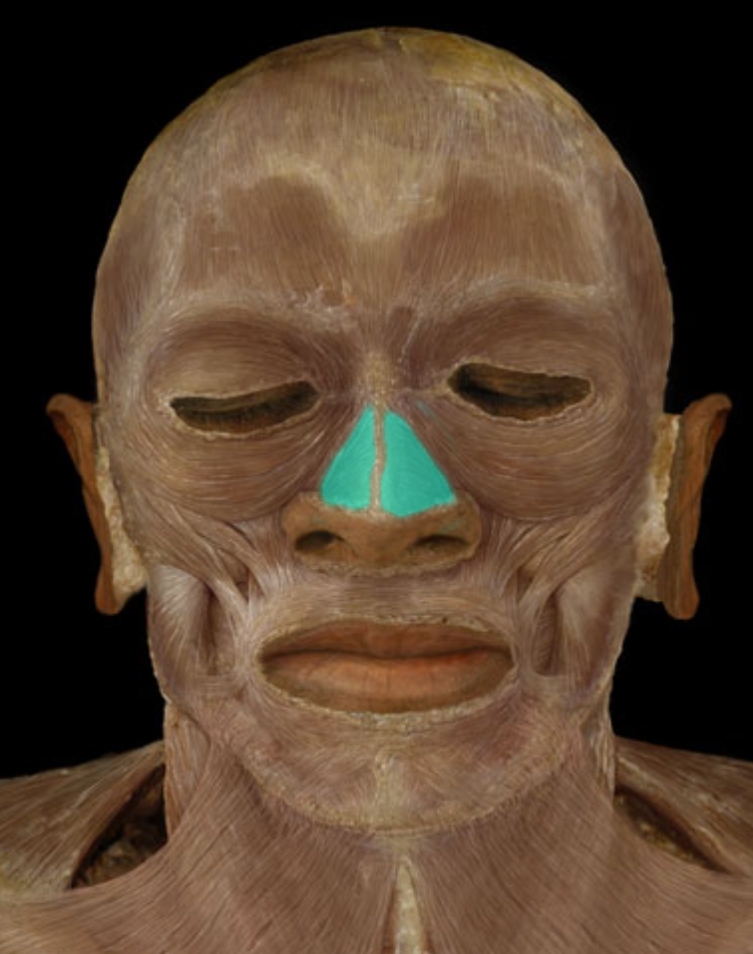
Nasalis m.
Action:
Widen naris (flares nostril)
Origin:
Maxilla
Insertion:
Alar cartilage (of nose)
Innervation:
Facial nerve (CN VII)
Orbicularis Oculi m.
Action:
Closes eyelids (blink/wink)
Origin:
Frontal bone
Maxilla
Insertion:
Fibers blend at lateral side of orbit
Innervation:
Facial nerve (CN VII)

Orbicularis oris m.
Action:
Closes lips
Protrudes lips (pucker)
Origin:
Skin around mouth
Mandible (midline)
Maxilla (midline)
Insertion:
Lips
Interdigitation with fibers from opposite side
Innervation:
Facial nerve (CN VII)

Platysma m.
Action:
Elevates and creases skin of neck (e.g., expression of "horror")
Depression of lower lip and angle of mouth
Origin:
Fascia of upper thorax and lower neck
Insertion:
Mandible (lower border)
Skin and fascia of lower face
Blends with muscles of lower lip and angle of mouth
Innervation:
Facial nerve (CN VII)

Scalene mm.
Action:
Anterior: elevation of rib 1; lateral flexion and rotation of neck
Middle: elevation of rib 1; lateral flexion of neck
Posterior: elevation of rib 2; lateral flexion of neck
Origin:
Transverse processes of C3-7 vertebrae
Insertion:
Anterior: rib 1
Middle: rib 1
Posterior: rib 2
Innervation:
Ventral rami of cervical spinal nerves
Comment:
Neck movements produced when rib is fixed
Important accessory muscles of respiration
Important anatomical relationship: roots of brachial plexus found between anterior and middle scalene muscles

Zygomaticus Major m.
Action:
Elevation of corner of mouth (smile)
Origin:
Zygomatic bone
Insertion:
Blends with muscles at angle of mouth
Innervation:
Facial nerve (CN VII)

Zygomaticus minor m.
Action:
Elevation of upper lip
Produces nasolabial furrow
Origin:
Zygomatic bone
Insertion:
Blends with orbicularis oris muscle
Innervation:
Facial nerve (CN VII)

Deprssor anguli oris m.
Action:
Depression of angle of mouth (grimace)
Origin:
Mandible (body)
Insertion:
Blends with muscles at angle of mouth
Innervation:
Facial nerve (CN VII)

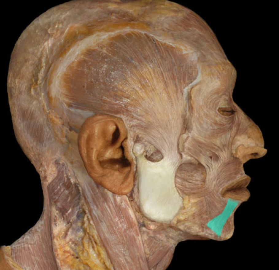
Depressor labii inferioris m.
Action:
Depression of lower lip (pout)
Origin:
Mandible (body)
Insertion:
Blends with muscles of lower lip
Innervation:
Facial nerve (CN VII)
Frontalis m.
Action:
Elevation of eyebrows
Creases skin of forehead
Moves scalp forward (works with occipitalis muscle)
Origin:
Epicranial aponeurosis
Insertion:
Skin of eyebrow and forehead
Blends with orbicularis oculi muscle
Innervation:
Facial nerve (CN VII)
Comment:
Part of occipitofrontalis muscle

Genioglossus m.
Action:
Protraction of tongue ("sticking tongue out")
Origin:
Mandible (mental tubercle)
Insertion:
Blends with musculature of tongue
Innervation:
Hypoglossal nerve (CN XII)
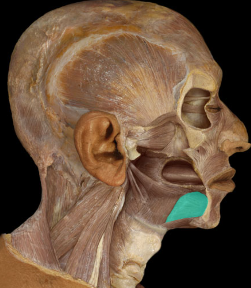
Hyoglossus m.
Action:
Depression of tongue
Origin:
Hyoid bone
Insertion:
Blends with musculature of tongue
Innervation:
Hypoglossal nerve (CN XII)

Inferior oblique m.
Action:
Elevation of eyeball (e.g., looking upward)
Abduction of eyeball (e.g., looking outward)
Lateral rotation of eyeball
Origin:
Maxilla (anterior medial floor of orbit)
Insertion:
Sclera (inferior lateral)
Innervation:
Oculomotor nerve (CN III)

Inferior recuts m.
Action:
Depression of eyeball (e.g., looking downward)
Adduction of eyeball (e.g., looking inward)
Lateral rotation of eyeball
Origin:
Common tendinous ring (at posterior of orbit)
Insertion:
Sclera (inferior surface of eyeball)
Innervation:
Oculomotor nerve (CN III)
Comment:
Common tendinous ring is a fibrous band inside the orbit that surrounds optic canal and part of superior orbital fissure

Lateral rectus m.
Action:
Abduction of eyeball (e.g., looking outward)
Origin:
Common tendinous ring (at posterior of orbit)
Insertion:
Sclera (lateral surface of eyeball)
Innervation:
Abducens nerve (CN VI)

Levator anguli oris m.
Action:
Elevation of angle of mouth (smile)
Origin:
Maxilla
Insertion:
Blends with muscles at angle of mouth
Innervation:
Facial nerve (CN VII)

Levator labii superioris m.
Action:
Elevation of upper lip (sneer)
Origin:
Maxilla
Zygomatic bone
Insertion:
Blends with muscles of upper lip
Innervation:
Facial nerve (CN VII)
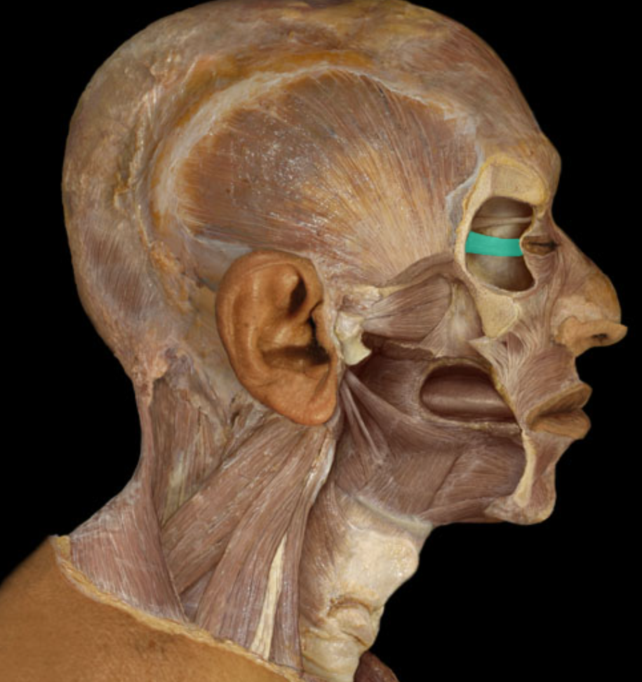
Levator palpebrae superioris m.
Action:
Elevation of upper eyelid
Origin:
Sphenoid bone (superior to common tendinous ring)
Insertion:
Upper eyelid
Innervation:
Oculomotor nerve (CN III)

Masseter m.
Action:
Elevation of mandible (e.g., closing mouth)
Protraction of mandible
Origin:
Zygomatic arch
Insertion:
Mandible (external surface of angle and ramus)
Innervation:
Trigeminal nerve (mandibular division - CN V3)
Comment:
Important muscle for mastication (chewing)

Medial pterygoid m.
Action:
Elevation of mandible (e.g., closing mouth)
Protraction of mandible
Side-to-side movement of mandible (e.g., chewing)
Origin:
Sphenoid bone (lateral pterygoid plate)
Insertion:
Mandible (medial surface of angle and ramus)
Innervation:
Trigeminal nerve (mandibular division - CN V3)

medial rectus m
Action:
Adduction of eyeball (e.g., looking inward)
Origin:
Common tendinous ring (at posterior of orbit)
Insertion:
Sclera (medial surface of eyeball)
Innervation:
Oculomotor nerve (CN III)
Comment:
Common tendinous ring is a fibrous band inside the orbit that surrounds optic canal and part of superior orbital fissure
Occipitalis
Action:
Elevation of eyebrows
Creases skin of forehead
Moves scalp backward (works with frontalis muscle)
Origin:
Occipital bone (superior nuchal line)
Insertion:
Epicranial aponeurosis
Innervation:
Facial nerve (CN VII)
Comment:
Part of occipitofrontalis muscle

occipitalsfrontalis m.
Action:
Elevation of eyebrows
Creases skin of forehead
Moves scalp backward and forward
Origin:
Occipitalis: occipital bone (superior nuchal line)
Frontalis: skin of eyebrow and forehead (blends with orbicularis oculi muscle)
Insertion:
Epicranial aponeurosis
Innervation:
Facial nerve (CN VII)

pharyngeal constrictor mm.
Action:
Produces a wave of contraction (peristalsis) during swallowing
Origin:
Pterygomandibular raphe (superior constrictor)
Hyoid bone (middle constrictor)
Thyroid and cricoid cartilages of larynx (inferior constrictor)
Insertion:
Posterior midline raphe
Base of skull (superior constrictor)
Innervation:
Vagus nerve (CN X)
Comment:
A raphe is a "seam" where two structures meet

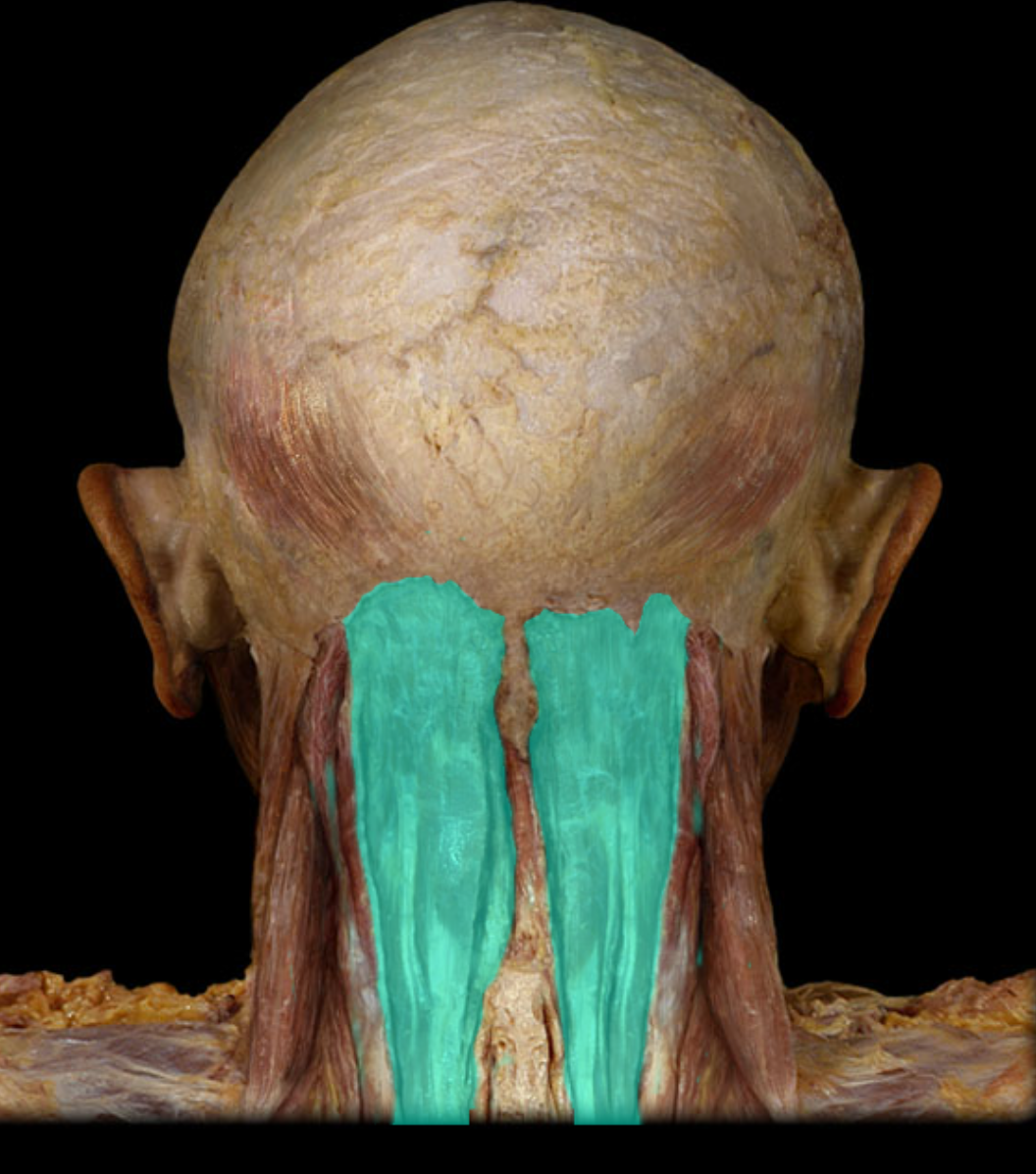
Semispinalis capitis m.
Action:
Unilateral: rotation of head to opposite side
Bilateral: extension of head and neck
Origin:
Transverse processes of C7-T6 vertebrae
Insertion:
Occipital bone
Innervation:
Cervical spinal nerves (dorsal rami)
Splenius capitis m.
Action:
Unilateral: rotation of head to same side
Bilateral: extension of head and neck
Origin:
Nuchal ligament
Vertebra prominens (C7 spinous process)
Spinous processes of T1-3 vertebrae
Insertion:
Temporal bone (mastoid process)
Occipital bone (superior nuchal line)

Splenius cervicis m.
Action:
Unilateral: rotation of head to same side
Bilateral: extension of head and neck
Origin:
Spinous processes of T3-6 vertebrae
Insertion:
Transverse processes of C1-3 vertebrae
Innervation:
Dorsal rami of C3-5 spinal nerves
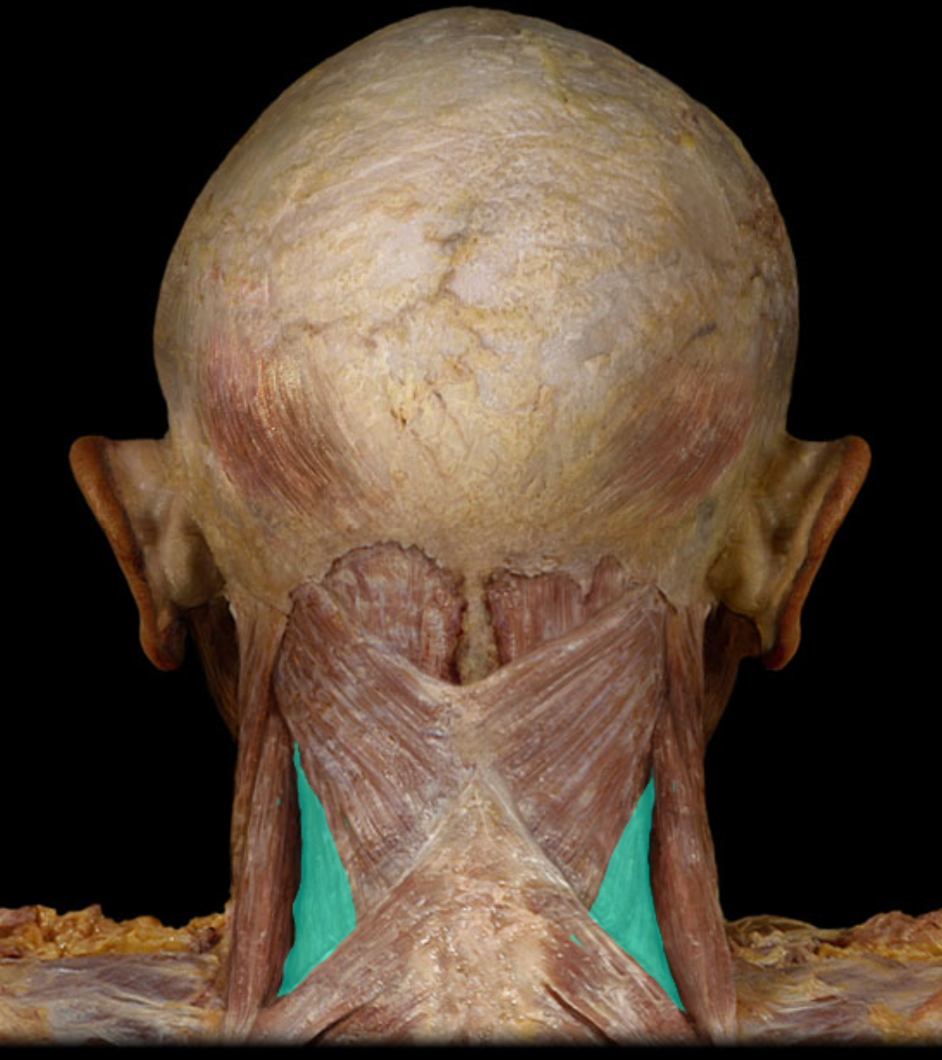
Manubrium
Location:
Sternum
Description:
Triangular shape
Superior part of sternum
Articulates with costal cartilages of ribs 1-2 and clavicles
Comment:
Provides attachment for sternocleidomastoid and pectoralis major muscles

Which vertebrae make up the cervical vertebra
C1-C7
Which vertebrae make up the thoracic vertebra?
L1 - L12
Which of the ribs are ture ribs?
ribs 1-7
Which of the ribs are false ribs?
8-10
Which vertebrae make up the lumbar vertabra
L1-L5
Inferior articular process of cervical vertebra

Superior articular process of vervical vertebra

Lamina of cervical vertebra

Pedicle of cervical vertebra

Spinous process of the cervical vertebra

Transverse foramen of the cervical vertebra.

Transverse process of the cervical vertebrae

Vertebral foramen of cervical vertebra

posterior arch of the atlas (C1)

Superior articular facet of the atlas (C1)

Dens of the axis (C2)
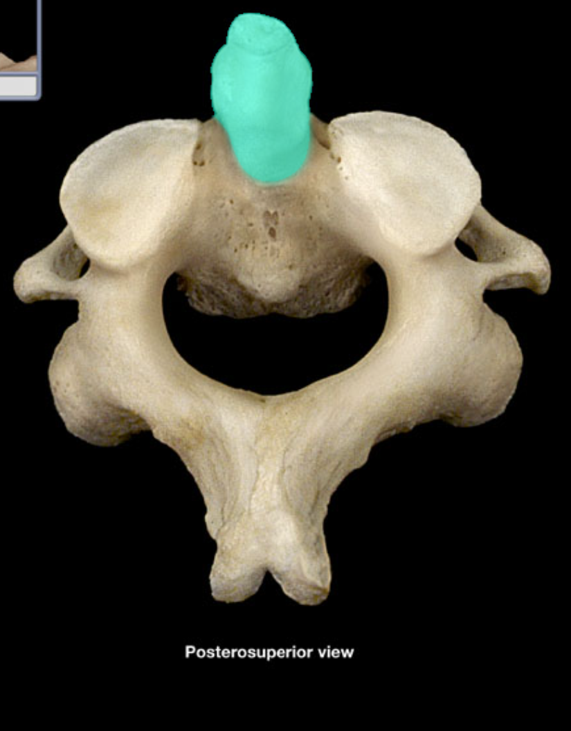
Pedicle of the axis

Thoracic vertebra
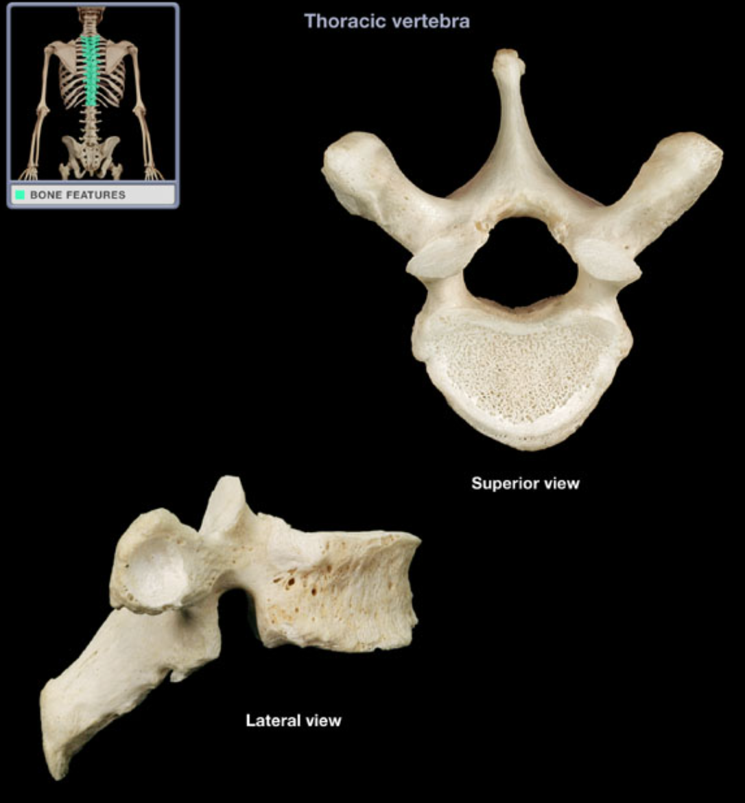
Lumbar vertebra

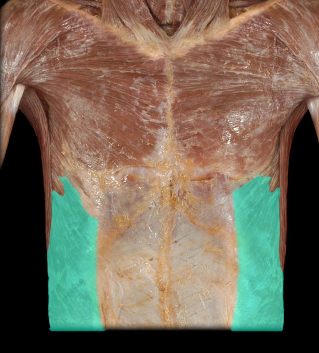
External abdominal oblique m.
Action:
Unilateral: rotation of trunk
Bilateral: flexion of trunk (sit-ups)
Compression of anterior abdominal wall and viscera (increases intra-abdominal pressure)
Origin:
Ribs and costal cartilages 5-12
Insertion:
Linea alba
Ilium (crest)
Innervation:
Ventral rami of T8-L1 spinal nerves
Comment:
Important in "straining" (e.g., lifting, defecation, urination, child-birth)
Functions in respiration ("abdominal breathing")
At right angle to internal abdominal oblique (strengthens abdominal wall)
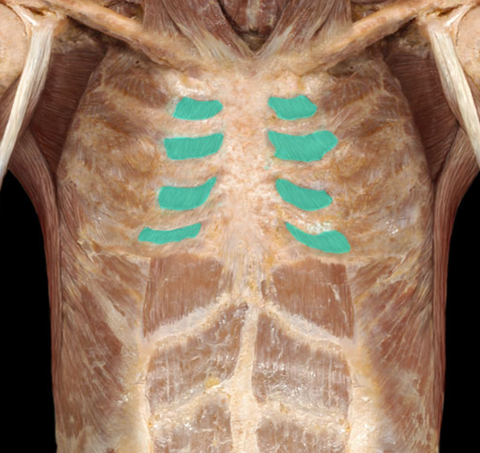
External intercostal m and membrane
Action:
Muscle of respiration
Elevation of ribs during inhalation to increase transverse dimensions (anterior-posterior, medial-lateral) of thoracic cavity
Origin:
Inferior border of rib (ribs 1-11)
Insertion:
Superior border of rib below (ribs 2-12)
Innervation:
Intercostal nerves (ventral rami of T1-11 spinal nerves)
Comment:
External intercostal muscle continuous with external intercostal membrane near sternum
In contemporary medical anatomy books, the origins of external and internal intercostal muscles are referred to as "superior attachments" and described for both muscles as the inferior border of the rib. Similarly, the insertions of these muscles are referred to as "inferior attachments" and described as the superior border of the rib below.

Internal intercostal m.
Action:
Muscle of respiration
Depression of ribs during forced exhalation to decrease transverse dimensions (anterior-posterior, medial-lateral) of thoracic cavity
Origin:
Superior border of rib (ribs 1-11)
Insertion:
Inferior border of rib above (ribs 2-12)
Innervation:
Intercostal nerves (ventral rami of T1-11 spinal nerves)
Comment:
Internal intercostal muscle continuous with internal intercostal membrane lateral to vertebral column
Muscle fibers run inferoposteriorly
In contemporary medical anatomy books, the origins of external and internal intercostal muscles are referred to as "superior attachments" and described for both muscles as the inferior border of the rib. Similarly, the insertions of these muscles are referred to as "inferior attachments" and described as the superior border of the rib below.
Latissimus dorsi m.
Action:
Adduction of arm
Extension of arm
Medial rotation of arm
Origin:
Sacral and lumbar spines via thoracolumbar fascia
Ilium (iliac crest) via thoracolumbar fascia
Ribs (lower four)
Spinous processes of T6-12 vertebrae
Scapula (inferior angle)
Insertion:
Humerus (floor of intertubercular sulcus)
Innervation:
Thoracodorsal nerve

Pectoralis majojr m.
Action:
Adduction of arm
Medial rotation of arm
Clavicular head: flexion of arm
Sternocostal head: extension of arm (from flexed position)
Origin:
Clavicular head: clavicle (medial part)
Sternocostal head: sternum, costal cartilages 1-6, and aponeurosis of external abdominal oblique muscle
Insertion:
Humerus (lateral lip of intertubercular sulcus)
Innervation:
Lateral pectoral nerve (C5-7)
Medial pectoral nerve (C8-T1)
Comment:
Pectoralis major and minor muscles also known as "pecs"
Pectoralis major active during forced inspiration

Pectoralis minor m.
Action:
Stabilizes scapula (holds it against thoracic wall)
Protraction (abduction) of scapula (assists serratus anterior)
Lateral rotation of scapula (e.g., depresses the acromion)
Origin:
Ribs 3-5
Insertion:
Scapula (coracoid process)
Innervation:
Medial and lateral pectoral nerves
Comment:
Pectoralis major and minor muscles also known as "pecs"
Pectoralis minor active during forced inspiration

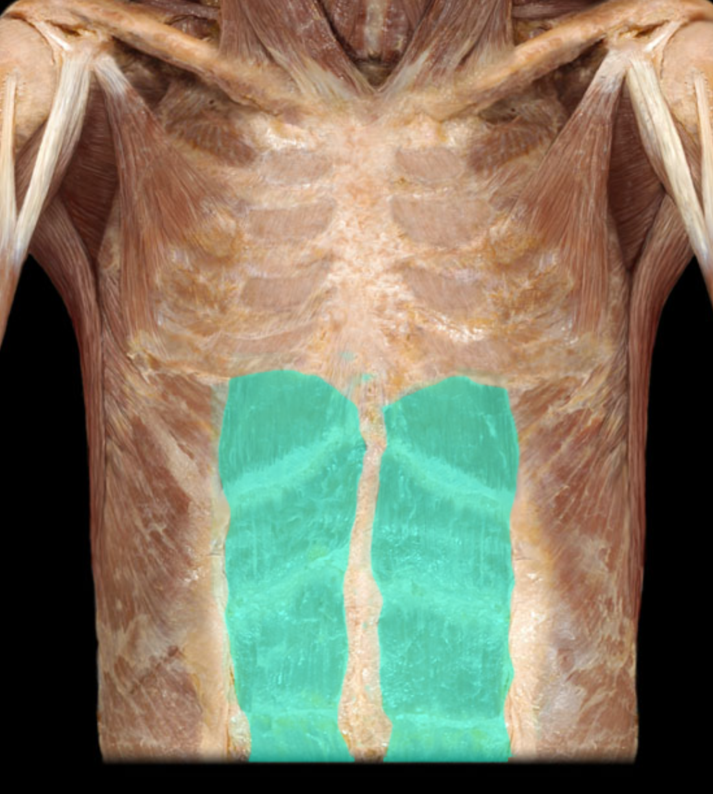
Rectus abdominis m.
Action:
Flexion of trunk (sit-ups)
Compression of anterior abdominal wall
Origin:
Pubis (crest and symphysis)
Insertion:
Sternum (xiphoid process)
Costal cartilages 5-7
Innervation:
Ventral rami of T7-12 spinal nerves
Comment:
Three or four bellies, separated by tendinous intersections
Encased in fibrous (rectus) sheath
Important in "straining" (e.g., lifting, defecation, urination, child-birth)
Functions in respiration ("abdominal breathing")
Serratus anterior m.
Action:
Protraction (abduction) of scapula
Superior rotation of scapula (directs glenoid cavity superiorly)
Stabilizes scapula (holds it against thoracic wall)
Origin:
Ribs 1-9
Insertion:
Scapula (medial border)
Innervation:
Long thoracic nerve
Comment:
Serratus anterior is a prime mover in all reaching and pushing movements

External abdominal oblique m.
Action:
Unilateral: rotation of trunk
Bilateral: flexion of trunk (sit-ups)
Compression of anterior abdominal wall and viscera (increases intra-abdominal pressure)
Origin:
Ribs and costal cartilages 5-12
Insertion:
Linea alba
Ilium (crest)
Innervation:
Ventral rami of T8-L1 spinal nerves
Comment:
Important in "straining" (e.g., lifting, defecation, urination, child-birth)
Functions in respiration ("abdominal breathing")
At right angle to internal abdominal oblique (strengthens abdominal wall)

Internal abdominal oblique m.
Action:
Unilateral: rotation of trunk
Bilateral: flexion of trunk (sit-ups)
Compression of anterior abdominal wall and viscera (increases intra-abdominal pressure)
Origin:
Inguinal ligament
Ilium (crest)
Thoracolumbar fascia
Insertion:
Linea alba
Costal cartilages 7-10
Innervation:
Ventral rami of T8-L1 spinal nerves
Comment:
Important in "straining" (e.g., lifting, defecation, urination, child-birth)
Functions in respiration ("abdominal breathing")
At right angle to external abdominal oblique (strengthens abdominal wall)

Rectus abdominis m.
Action:
Flexion of trunk (sit-ups)
Compression of anterior abdominal wall
Origin:
Pubis (crest and symphysis)
Insertion:
Sternum (xiphoid process)
Costal cartilages 5-7
Innervation:
Ventral rami of T7-12 spinal nerves
Comment:
Three or four bellies, separated by tendinous intersections
Encased in fibrous (rectus) sheath
Important in "straining" (e.g., lifting, defecation, urination, child-birth)
Functions in respiration ("abdominal breathing")
