Anatomy Lower Extremity
 Call Kai
 Learn
Learn Practice Test
 Spaced Repetition
 Match
Match Flashcards
 Knowt Play
1/256

There's no tags or description
Looks like no tags are added yet.
Name | Mastery | Learn | Test | Matching | Spaced | Call with Kai | Chat |
|---|
No analytics yet
Send a link to your students to track their progress
257 Terms
Lower limb stability
mobility has been sacrificed
▪ Atmospheric pressure: maintains joint congruency
▪ Gravity in the erect position
▪ Acetabular labrum: increases the contact btwn. the acetabulum and the head of the femur
▪ Capsule and ligaments: Improves stability by keeping the ball in the socket – based on the attachments
hip region
most are referring to both the hip and the pelvis
▪ Encompasses the articulations of the pelvis and their interplay with the actual hip joint
• Pelvis is the articulations between the ilia and the sacrum
• Hip is the articulation of the femoral head with the acetabulum
Hip joint
o Multi-Axial Ball in socket; Three Degrees of Freedom
o Movements:
▪ Flexion/extension- sagittal plane
▪ Abduction/adduction- frontal plane
▪ Internal/external rotation- transverse plane
proximal femur
the portion that articulates with the acetabulum: Head
▪ Femoral head is covered with articular cartilage
▪ Fovea Capitis: Pit for the ligament to the head of the femur
▪ The convex surface of the ball & socket joint fits into the acetabulum
femoral neck
▪ Continuation of the shaft of the femur running anterior superior and medially
▪ Angulation of the neck is 125
▪ Joins with the upper femur at the greater trochanter
femoral neck: angle of inclination
o Angulation of the femoral neck in the frontal plane
o Normal Value in adult= 125
o Coxa Valga= angle of inclination > 125: Coxa Valga can occur in cases of congenital hip dislocation – clinically, in this condition there will be a limitation in adduction of the hip
o Coxa Vara= angle of inclination < 120: Coxa Vara – Fractures in neck of the femur and slipping of the epiphysis – clinically there will be a limitation in abduction of the hip
o Changes the stresses placed on the distal joints
Shenton’s Line
o Normal hip radiograph demonstrates Shenton’s line
o Break in Shenton’s line: indicates Hip Fracture – Neck; Ilia Fracture – Acetabular

femoral neck: angle of declination
o Angulation of the femoral neck in the transverse plane
o Normal value in adult= 14 degrees of anteversion
o Increase in angle is called anteversion (typically toe-in posturing)
o Decrease in angle is called retroversion (typically toe-out posturing)
trabecular pattern of the femur
o Internal architecture of the femur and pelvis adapts to mechanical stresses
o Most of the weight-bearing lines in the pelvis pass into the acetabulum
o Areas of trabecular overlap offer the greatest resistance to stress and strain
o Zone of weakness (Ward’s triangle): Trabeculae are weak and non-overlapping

Inominate bone
o Components
▪ Ilium
▪ Ischium
▪ Pubis
o Fuse in the acetabulum to form a solid mass of bone
acetabulum
o Oriented lateral, anterior and inferior
o Acetabular notch- inferior interruption of the acetabulum, spanned by the transverse acetabular ligament
o Acetabular fossa- deepest portion, not covered by articular cartilage; contains a fat pad
articular cartilage in the hip joint
o Acetabulum: Horseshoe shaped; Avascular; Thickest superiorly
o Femoral: Thickest superior and posterior; Thinnest inferiorly
o Acetabular Fossa is the non-articular part of the acetabulum that does not have any cartilage
acetabular labrum
o Fibrocartilaginous ring attached to the periphery of the acetabulum which continues across the acetabular notch as the transverse acetabular ligament
o Deepens hip socket and increases congruity
o Increases stability

hip joint capsule
o Thick and strong and reinforced by strong ligaments
o Dense, inelastic and fibrous
o Attaches to the periphery of the acetabulum blending with the labrum medially
o Encloses the entire joint
o Thickest anterosuperior; Thinnest posteroinferior
o Joint capsule has two layers: outeer layer which is generally fiberous and an inner layer which is a synovial / secreting layer.
o It surrounds the neck of the femur, and is attached, in front, to the intertrochanteric line; above, to the base of the neck; behind, to the neck, about 1.25 cm. above the intertrochanteric crest; below, to the lower part of the neck, close to the lesser trochanter.
o Has to be strong to provide both the stability and the mobility needed in the lower extremity
o Encloses most of the hip joint and the neck of the femur
o Reinforced by 3 ligaments (iliofemoral, pubofemoral and ischiofemoral)
o Anterior capsule also reinforced by rectus femoris tendon
hip joint ligaments
o Anterior
▪ Iliofemoral
▪ Pubofemoral
o Posterior
▪ Ischiofemoral
iliofemoral ligament
o Strongest ligament of the hip
o Sometimes referred to as the “Y ligament of Bigelow”
o Attaches to AIIS with two arms fanning out to the intertrochanteric line
o Limits extension primarily and external rotation secondarily
o Prevents overextension in standing
o Permits person to stand with the joint in extension using minimal muscle action

pubofemoral ligament
o Strengthens the anterior and the inferior capsule
o Runs from pubis, near the acetabulum, to the femur, just anterior to the lesser trochanter
o Limits extension, abduction and external rotation

ischiofemoral ligament
o Attaches to the posterior-inferior surface of the acetabulum to the posterior-superior aspect of the neck of the femur (where neck meets the greater trochanter)
o Limits extension and internal rotation
o Reinforces the hip posterior

intracapsular ligament- ligamentum teres
o Triangular-shaped band arising from the acetabular fossa and transverse acetabular ligament to the fovea of the femoral head. Covered by synovial membrane. Approx. 3.5cm long and very weak
o No significant role in joint stability
o Contains the artery to the head of the femur, a branch of the obturator artery
mechanics of hip joint capsule and ligaments
o In standing position, ligaments are under moderate tension
o Extension causes a twisting of all the capsular ligaments forcing the femur into the acetabulum
o Flexion relaxes all the ligaments
pelvic ligaments
iliolumbar
sacrospinous
sacrotuberous
iliolumbar ligament
▪ Arises at tip of the L4 and L5 transverse process
▪ Connects L4 and L5 to the ilium
stabilizes spine and prevents excessive movements
sacrospinous ligament
▪ Attaches sacrum to the ischial spine
▪ Prevents upward rotation of the inferior sacrum
sacrotuberous ligament
▪ Attaches sacrum to ischial tuberosity
▪ Bind the sacrum to the ischium
▪ Prevents upward rotation of the inferior sacrum
▪ The sacrotuperous and the sacrospinous also create foramina
hip joint bursae
o Bursae are closed sacs lined with a synovial membrane and contain synovial fluid
o Purpose is to reduce friction
o Area's subject to friction: Exposed area or where a tendon passes over a bone or between other structures where friction is likely to occur
gluteal, trochanteric, and ischial
gluteal bursae
▪ Above and behind the greater trochanter
▪ Underneath the gluteus maximus and medius
trochanteric bursae
▪ Superficial
▪ Between the greater trochanter and the iliotibial band
ischial bursae
Overlies the ischial tuberosity. Covered by the edge of the gluteus maximus
flexors of the hip
sartorius
rectus femoris
iliopsoas: iliacus and psoas major
sartorius
Part of the anterior compartment of the thigh; most superficial muscle of the ant thigh; Straplike muscle; Not affected by the position of the knee.
▪ O: ASIS
▪ I: Pes anserine
▪ A: Main action is hip flexion; Also produces abduction and external rotation
▪ N: Femoral nerve

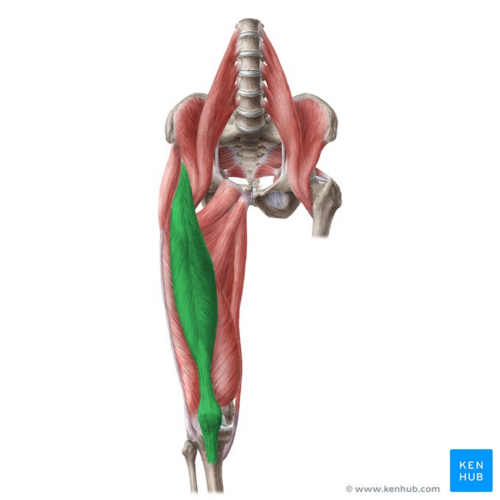
rectus femoris
Power as a hip flexor; Only portion of the quad that passes the hip and the knee; Optimal length for hip flexion occurs when the knee is flexed
▪ O: AIIS
▪ I: common tendon to tibial tuberosity
▪ A: assist illiopsoas in hip flexion and extends the knee with the quads
▪ N: femoral nerve
iliopsoas
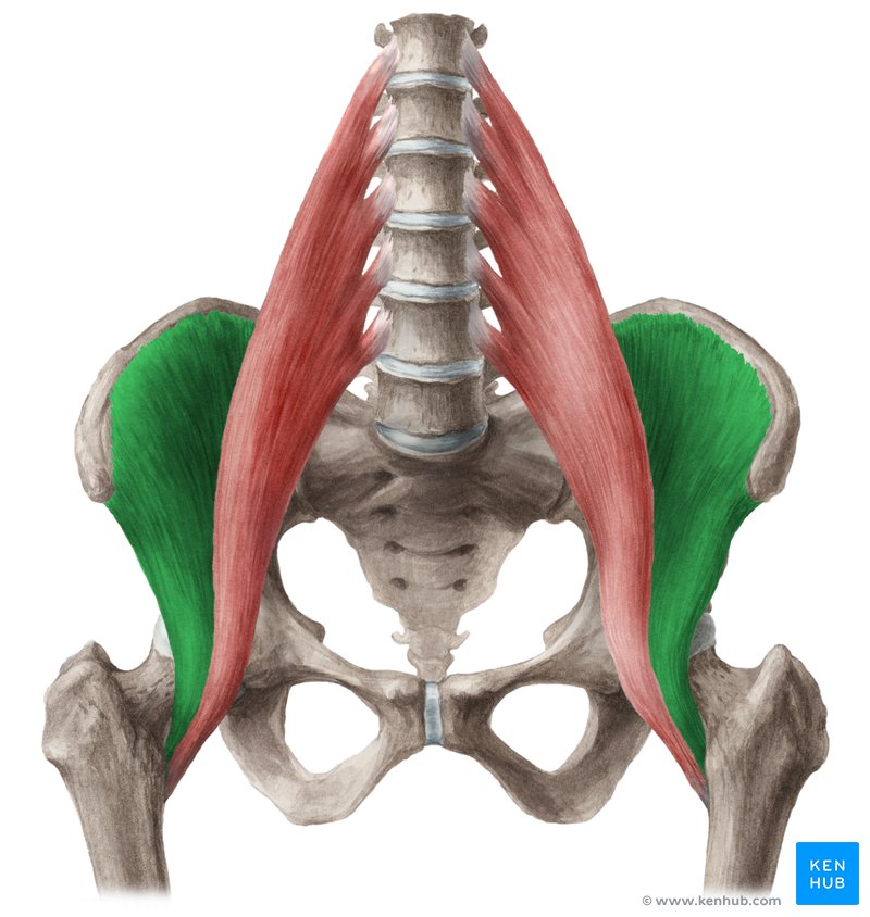
Actually 2 muscles with the same insertion points – iliacus – Psoas major; Also stabilizes the joint.

iliacus
• O: Iliac fossa, illiac crest, ala of sacrum
• transverse processes of lumbar vertebrae
• I: Lesser trochanter of the femur
• A: Hip flexion
• N: Femoral nerve
psoas major
• O:transverse processes of lumbar vertebrae T12-L5
• I: Lesser trochanter of the femur
• A: Hip flexion, action has been noted in lateral rotation
• N: Anterior Rami of the lumbar nerves L1, L2, L3

extensors of the hip
Muscles of the Gluteal Region and Posterior Compartment of the Thigh
o Primary:
▪ Gluteus Maximus
▪ Hamstrings
o Secondary:
▪ Gluteus Medius
▪ Adductor Magnus
▪ Piriformis
gluteus maximus
▪ O: Posterior ilium and sacrum and associated ligaments
▪ I: Superior fibers insert into ITB Inferior fibers into gluteal tuberosity on the femur
▪ A: Hip extension and external rotation
▪ N: Inferior gluteal nerve

hamstrings
▪ Biceps Femoris: Two Heads
▪ Semitendinosus
▪ Semimembranosus
▪ As a whole the hamstrings are 2/3 as powerful as the Maximus
biceps femoris
▪ O: Long head- ischial tuberosity
▪ O: Short head- lateral linea aspera
▪ I: Fibular head
▪ A: Flexes knee and extends hip,
▪ N: Long Head:Tibial
▪ Short Head: Common Peroneal

semimembranosus
▪ O: Ischial tuberosity
▪ I: Medial condyle of the tibia
▪ A: Flexes knee and extends the hip
▪ N: Sciatic nerve

semitendinosus
Usually can be found on top of the semimembranosus
▪ O: Ischial tuberosity
▪ I: Pes anserine
▪ A: Flexes knee and extends hip
▪ N: Sciatic nerve

abductors of the hip
tensor fascia latae
gluteus medius
gluteus minimus
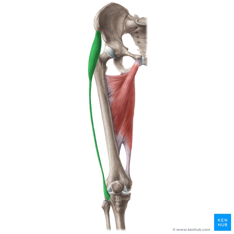
tensor fascia latae
Sometimes considered one of the muscles of the gluteal region; Also helps to keep the knee extended during upright posture. That’s why hip pain sometimes refers to the knee; Keeps pelvis level when opposite leg is raised.
▪ O: anterior external lip of iliac crest and the ASIS
▪ I: IT band which attaches to the lateral condyle of the tibia
▪ A: Assists in abduction and medial rotation of the thigh
▪ N: Superior gluteal nerve
gluteus medius
most powerful abductor
▪ O: external surface of ilium
▪ I: greater trochanter of femur
▪ A: hip abduction
▪ N: superior gluteal nerve

gluteus minimus
▪ O: external surface of the ilium
▪ I: greater trochanter of femur
▪ A: hip abduction
▪ N: superior gluteal nerve

adductors of the hip
Medial compartment of the thigh
obturator externus
pectinius
adductor brevis
adductor longus
adductor magnus
gracilis
obturator externus
▪ O: Margins of the obturator foramen
▪ I: Trochanteric fossa
▪ A: Lateral rotation of the thigh
▪ N: Obturator Nerve

pectinius
▪ O: Superior ramus of pubis
▪ I: Pectineal line of femur inferior to lesser trochanter
▪ A: Adduction and flexion of the thigh. Assists with medial rotation
▪ N: Femoral Nerve

adductor brevis
▪ O: Body and inferior ramus of pubis
▪ I: Pectineal line of femur, proximal linea aspera
▪ A: Adduction and flexion of the thigh. Assists with medial rotation
▪ N: Obturator Nerve

adductor longus
▪ O: Body of pubis inferior to crest
▪ I: Middle Third of linea aspera
▪ A: Adduction and flexion of the thigh. Assists with medial rotation
▪ N: Obturator Nerve

adductor magnus
▪ O: Inferior ramus of pubis and ramus of ischium
▪ I: Adductor Part: Gluteal Tuberosity, linea aspera, and medial supracondylar line.
Hamstring Part: Adductor Tubercle
▪ A: Adduction and flexion of the thigh. Assists with medial rotation
▪ N: Adductor Part: Obturator Nerve.
Hamstring Part: Tibial portion of the Sciatic Nerve

gracilis
▪ O: Body and inferior ramus of pubis
▪ I: Pes Anserine
▪ A: Adduction and flexion of the thigh. Assists with medial rotation
▪ N: Obturator Nerve

external rotators of the hip
piriformis
gemellus superior
obturator internus
gemellus inferior
quadratus femoris
piriformis
▪ Origin: Anterior sacrum
▪ Insertion: Greater trochanter of femur
▪ Action: Laterally rotates extended thigh, Abducts a flexed thigh
▪ Nerve: Nerve to Piriformis

gemellus superior
▪ Origin: Ischial spine
▪ Insertion: Greater trochanter of femur
▪ Action: Laterally rotates thigh, Abducts thigh when flexed (2o)
▪ Nerve: Nerve to Obturator Internus

obturator internus
▪ Origin: Posterior margins of obturator foramen, obturator membrane
▪ Insertion: Greater Trochanter of femur
▪ Action: Laterally rotates thigh, Abducts thigh when flexed
▪ Nerve: Nerve to obturator internus

gemellus inferior
▪ Origin: Ischial tuberosity
▪ Insertion: Greater trochanter of femur
▪ Action: Laterally rotates thigh
▪ Nerve: Nerve to Quadratus Femoris

quadratus femoris
▪ Origin: Lateral aspect of ischial tuberosity
▪ Insertion: Greater trochanter of femur
▪ Action: Laterally rotates thigh
▪ Nerve: Nerve to Quadratus Femoris

iliotibial band
o Continuous with deep fascia throughout leg.
o Tensor fasciae lata line of pull through a thickening of the deep fascia.
o Iliotibial band/tract
o I : Gerdy’s tubercle

blood supply to hip
o The intra-articular isolation of the femoral head and most of the neck restrict their blood supply to two sources
o Ascending arteries: Major blood supply is derived from the medial and lateral circumflex arteries which pass posterior and anterior respectively and give off branches piercing the capsule
o Ligamentum teres: Carries a small branch of the obturator artery and may vascularize a small area of the femoral head
nerve supply to hip
o Deep branches of the sciatic nerve supply the posterior capsule
o Deep branches of the femoral nerve and obturator nerve supply the anterior and inferior capsule
o These nerves represent levels L2-S2
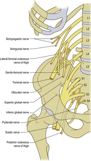
lumbar plexus
sacral plexus
lumbar plexus
▪ Iliohypogastric (L1)- muscular branches to the abdominals and the cutaneous innervation to the skin of the gluteal region and hypogastric area
▪ Ilioinguinal (L1)- muscular branches to the abdominals and the cutaneous innervation to the skin of the anterior groin
▪ Genitofemoral (L1-L2)- Femoral branch to cremaster muscle and genital branch supplying cutaneous innervation to a small area of the anterior thigh
▪ Lateral Femoral Cutaneous (L2-L3)- runs on top of the iliacus muscle, crosses under the inguinal ligament to supply skin of the anterior/lateral thigh
▪ Obturator (L2-L4)- enters the thigh through the obturator foramen into the medial thigh to supply the adductor longus, brevis, part of magnus, gracilis, obturator externus, and a small area of skin on the medial thigh
▪ Femoral (L2-L4)- enters the anterior thigh under the inguinal ligament to supply the quadriceps, sartorius, pectineus, iliacus and the skin of the anterior/medial thigh

iliohypogastric nerve
L1
muscular branches to the abdominals and the cutaneous innervation to the skin of the gluteal region and hypogastric area
ilioinguinal nerve
L1
muscular branches to the abdominals and the cutaneous innervation to the skin of the anterior groin
genitofemoral nerve
L1-L2
Femoral branch to cremaster muscle and genital branch supplying cutaneous innervation to a small area of the anterior thigh

lateral femoral cutaneous nerve
L2-L3
runs on top of the iliacus muscle, crosses under the inguinal ligament to supply skin of the anterior/lateral thigh
obturator nerve
L2-L4
enters the thigh through the obturator foramen into the medial thigh to supply the adductor longus, brevis, part of magnus, gracilis, obturator externus, and a small area of skin on the medial thigh
femoral nerve
L2-L4
enters the anterior thigh under the inguinal ligament to supply the quadriceps, sartorius, pectineus, iliacus and the skin of the anterior/medial thigh
sacral plexus
▪ The plexus forms on the anterior surface of the piriformis muscle
▪ All branches exit through the greater sciatic foramen with the piriformis
▪ Nerve to quadratus femoris and gemellus inferior (L4-S1)
▪ Nerve to Piriformis (S1-S2)
▪ Nerve to Obturator Internus and Gemellus Superior (L5-S2)
▪ Superior Gluteal (L4-S1)- exists the greater sciatic foramen superior to piriformis to supply the gluteus medius, minimus, and tensor fascia latae
▪ Inferior Gluteal (L5-S2)- exits the greater sciatic foramen inferior to the piriformis to supply the gluteus maximus
▪ Posterior Femoral Cutaneous (S1-S3)- exits with the inferior gluteal nerve supplying the skin of the posterior thigh and calf
▪ Sciatic (L4-S3)- exits the greater sciatic foramen inferior to the piriformis; composed of two nerves (tibial and common peroneal)
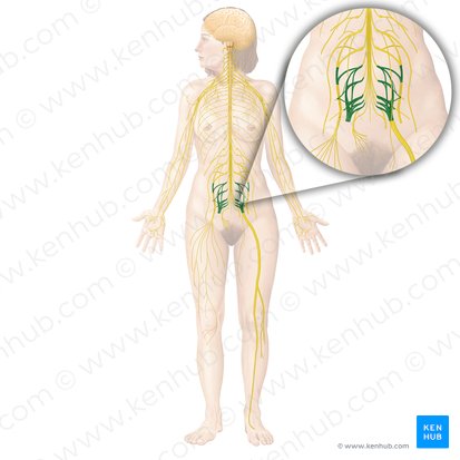
superior gluteal nerve
L4-S1
exists the greater sciatic foramen superior to piriformis to supply the gluteus medius, minimus, and tensor fascia latae
inferior gluteal nerve
L5-S2
exits the greater sciatic foramen inferior to the piriformis to supply the gluteus maximus
posterior femoral cutaneous nerve
S1-S3
exits with the inferior gluteal nerve supplying the skin of the posterior thigh and calf
sciatic nerve
L4-S3
exits the greater sciatic foramen inferior to the piriformis; composed of two nerves (tibial and common peroneal)
usually ends at the superior angle of the popliteal fossa by dividing into tibial and common fibular nerves. The tibial n. is the larger more medial branch. The common fibular nerve is more lateral and winds around the head of the fibula where it divides into superficial and deep fibular branches
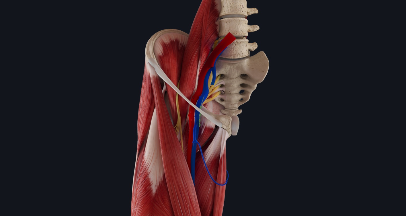
femoral triangle (boundaries and contents)
o Boundaries
▪ Inguinal ligament: Superior
▪ Adductor longus: Medially
▪ Sartorius: Laterally
o Floor: Iliopsoas and Pectineus
o Roof: Skin and Fascia of the thigh
o Contents
▪ Femoral nerve
▪ Femoral artery
▪ Femoral vein
adductor canal (boundaries, contents, etc.)
▪ Sometimes referred to as Hunter’s or the Subsartorial canal
▪ Deep to Sartorius
▪ Transmits vessels to the Adductor Hiatus
▪ Contents:
• Femoral artery and vein
• Saphenous nerve
• Nerve to vastus medialis
▪ Boundaries:
• Anteriolaterally by vastus medialis
• Posteriorly by adductors longus and magnus
• Anteriorly by Sartorius

adductor hiatus
▪ Is an opening or gap between the aponeurotic distal attachment of the adductor part of the adductor magnus and the tendinous distal attachment of the hamstring part.
▪ Put simply: an opening in the tendon of the adductor magnus in the distal third of the thigh
▪ Transmits the femoral artery and vein from the adductor canal in the thigh to the popliteal fossa posterior to the knee

popliteal fossa (boundaries, contents, etc.)
o When the knee is extended the popliteal fossa appears as a diamond-shaped impression posterior to the knee joint.
o Boundaries of the Fossa:
▪ Superolaterally: biceps femoris m.
▪ Superomedially: Semimembranosus, lateral to which is the semitendinosis
▪ Inferolaterally: lateral head of the gastrocnemius (major calf muscle)
▪ Inferomedially: medial head of the gastronemius
o Contents of the Popliteal Fossa
▪ Termination of the small saphenous vein
▪ Popliteal arteries and veins and their branches and tributaries
▪ Tibial and common fibular nerves (sciatic)
▪ Posterior Cutaneous nerve of the thigh
▪ Popliteal lymph nodes and lymphatic vessels.
o The sciatic nerve usually ends at the superior angle of the popliteal fossa by dividing into tibial and common fibular nerves. The tibial n. is the larger more medial branch. The common fibular nerve is more lateral and winds around the head of the fibula where it divides into superficial and deep fibular branches.
o When the femoral artery passes through the adductor hiatus of the medial thigh it changes its name to the popliteal artery.
iliac arteries
o Descending Aorta divides into Common Iliac Arteries
o Common Iliac Arteries divide into Internal and External Iliac Arteries
o External Iliac Artery crosses under inguinal ligament to become Femoral Artery in the Femoral Triangle
o Femoral Artery can palpate the femoral pulse. Located posterior to the deep fascia. Primary blood supply to the LE.
o Femoral then runs deep to sartorius and within the adductor canal
femoral arteries
o Approximately 4 cm into Femoral Triangle the Femoral Artery gives off the Profunda (deep) Femoris artery
o Profunda Femoris is the major artery to the thigh. Runs lateral to the femoral artery and then passes posterior to it
o The Femoral Artery then leaves the Femoral Triangle, travels down the antero-medial thigh and enters the Adductor Canal.
o Femoral Artery then pierces the Adductor Magnus muscle at the Adductor Hiatus
popliteal arteries
o After exiting the Adductor Hiatus, the femoral artery travels into the popliteal fossa and becomes the popliteal artery.
o The popliteal artery gives out five branches: medial superior and inferior, lateral superior and inferior and middle genicular arteries supply the articular capsule and ligaments of the knee.
o At the lower border of Popliteus the Popliteal Artery divides into the Anterior and Posterior Tibial Arteries
anterior tibial artery
▪ The smaller of the 2 communicating branches of popliteal
▪ Passes through the interosseus membrane and then travels along the ant. surface of the membrane btwn. Extensor Hallucis Longus and Tibialis Anterior
▪ Supplies muscles in the ant. compartment
▪ The Anterior Tibial Artery turns into the Dorsalis Pedis Artery distal to Extensor Retinaculum
posterior tibial artery
▪ Larger of the 2 branches of the tibial artery
▪ Passes deep to the origin of the Soleus
▪ Descends with the Tibial nerve
▪ Gives off Nutrient Artery and Peroneal Artery.
▪ Posterior Tibial Artery passes behind medial malleolus and distal to flexor retinaculum and divides into the Medial and Lateral Plantar Arteries
▪ Medial plantar deep to the retinaculum – passes medial side of the foot btwn AbH. and FDB – muscular and cutaneous branches
▪ Lateral plantar deep to the retinaculum and passes to the lateral side obliquely across the foot – muscular and cutaneous branches
veins of lower limb
The veins of the lower limb drain deoxygenated blood and return it to the heart. They can be divided into two groups – deep and superficial: Deep veins are located underneath the deep fascia of the lower limb, accompanying the major arteries. Superficial veins are found in the subcutaneous tissue. They eventually drain into the deep veins
ankle and foot deep veins
▪ The main venous structure of the foot is the dorsal venous arch, which mostly drains into the superficial veins. Some veins from the arch penetrate deep into the leg, forming the anterior tibial vein.
▪ On the plantar aspect of the foot, medial and lateral plantar veins arise. These veins combine to form the posterior tibial and fibular veins. The posterior tibial vein accompanies the posterior tibial artery, entering the leg posteriorly to the medial malleolus
dorsal venous arch
mostly drains into the superficial veins. Some veins from the arch penetrate deep into the leg, forming the anterior tibial vein
posterior tibial and fibular veins
On the plantar aspect of the foot, medial and lateral plantar veins arise. These veins combine to form the posterior tibial and fibular veins. The posterior tibial vein accompanies the posterior tibial artery, entering the leg posteriorly to the medial malleolus
lower leg and knee region deep veins
On the posterior surface of the knee, the anterior tibial, posterior tibial and fibular veins unite to form the popliteal vein. The popliteal vein enters the thigh via the adductor canal.
popliteal vein
On the posterior surface of the knee, the anterior tibial, posterior tibial and fibular veins unite to form the popliteal vein. The popliteal vein enters the thigh via the adductor canal.
thigh and pelvis deep veins
▪ Thigh:
• Once the popliteal vein has entered the thigh, it is known as the femoral vein. It is situated anteriorly, accompanying the femoral artery.
• The deep vein of the thigh (profunda femoris vein) is the other main venous structure in the thigh. Via perforating veins, it drains blood from the thigh muscles. It then empties into the distal section of the femoral vein. (Typically in the femoral triangle)
• The femoral vein leaves the thigh by running underneath the inguinal ligament, at which point it is known as the external iliac vein.
▪ Gluteal Region: The gluteal region is drained by inferior and superior gluteal veins. These empty into the internal iliac vein
deep vein of the thigh (profunda femoris vein)
main venous structure in the thigh. Via perforating veins, it drains blood from the thigh muscles. It then empties into the distal section of the femoral vein. (Typically in the femoral triangle)
femoral vein
leaves the thigh by running underneath the inguinal ligament, at which point it is known as the external iliac vein
inferior and superior gluteal veins
The gluteal region is drained by inferior and superior gluteal veins. These empty into the internal iliac vein
superficial veins of lower extremity
The superficial veins of the lower limb run in the subcutaneous tissue. There are two major superficial veins – the great saphenous vein, and the small saphenous vein
great saphenous vein
The great saphenous vein is formed by the dorsal venous arch of the foot, and the dorsal vein of the great toe. It ascends up the medial side of the leg, passing anteriorly to the medial malleolus at the ankle, and posteriorly to the medial condyle at the knee. As the vein moves up the leg, it receives tributaries from other small superficial veins. The great saphenous vein terminates by draining into the femoral vein immediately inferior to the inguinal ligament (Femoral Triangle). Surgically, the great saphenous vein can be harvested and used as a vessel in coronary artery bypasses
small saphenous vein
The small saphenous vein is formed by the dorsal venous arch of the foot, and the dorsal vein of the little toe. It moves up the posterior side of the leg, passing posteriorly to the lateral malleolus, along the lateral border of the calcaneal tendon. It moves between the two heads of the gastrocnemius muscle and empties into the popliteal vein in the popliteal fossa.
varicose veins
In the lower limbs, venous blood flows from the skin to superficial veins, which drain into the deep veins. These valves become incompetent, blood can flow back into the superficial veins. This results in an increased intra-luminal pressure, which the veins cannot withstand, causing them to become dilated and tortuous. This condition is known as varicose veins. There are various soft tissue changes that can occur with chronic varicose veins. Due to the incompetence of the valves, the pressure in the venous system rises. This damages the cells, causing blood to extrude into skin. Further complications can produce a brown pigmentation and ulceration of the surrounding tissue.
lumbar plexus
femoral nerve
Cutaneous Nerves: Lateral Femoral Cutaneous Nerve; sensory branches of Femoral Nerve (including saphenous nerve)
obturator nerve
sacral plexus
o Nerve to Piriformis
o Nerve to Quadratus Femoris
o Nerve to Obturator Internus
o Posterior Femoral Cutaneous Nerve
o Superior Gluteal Nerve
o Inferior Gluteal Nerve
o Sciatic Nerve
▪ Tibial
▪ Common Peroneal
femoral nerve
o Arises from the lumbar plexus L2-L4
o Pierces psoas and runs inferolaterally and emerges between the psoas major and the iliacus
o Innervates the extensors of the knee and the flexors of the hip
o Muscular branches to the Psoas (L1, L2, L3) Iliacus (L2, L3) Pectinius (L2, L3) Sartorius (L2, L3) all quads (L2, L3, L4)
o Means the femoral nerve is responsible for Hip flexion with contribution to abduction, adduction, and knee extension
o Grossly it supplies most of the anterior thigh musculature
o Passes under the inguinal ligament to supply the the iliacus, pectineus and sartorius and quadriceps femoris
femoral nerve clinical relevance
▪ Loss of Hip Flexion
▪ Loss of Knee Extension
▪ Weakened Hip Abduction
▪ Weakened Hip Adduction
lumbar plexus cutaneous nerves
o Sensory innervation to the thigh is provided by the
▪ Lateral Femoral Cutaneous Nerve: Injury causes Meralgia Paresthetica
▪ Cutaneous Branches of the Femoral and the Saphenous Nerve
• Superior to the knee are the Femoral branches
• Inferior to the knee are the Saphenous branches