Week 8: Body Fluids
 Call Kai
 Learn
 Practice Test
 Spaced Repetition
 Match
 Flashcards
 Knowt Play
1/195

There's no tags or description
Looks like no tags are added yet.
196 Terms
components of routine BF analysis
gross exam
total cell count
differential count
micro
chem
cytology
CSF function
brain & spinal cord
cushion for nerves
waste collection
nutrient circulation
lubrication
intracranial pressure
chemical environment maintenance
CSF circulates between which layers?
pia and arachnoid
meninges
dura mater = outer
arachnoid = middle
pia = inner
CSF volume in adults
90-150 mL
CSF volume in neonates
10-60 mL
the CSF turns over how much each day?
500-600 mL (20 mL per hour)
hydrocephalus
increased volume of CSF
caused by blocked circulation or impaired reabsorption
where is lumbar puncture performed?
L3-L4 = adults
L4-L5 = neonates
how much CSF can be safely removed during lumbar puncture?
10-20 mL
the total ___________ must be recorded of the tap in all tubes of CSF
volume
CSF collection tubes
chemistry
microbiology
hematology
storage
CSF stability
within 1 hour, no clots
hematology → 4 hours refrigerated
chemistry → freeze
microbiology → RT
CSF cell count is usually performed how?
hemocytometer or automation
what can be indicated with abnormal results from CSF?
meningeal infection
subarachnoid hemorrhage
CNS malignancies
demyelinating disorders
what is observed during gross examination of CSF?
3 Cs
color
clarity
clotting
what is evaluated during microscopic examination of CSF?
RBC count
WBC count
differential
what is analyzed during chemical analysis of CSF?
glucose
protein
when determining a traumatic tap vs. an intracranial hemorrhage, observe CSF for _____________ from tube to tube
clearing
what is indicated from the following?
first CSF tube contains blood but the remaining are clear
supernatant clear
traumatic tap
what is indicated from the following?
all CSF tubes are uniformly bloody
supernatant yellow/pink
subarachnoid hemorrhage
after CSF centrifugation, observe for ______________
xanthochromia
a cloudy CSF supernatant indicates what?
bacteria
a milky CSF supernatant indicates what?
lipids
an oily CSF supernatant indicates what?
X ray media
what is indicated if CSF supernatant is pale pink?
subarachnoid hemorrhage within 1-4 hours
oxyhemoglobin
erythrocytes, neutrophils, lymphocytes
what is indicated if CSF supernatant is yellow?
subarachnoid hemorrhage within 12 hours
bilirubin
peaks 2-4 weeks
macrophages engulf RBCs or stored iron (hemosiderin)
siderophages 1-8 weeks
there should be no _____________ in normal CSF, since it lacks fibrinogen
clotting
is this a traumatic tap or subarachnoid hemorrhage?
blood decreasing amounts
clot formation
colorless supernatant
negative D-dimer
traumatic tap
is this a traumatic tap or subarachnoid hemorrhage?
blood equal amounts
does not clot
xanthochromia
positive D-dimer
hemosiderin
subarachnoid hemorrhage
normal cell count in CSF
adult = 0-5 uL
neonate = 0-30 uL
normal cells in CSF
lymphocytes (60%) and monocytes (30%)
normal range of glucose in CSF
50-80 mg/dL
normal range of protein in CSF
15-45 mg/dL
cell count formula for CSF using hemocytometer
(number of cells x DF) / (number of squares x volume of 1 square)
most serum proteins are ___________ in CSF
present
_________ is the most abundant protein in CSF, followed by ___________
albumin, prealbumin (transthyrein)
fibrinogen should NOT be found in _______
CSF
_________ have higher protein CSF levels, and after the age of ______ protein increases
neonates, 40
what can increased protein in CSF indicate?
traumatic tap
increased BBB permeability
infections
subarachnoid hemorrhage
increased IgG production
neurosyphilis
MS
what does decreased glucose in CSF indicate?
impaired glucose transport
increased glycolysis
infection (utilization by bacteria)
CSF lactate indications
bacterial meningitis > 35 mg/dL
viral meningitis < 25 mg/dL
monitors head injuries
tissue destruction
pleocytosis
increased WBCs in body fluid
what are neutrophils in body fluid associated with?
bacterial infections
what are lymphocytes in body fluid associated with?
viral infections
Guillain-Barre
what are plasmacytes in body fluid associated with?
multiple sclerosis
what are eosinophils in body fluid associated with?
allergic reactions (rare finding)
what are monocytes and macrophages in body fluid associated with?
phagocytosis
what type of cells can be found in CSF potentially?
WBCs
RBCs
ependymal
choroid plexus
germinal matrix cells
macrophages
malignant cells
which type of meningitis is indicated?
> 50,000 WBC/uL
90% neutrophils
increased protein
decreased glucose
bacterial meningitis
what species are associated with bacterial meningitis?
H. influenzae
S. pneumoniae
N. meningitidis
group B strep
which type of meningitis is indicated?
mild to severe leukocytosis (mainly lymphocytes)
large and reactive lymphocytes
viral meningitis
main cause of viral meningitis
enteroviruses
what is associated with fungal meningitis?
Cryptococcus neoformans
india ink prep
low-normal glucose
elevated protein
intracranial shunts for _______________ increase monocytes, macrophages, and eosinophils
hydrocephalus
where is synovial fluid located?
joint cavities
function of synovial fluid
supplies nutrients to cartilage
lubricant
debris removal
normal synovial fluid has the same chemical composition as __________
plasma
normal color and texture of synovial fluid
straw and viscous
what is the principle mucin for lubrication in synovial fluid?
hyaluronic acid
normal volume of synovial fluid
1-4 mL in knee, hip, elbow
normal WBC count in synovial fluid
< 200 uL
most WBCs in synovial fluid are normally what?
lymphocytes (< 25% neutrophils)
should there be RBCs and crystals in synovial fluid
no
how is synovial fluid collected?
arthrocentesis with a heparinized needle
patient should fast for 6 hours
synovial fluid collection tubes
sterile for microbiology
heparin (Na heparin preferred) or EDTA for microscopy
plain for clot formation, gross exam, chemical exam
_______ tube is recommended for cell counting in synovial fluid
EDTA
routine examination components for synovial fluid
physical appearance
cell count — blood cells
differential
crystal examination
chemical analysis — glucose and protein
synovial fluid is examined for what physical properties?
color
viscosity
clarity
what does cloudy synovial fluid indicate?
inflammation
viscosity string test for synovial fluid
4-6 cm = normal
small discrete droplets → inflammation
decreased viscosity
clotting in synovial fluid indicates excess ______________ is present
fibrinogen
mucin clot or rope’s test
synovial fluid is treated with acetic acid to polymerize the hyaluronic acid and forms a clot
normal = tight ropy clot
neutrophils in synovial fluid indicate what?
bacteria or crystals
lymphocytes in synovial fluid indicate what?
non septic inflammation
macrophages in synovial fluid indicate what?
normal or viral infection
what is indicated with a synovial lining cell resembling a mesothelial cell in synovial fluid?
normal
LE cell
lupus erythematosus cell
found in synovial fluid
neutrophil with a round body ingested
reiter cell
macrophage found in synovial fluid
contains ingested neutrophil
associated with RA
RA cell
neutrophil with dark granules (immune complexes)
rice bodies
pieces of cartilage surrounded by fibrin
found in synovial fluid
most common with RA
fat droplets in synovial fluid
refractile globular
stains with sudan black
associated with chronic inflammation
hemosiderin
inclusion of iron
which crystal is described?
most common
needle shaped uric acid crystal
gout
arthritis
monosodium urate
which crystal is described?
rhomboid shape
pseudogout
calcification
calcium pyrophosphate dehydrate (CPPD)
which crystal is described?
flat with one corner punched out
RA
osteoarthritis
cholesterol crystal

what crystal is this?
monosodium urate
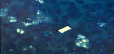
what crystal is this?
calcium pyrophosphate (CPPD)
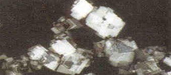
what crystal is this?
cholesterol crystal
lipid crystals showing a ______________ formation with polarized light can cause acute arthritis
Maltese cross
corticosteroid crystals are ____________ shaped
needle
monosodium appears __________ with a red compensator
yellow
pyrophosphate appears ________ with a red compensator
blue
non inflammatory joint disorders
osteoarthritis
sickle cell disease
inflammatory joint disorders
RA
LE
reiter’s
rheumatic fever
what can the synovial fluid described be classified as?
clear
yellow
good viscosity
WBC < 5000
neutrophil < 30%
normal glucose
no crystals
non inflammatory
what can the synovial fluid described be classified as?
cloudy
poor viscosity
WBC 2000-25,000
neutrophils > 50%
decreased glucose
inflammatory
what can the synovial fluid described be classified as?
cloudy
green
poor viscosity
WBC 10,000-50,000
neutrophils >90%
decreased glucose
positive culture
septic
what can the synovial fluid described be classified as?
cloudy
poor viscosity
WBC 500-50,000
neutrophils >90%
decreased glucose
crystals
crystal-induced
what can the synovial fluid described be classified as?
cloudy
red
poor viscosity
WBC <5,000
neutrophil <50%
normal glucose
RBCs present
hemorrhage