The Heart Anatomy
 Call Kai
 Learn
Learn Practice Test
 Spaced Repetition
 Match
Match Flashcards
 Knowt Play
1/29

There's no tags or description
Looks like no tags are added yet.
Name | Mastery | Learn | Test | Matching | Spaced | Call with Kai | Chat |
|---|
No analytics yet
Send a link to your students to track their progress
30 Terms
Where is the heart located?
It’s enclosed within the meda stinum, the medial cavity of the thorax.
What is it called when the point, in which the apex of the heart contacts the chest wall?
Point of maximal intensity
What is the pericardium of the heart?
The membrane that surrounds and protects the heart.
Describe the outer layer of the pericardium texture?
The pericardium has a fibrous dense irregular connective tissue
Describe the inner layer of the perciardium texture?
The inner layer of the pericardium is thinner and delicate.
How many layers does the heart wall have? Desribe each layer.
3 Heart walls:
Superficial epicardium
Medial myocardium
Deep Endocardium
What is the endocardium of the heart?
It’s the internal chambers of the heart.
What is the blood supply of the living heart tissue?
Coronary Circulation
Describe the vessels involved coronary circulation.
Right Coronary Artery
Anterior Interventricular Artery
Anterior Cardiac Veins
Right Marginal Vein
Great Cardiac Vein
Descrie the heart chambers.
Right atrium
Right Ventricle
Left Ventricle
Left Atrium
What is the trabecular larnera?
Ridges created by raised bundles of cardiac muscle fibers.
What’s present in the right ventricle?
The chordae tendineae
What are the chordae tendinaea?
Tendon-like cords connected to the cusps of the tricuspid valve and to cone-shaped extensions of the trabeculae carneae called papillary muscles.
What are papillary muscles?
Cone-shaped extensions of the trabecular carnae.
What prevents eversion of the atrioventricular valves during systole?
The chordae tendinaea.
What form of blood is pumped through the tricupsid valve into the ventricle?
Deoxygenated Blood is pumped through the T valve.
Where does the blood pump through when the right ventricle is contracting?
It pumps the blood out through the pulmonary valve into the pulmonary trunk on its way to the lungs, where gas exchange occurs.
How does the blood pump through the bicuspid valve?
Blood returns from the lungs into the left atrium which pumps it into the left ventricle through the bicuspid (mital) valve.
How does the blood pump into the general system circulation and get into the ascending aorta?
The left ventricle them pumps blood out into general systemic circulation through the aortic valve into the ascending aorta.
Which ventricle has a stronger myocardium? Describe it.
The myocardium of the left ventricle is thicker, stronger, and requires a constant supply of nutrient rich oxygenated blood.
P.S. Some blood is diverted immediately from the aortic arch into the coronary circulations.
Describe the function of the ligamentum arteriosum.
Connects the arch of the aorta and the pulmonary trunks.
What artery helps pumps deoxygenated blood to the lungs with the right ventricle?
The pulmonary artery
What happens when the pulmonary artery pumps blood to the lungs, and how does it return to the left atrium?
The blood becomes oxygenated, and it returns to the left atrium via the pulmonary veins.
What does the pulmonary veins and arteries conduct?
These veins and arteries conducts blood with deoxygenated/ oxygenated from regular systemic circulation.
What provides oxygenated blood to the myocardium?
The Coronary artery
How many branches from the aorta? Describe them.
There are 3 branches:
Brachiocephalic trunk
Left common carotid artery
Left subclavian artery

Whay layer of the heart is this?
The pericardium.
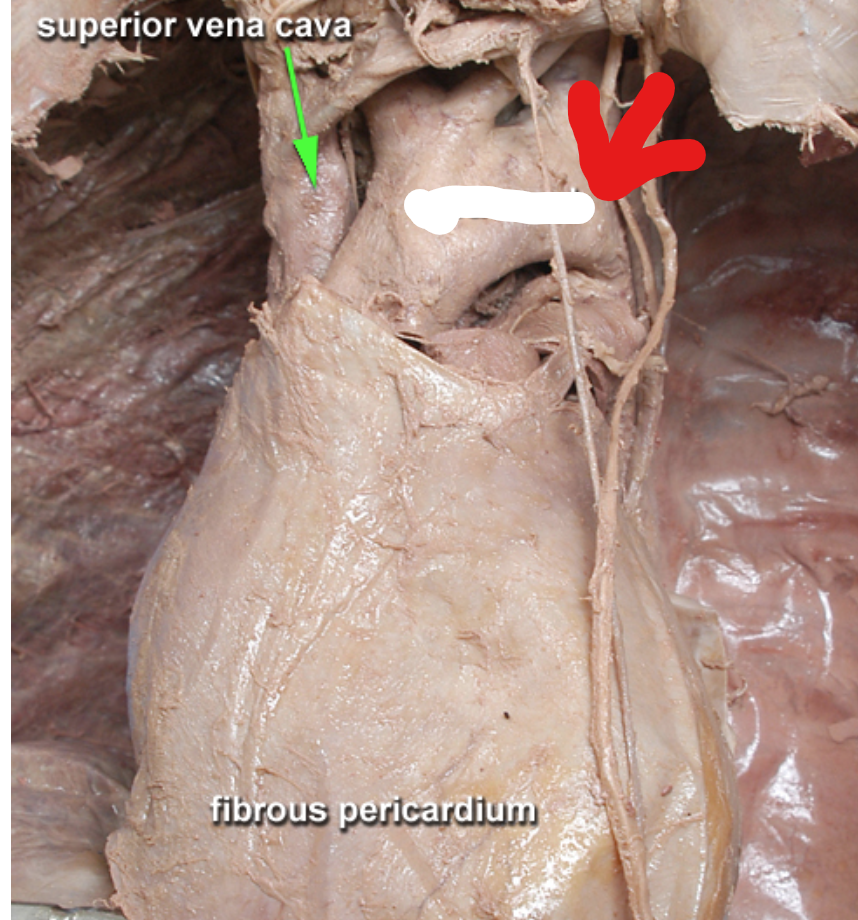
What part of the heart is this?
The Aortic arch

What part of the heart is this?
The superior vena cava

Label each segment around the heart.
Pulmonary trunk
left coronary artery