CT San Exam Practice
 Call Kai
 Learn
 Practice Test
 Spaced Repetition
 Match
 Flashcards
 Knowt Play
1/48

There's no tags or description
Looks like no tags are added yet.
Name | Mastery | Learn | Test | Matching | Spaced | Call with Kai | Chat |
|---|
No analytics yet
Send a link to your students to track their progress
49 Terms
What is the name of the mathematical set of all defined instructions for accomplishing a task?
Algorithm
What is the principle called where an X-ray beam passes through tissue and is affected?
Attenuation
In what format are CT images displayed?
Cross sectional format
According to the lecture, what year is emphasized for the invention of CT?
1972
Which two generations of CT machines only ever rotated 180 degrees and back 180 degrees?
First and second generation
Why did the first generation CT machine take a long time to process information?
It had to process the information off-site because there was no computer system built into the machine.
Why did it not make a difference whether patients could hold their breath during the earliest first-generation CT scans?
Because the only part of the body being scanned was the head.
What was the twofold purpose of every successive generation of CT machines?
To scan faster and get better images.
How do the detectors in a fourth-generation CT machine differ from earlier designs?
The detectors are stationary all the way around the machine while the X-ray tube rotates all the way around.
What are the defined attenuation parameters for bone, air, and water in CT scanning?
Bone is +1000, air is −1000, and water is 0.
What scan is performed daily to ensure the CT machine is calibrated to the correct density parameters?
A phantom scan (placing a phantom in the machine)
What two elements always combine to help increase CT image resolution?
The number of detectors (more detectors) and the algorithm that is run.
A CT image/information is based on a principle of the x-ray beam going thru tissue and being affected, what is this called?
Attenuation
Tomo
to cut
Graphia
to display
Gustave Grossman “1934” was a pioneer in the development
of computed tomography (CT) technology, contributing to the advancement of X-ray imaging techniques that allow for detailed cross-sectional views of the body.
William Oldendorf “1960” was a key figure in the invention
of the first commercially available CT scanner in 1971, revolutionizing diagnostic imaging by enabling precise visualization of internal structures.
CT was invented in 1972 by
British engineer Godfrey Hounsfield of EMI Laboratories, England, and independently by South
African born physicist Allan Cormack of Tufts
University, Massachusetts
Tomographic Image
a picture of a slab of the patient’s anatomy

CT slice thickness is very
thin (1 to 10 mm) and is approximately uniform
Tomographic acquisition is the method
of obtaining cross-sectional images by using X-rays from multiple angles.
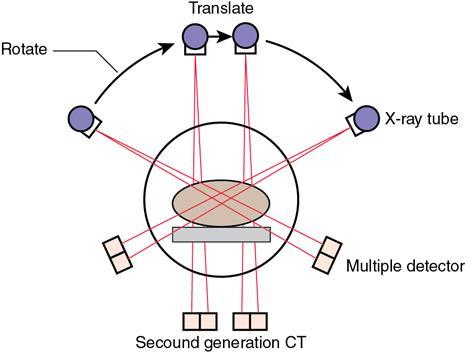
Second Generation CT Scan
utilizes two detectors and can produce images faster than the first generation, allowing for improved patient comfort and reduced scan time.

Third Generation CT Scan
uses a curved array of detectors and rotates around the patient, which allows for faster imaging and enhanced image quality.

CT 3rd Generation size and shape
fan-shaped beam 30-60 degrees
How many detectors were in the CT 3rd Generation?
288-700 detectors needed to read larger fan-shaped beam

Fourth Generation CT Scan
fixed detector ring source-rotation large fan beam
CT 4th Generation fan shape
same as 3rd generation
How many detectors are in the CT 4th Generation?
It typically has a fixed ring of detectors, usually fewer than in 3rd generation systems, generally around 200-400 detectors.

Cross-sectional Imaging
A section formed by a plane cutting thru an object, usually at right angles to an axis. Thus, allowing the visualization of 2 (TRUE) dimensions displayed on one image.
Algorithm
A step-by-step procedure used for calculations, data processing, or automated reasoning tasks in CT imaging.
The Next Generation is
Helical/ Spiral Scanning
Helical Simultaneous
translation, data acquisition & table movement and can obtain overlapping images (hundreds)
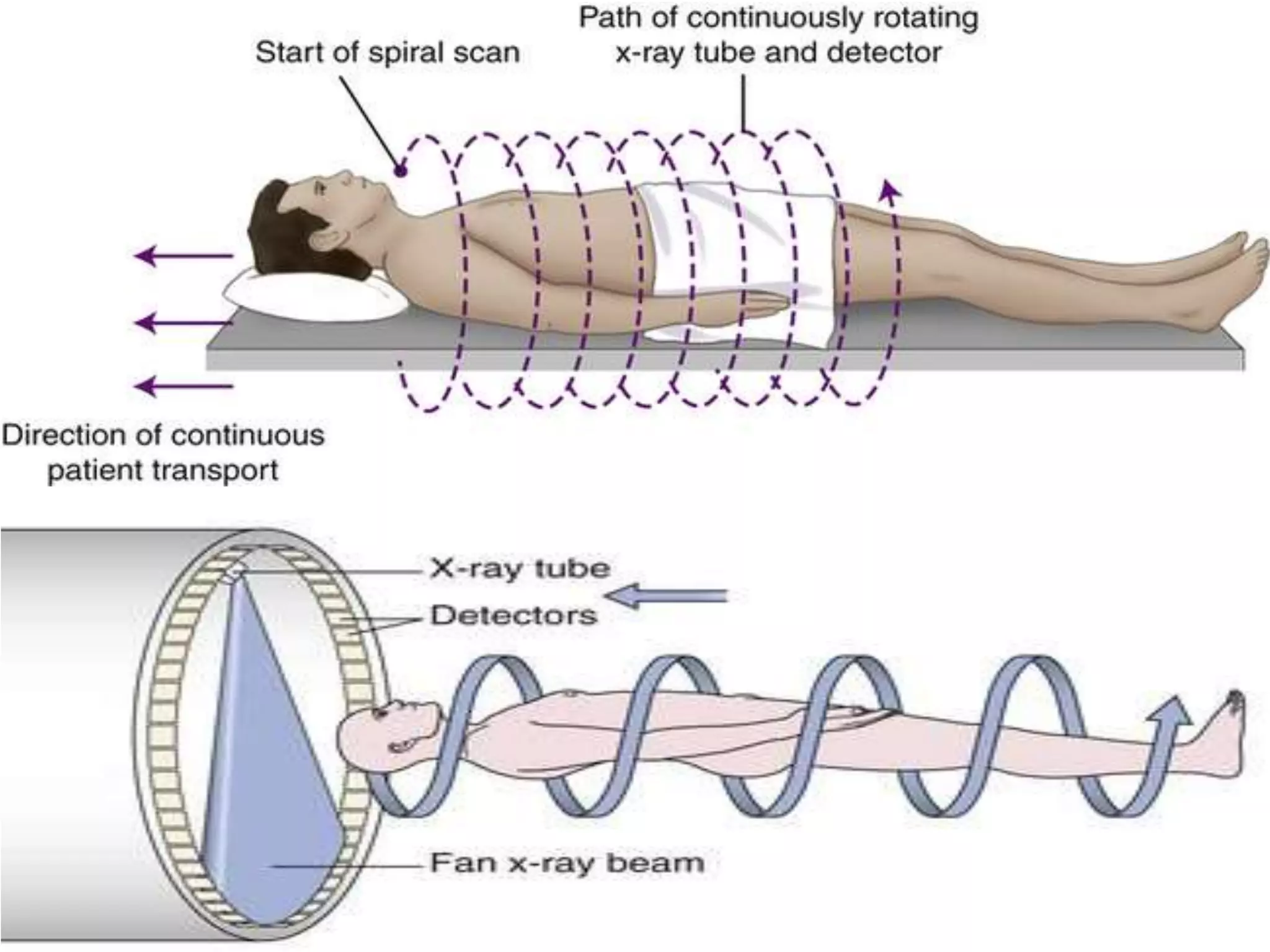
5th Generation “1990”
Spiral/ helical scanning, source of rotation, table translation, data acquisition
6th Gen Helical Multi-slice CT
Multi slice detectors, introduced 1998, allow acquisition of multiple slices in a single rotation.
As x-rays pass through the body they are ________ _ ________ at differing levels creating a matrix or profile of
x-ray beams of different strength.
absorbed or attenuated
Voxel
3D analog of a pixel
Pixel
2D picture element
The linear attenuation coefficient (μ) of each pixel is determined by
Composition of the voxel, thickness of the voxel, and quality of the radiation beam.
The numerical value stored in each pixel is a CT
number or
Hounsfield unit
CT scan subjects the human body to between _______ ______ times the radiation of a conventional x-ray, or around a year's worth of exposure to radiation from
both natural and artificial sources in the environment.
150 and 1,100
Cross Sectional Imaging CT
is a medical imaging technique that produces detailed images of the body's internal structures in slices. It combines X-ray technology and computer processing to create cross-sectional views of organs and tissues.
What x-ray modality was the precursor to CT?
Plain film x-ray
What devices allowed the processing of information that was gathered?
Computer systems and algorithms
What is the name of the mathematical set of well-defined
instructions for accomplishing a task?
algorithm
A CT image/information is based on a principle of the x-ray beam going thru tissue and being affected, what is this called?
attenuation
CT images are displayed in what format?
Digital slice format, typically displayed in cross-sectional images.
CT images are displayed in shades of?
gray
What allows the formation of 3D images?
Digital reconstruction algorithms that combine multiple 2D slices.
1st Generation CT
one x-ray source and one x-ray detector
