Chapter 9 Neurons: Cellular and Network Properties
 Call Kai
 Learn
Learn Practice Test
 Spaced Repetition
 Match
Match Flashcards
 Knowt Play
1/89

There's no tags or description
Looks like no tags are added yet.
Name | Mastery | Learn | Test | Matching | Spaced | Call with Kai | Chat |
|---|
No analytics yet
Send a link to your students to track their progress
90 Terms

Nervous System
• Network of billions of cells linked
together in a highly organized manner to
form the rapid control system of the
body

Introduction: Disorders/Health/Development
Living With The World's Most Painful Disease | MSNBC
Trigeminal Neuralgia
• Compression of the trigeminal nerve by an
adjacent blood vessel causes degeneration of the
myelin sheath that surrounds the sensory nerve
fibers
• Loss of insulation causes impulses that carry touch
sensations to misfire (brain perceives pain)

Infant Sensory Stimulation & Development
• Neglect or sensory deprivation in infancy can lead to
delayed development ("failure to thrive")
• Lack of nervous system stimulation is a key factor in
developmental delays
• Extra stimulation (e.g., art, music, foreign
languages) has no proven effect on enhancing
intellectual development
• Popular trends promoting early enrichment lack scientific
support

9.1 Organization of the Nervous System
“outline”
• Neurons w/ processes secrete
neurotransmitters
• Emergent properties
• Central nervous system: brain & spinal cord
• Peripheral nervous system (PNS)
• Sensory (afferent) neurons
• Efferent neurons: somatic motor & autonomic
divisions
—Autonomic divided into sympathetic &
parasympathetic branches
• Enteric nervous system
• Network of neurons in the walls of the digestive tract
• Controlled by ANS but is able to function autonomously

“summary”- information flow
through the nervous system
follows a basic reflex pattern
stimulus-→ sensor-→ input signal-→
integrating center-→ output signal-→
target-→ response

9.2 Cells of the Nervous System
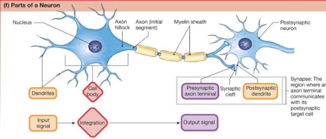
• Neurons are the functional unit of
the nervous system
• Specialized to transmit electrical and
chemical signals
• A single nerve cell
• Has three main parts: dendrites, cell
body (soma), and axon
• Found in the brain, spinal cord, and
throughout the nervous system
Neuron structure
1. Cell body- the control center
2. Dendrites (incoming signals) vs. axons (outgoing signals)
3. Collaterals, axon terminals vs. varicosities
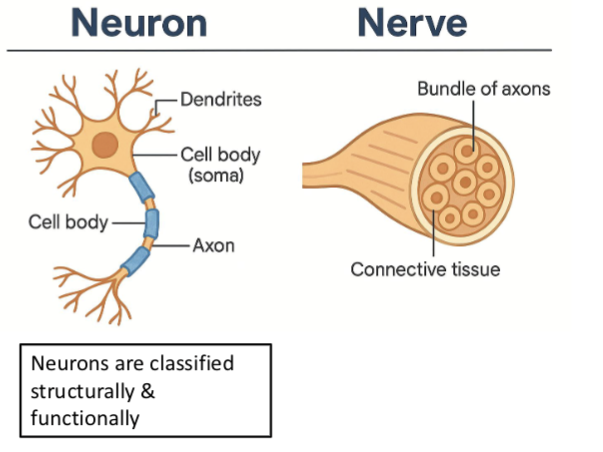
Are neurons and nerves the same thing?
Nerves- a bundle of axons (which are
parts of neurons)wrapped in connective
tissue
• Found in the PNS (outside the brain and
spinal cord)
• Functions like a communication cable,
carrying signals between the
brain/spinal cord and the rest of the
body
• Sensory nerves, motor nerves, mixed
nerves
Neuron classification:
1. Function
• Sensory (afferent) neurons, efferent neurons, &
interneurons
2. Structure – number of
processes
• Multipolar, pseudounipolar, bipolar, anaxonic
neurons

(a) Pseudounipolar
Pseudounipolar neurons have a single process called the axon. During development, the dendrite fused with the axon.
Key idea:
Pseudounipolar = one process

(b) Bipolar
Bipolar neurons have two relatively equal fibers extending off the central cell body.
Key idea:
Bipolar = two processes

c) Anaxonic
Anaxonic CNS interneurons have no apparent axon.
Key idea:
Anaxonic = no apparent axon

(d) Multipolar — CNS interneurons
Multipolar CNS interneurons are highly branched but lack long extensions.
Key idea:
Multipolar CNS interneurons = highly branched, no long extensions
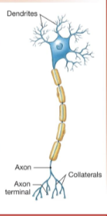
(e) Multipolar — Typical efferent neuron
A typical multipolar efferent neuron has five to seven dendrites, each branching four to six times. A single long axon may branch several times and end at enlarged axon terminals.
Key ideas:
5–7 dendrites
Each dendrite branches 4–6 times
Single long axon
Axon may branch several times
Ends at enlarged axon terminals

Axons Carry Outgoing Signals
Most peripheral neurons have a single axon starting at the axon
hillock.
• Axons may form collaterals, each ending in an axon terminal
• Terminals contain mitochondria & vesicles filled w/ neurocrine
molecules
• transmits electrical signals from the neuron’s integrating center
to target cells.

Signal Transmission (from neuron to target)
• Electrical signal usually
triggers chemical
messenger release
• In some CNS neurons,
signals pass via gap
junctions

Axons are Specialized to Convey Chemical
and Electrical Signals
Axonal Transport and Protein Synthesis
Axons transmit chemical and electrical signals.
• The axon cytoplasm contains fibers & filaments
but lacks ribosomes and ER.
• Protein synthesis occurs in the cell body on
the rough ER.
• Proteins are packaged into vesicles & moved
down the axon via axonal transport
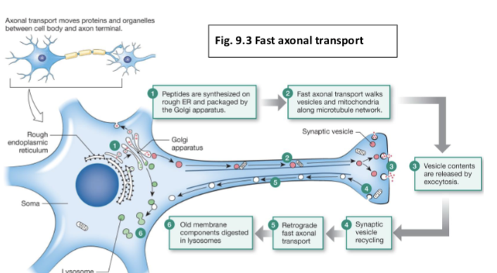
Proteins & Organelles are Moved via Axonal Transport
Anterograde (forward): Cell
body → Axon terminal (e.g.,
vesicles, mitochondria).
Retrograde (backward):
Axon terminal → Cell body
(e.g., old components,
growth factors, viruses).
Fast axonal transport- moves organelles at rates of up to 400
mm/day
Slow axonal transport- moves material by axoplasmic
(cytoplasmic) flow at 0.2–2.5 mm/day; (“stop and go”)
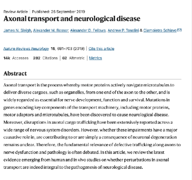
Disruption of Axonal Transport
• Mutations or alterations in proteins
associated w/ axonal transport have been
linked to a variety of inherited & acquired
disorders
• Congenital defects include microcephaly
(small head due to underdevelopment of
the brain)
• fragile X syndrome (common cause of
inherited intellectual disability)
—roles of defective axonal transport in
Alzheimer’s disease & other
neurodegenerative diseases?

Synapses
Synapse- region where an axonal
terminal meets its target cell
• Presynaptic cell vs. postsynaptic cell
• Synaptic cleft
how can billions of neurons in the brain
find their correct targets and make
synapses (during development?)
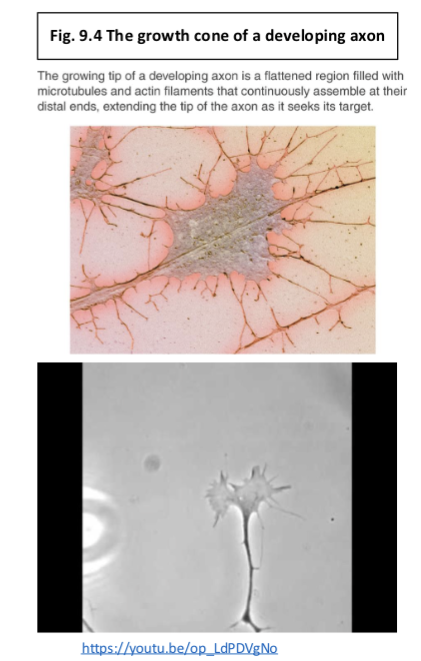
Establishing Synapses Depends
on Chemical Signals
• Growth cones & neurotrophic factors allows
developing neurons to find their targets
• Neurotrophic factors--chemicals secreted by
Schwann cells that keep damaged neurons alive
• Synapses are not fixed for life, can be rearranged
• Older adults are urged to keep learning

Glial Cells
Provide Support for Neurons
Cell Types in Nervous Tissue: Glial Cells
• 4 in the CNS (make up half of the brain’s mass, divide
throughout life) & 2 in the PNS
• Each has a unique function, but also share general
functions:
– provide a supportive scaffolding for neurons
– cover all nonsynaptic parts of neurons
– provide insulation for the neurons (prevents other neurons
from interfering w/ electrical impulses)
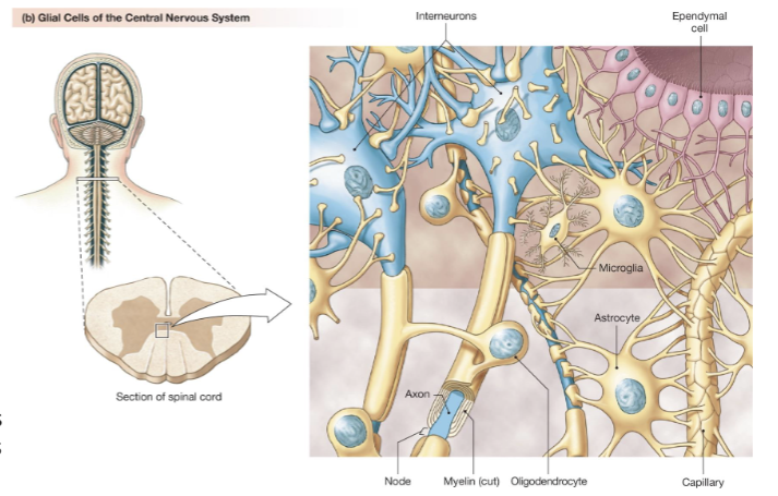
Glial Cells of the CNS (4)
Astrocytes-most abundant CNS neuroglia;
multiple functions (regulate blood flow)
Microglial cells are defensive cells in the
CNS
Ependymal cells line cerebrospinal
fluid-filled cavities; one source of neural
stem cells
Oligodendrocytes have processes that
form myelin sheaths around long axons in
the CNS.
In the CNS, one oligodendrocyte branches
and forms myelin sheaths around portions
of several axons

Glial Cells of the PNS (2)
• Schwann cells- wrap around PNS
axons to form myelin sheath
• Satellite cells- support neurons in
sensory, sympathetic, &
parasympathetic ganglia
• “non-myelinating” Schwann cells
• Regulate the extracellular
environment, including ion balance &
neurotransmitter levels.
• Protective roles
• Respond to injury and may play a
role in pain signaling.

Schwann cells (PNS)
• Myelin sheaths made by Schwann Cells
in the PNS
• one Schwann cell associates w/ one axon
• provide insulation that prevents
leakage of electrical signals
• myelin sheath have gaps (nodes of
Ranvier)
• gaps along axon that speed up
nerve transmission
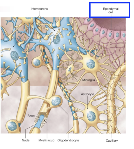
Ependymal Cells (CNS)
• Ependymal cells line cerebrospinal
fluid-filled cavities
• one source of neural stem cells
—immature cells that can differentiate into
neurons or glial cells
—when neural stem cells receive the correct
signals, they transform into neurons &
glial cells
Opportunity for reversing degenerative
neurological diseases?

Enzyme Replacement Therapy and Batten Disease
• fatal lysosomal enzyme disorder (similar to Tay-Sachs)
• abnormal accumulation of cell waste (proteins & lipids)
• waste buildup leads to the progressive death of neurons; severe
nervous system symptoms


Can Stem Cells Repair Damaged Neurons?
• After the fetal period there is no replacement of
dead or damaged neurons
——--• neural injuries are usually permanent
• If the cell body dies, the neuron dies
• If axon is severed, then cell body & attached
segment survives; severed portion degenerates
• If motor neuron, then target muscle results in
permanent paralysis
• If sensory neuron, then experience loss of
sensation from innervated area
Regeneration may occur in PNS; less likely in CNS
Why is Regeneration less likely in the CNS?

Steps of
Neuronal Repair
• If only axons are damaged, cell
bodies can survive & the axon can
regenerate
• This only occurs in the PNS, in the
CNS the neuroglia never forms
bands/regeneration tube to guide
the regrowing axons
• Glial cells secrete growth inhibiting
chemicals which limit axon growth
(next slide-axon inhibitors)
thus, no regeneration after injury to
spinal cord or brain
1. Axoplasm leaks out & seals the damaged end & attached segment of axon swells
2. Schwann cells release chemical signals alerting of tissue damage
• Degeneration of the distal axon segment
• Myelin sheath unravel
• Cellular debris removed
Schwann cells secrete neurotropic factors to keep cell body alive & encourage axon
regeneration
Axon behaves like growth cone
Strategies to Promote Regeneration in the CNS
We do not understand the mechanism of this inhibition (axon inhibitors)
forms the basis of strategies to promote regeneration in the CNS
• Induce degradation of inhibitors at the site of injury?
• stem cell transplantation?
• “trophic factors”- molecules that support the survival of cells

Biotechnology
Neurotrophic Factors
• regulate the proliferation, survival,
migration, & differentiation of cells in the
nervous system
• “neuroprotective functions” in addition
to regenerative roles
• delivery of neurotrophic factors to the
CNS is problematic---> large molecules
cannot pass through the blood-brain
barrier
• Genetically engineered cells/gene
therapy
9.3 Electrical Signals
in Neurons
Nerve and muscle are
excitable tissues
—propagate electrical signals
rapidly in response to a stimulus
basic reflex pattern w/in the nervous system
stimulus-→ sensor-→ input signal-→
integrating center-→ output signal-→
target-→ response
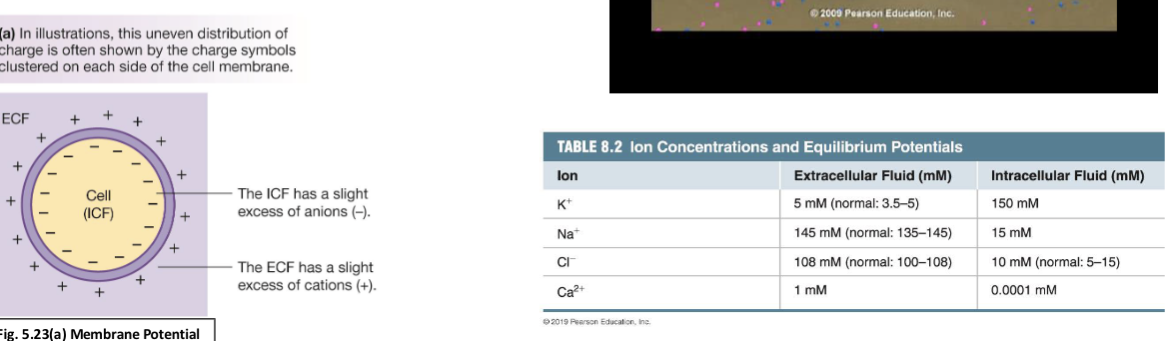
Resting Membrane Potential
Separation of electrical charge across a cell
membrane
Influenced by 2 factors:
1. The uneven distribution of ions across the cell
membrane
2. Differing membrane permeability to those ions

Ion Movement Creates Electrical Signalssac
• addition of Na+ to the
intracellular fluid
depolarizes the cell
membrane & creates an
electrical signal
• cells can become more
negative (hyperpolarizes)
(ex: loss of K+ or entry of
Cl-)
A Significant Change in Membrane Potential Occurs w/ the Movement of Very Few
Ions
• change in membrane potential does not mean that ion concentration gradients have
reversed

How does a cell change its ion permeability?
Gated Channels Control the Ion
Permeability of the Neuron
Most gated channels fall into 1 of 3 categories
1. Mechanically gated ion channels are found in sensory neurons &
open in response to physical forces such as pressure or stretch.
2. Chemically gated ion channels in most neurons respond to a variety
of ligands, such as extracellular neurotransmitters & neuromodulators
or intracellular signal molecules.
3. Voltage-gated ion channels respond to changes in the cell’s
membrane potential. Voltage-gated Na+ and K+ channels play an important role in the initiation & conduction of electrical signals along the axon.
The ease with which ions flow through a channel is called the
channel’s conductance

Long Q-T Syndrome (LQTS)
• Channelopathies- are inherited
diseases caused by mutations in ion
channel proteins
• LQTS named for changes in the
electrocardiogram test
• is a cardiac problem characterized by
an irregular heartbeat, fainting, and
sometimes sudden death
• 8 different mutations in various ion
channels result in subtypes of LQTS
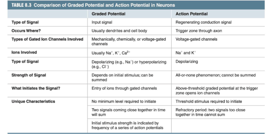
Voltage Changes Across the Membrane: Graded
Potentials & Action Potentials
Graded Potentials (reflect
stimulus strength)
–Variable strength
–Used for short-distance
communication
Action Potentials
–Very brief, large
depolarizations
–Rapid signaling over long
distance

Graded Potentials Reflect Stimulus
Strength
Local current flow is a wave of
depolarization that moves
through the cell
• Graded potentials lose strength
as they move through the cell
due to current leak &
cytoplasmic resistance
• If strong enough, graded
potentials reach the trigger
zone in the axon hillock & initial
segment


Action Potential Basics (1)
Neurons are Excitable
Action Potential- change in electrical potential
associated w/ the passage of an impulse along the
membrane of a muscle cell or nerve cell
• rapid change in mp in response to a signal
• these changes in mp act as electrical signals
• membrane depolarization- mp becomes “less negative”
• membrane repolarization- mp returns to resting value
• membrane hyperpolarization- mp becomes more
negative than resting value
• AP’s Travel long distances; occur when a neuron sends
information down an axon, away from the cell body

AP’s Travel Long Distances (2)
• Conduction is the
high-speed
movement of an
AP along an axon.
• All-or-none
• Wave of electrical
signal at constant
amplitude
• changes in MP are the result of opening & closing
of specific ion channels

Na+ and K+ Move across the
Membrane during AP’s (3)
AP begins when graded potential
reaching trigger zone depolarizes to
threshold
• Rising phase of the AP→ voltage-
gated Na+ channels open & Na+ entry
depolarizes the cell
• Falling phase of the AP-→ at peak,
Na+ channels close, slower voltage-
gated K+ channels open
• K+ exit repolarizes then
hyperpolarizes cell
• Voltage-gated K+ channels close, less
K+ leaks out of the cell
• Cell returns to resting membrane
potential of -70 mV


Axonal Na+ Channels Have Two Gates
Activation vs. inactivation gates
● If the membrane is highly permeable to an ion, that
ion makes a large contribution to the mp
● Thus, neurons depolarize or hyperpolarize by
selectively altering the permeability of their
membranes to ions
● Accomplished by opening gated ion channels in the
membrane--> alters mp--> generates electrical
signals
--Gated ion channels open in response to
neurotransmitters, changes in membrane
potential, or other stimuli
An AP is a classic example of
a positive feedback loop

AP Positive Feedback Loop
• increased depolarization causes more Na⁺
channels to open, allowing even more Na⁺
to enter
Depolarization → Na⁺ channel opening
→ More Na⁺ influx → More
depolarization
• amplification continues until a cutoff
mechanism (Na⁺ channel inactivation)
stops it


A.P. ’s Travel Long Distances w/out Losing Energy —
A Process Called Conduction
The A.P. at the end of the axon is identical to the one that
started at the trigger zone.
• Depolarization of an axon section causes local current flow:
• Positive current spreads through the cytoplasm in all directions.
• Simultaneously, current flows back toward the depolarized
section on the outside of the membrane.
• Local current flow diminishes over distance due to energy
dissipation.
• Voltage-gated Na⁺ channels reinforce the signal:
• When depolarization reaches these channels, they open, allowing
more Na⁺ to enter.
• This creates a positive feedback loop, sustaining the A.P.
• This mechanism ensures continuous propagation of the A.P.
along the axon.

AP’s Cannot Travel
Backwards
The refractory period
prevents backward
conduction
• delay of 1-2 msec
between AP’s,
independent of intensity
of trigger
• due to voltage-gated Na+
channels resetting
AP’s Will Not Fire during the
Absolute Refractory Period

Larger Neurons Conduct APs Faster
Electrical Signals: Speed of AP
• Larger neurons conduct AP’s
faster
• Conduction is faster in
myelinated neurons
–Resistance of axon membrane to
ion leakage out of the cell
• Saltatory conduction between
nodes of Ranvier

Saltatory
Conduction
● only nodes have voltage
gated ion channels
● Depolarization at one node
of Ranvier is transmitted
quickly via electric current
spread through the
internodes
● This triggers an action
potential at the next node
of Ranvier
● “appearance of jumping” or
“saltaring”

Demyelinating Diseases- Cause Loss of Myelin
• multiple sclerosis (MS)- chronic autoimmune
disease affecting the CNS
• immune system mistakenly attacks the myelin
sheath, the protective covering of nerve fibers
• damage disrupts nerve signals
• numbness, weakness, vision problems, balance
issues, and cognitive difficulties
• currently incurable, treatments exist to
manage the condition and its symptoms
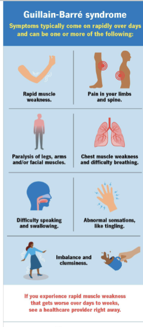
Guillain-Barré Syndrome (GBS)
• rare, acute autoimmune disorder
• body's immune system attacks the PNS
• leading to nerve inflammation and damage
Clinical Features:
• Rapidly progressive symmetrical weakness (usually ascending
from legs)
• tingling, numbness, loss of reflexes
• Cranial nerve involvement
• facial droop, dysphagia, ophthalmoplegia
• Autonomic dysfunction
• BP fluctuations, arrhythmias, bladder issues
• Respiratory compromise in severe cases
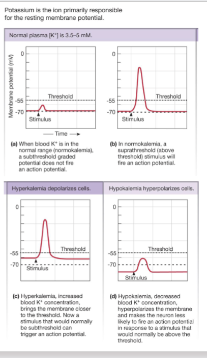
Chemical Factors Alter Electrical
Activity
• Variety of chemicals alter the conduction of
A.P.’s by binding to the channel
• Alterations in ECF concentration of ions also
affects effects electrical activity
• Ca2+ and K+
• Hyperkalemia brings neuron closer to
threshold
• Hypokalemia moves neuron further from
threshold
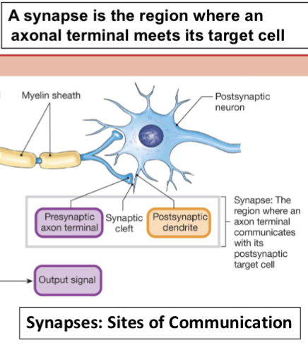
9.4 Cell-to-Cell Communication in the Nervous System
• Neurons communicate at synapses; “union”;
transmit information via chemical messengers
(usually)
• Some neurons of the CNS transmit signals
electrically through gap junctions
• b/c signals pass across most synapses in
one direction only, synapses determine the
direction of information flow through the
nervous system

Structure & Function of the Axon Terminal
• Synaptic Vesicles: contain neurotransmitters; some are docked at active zones ready for release, others form a reserve pool.
• Protein Synthesis: Occurs in the cell body; axon terminals lack necessary organelles.
• Polypeptide Transport: Propeptides & modifying enzymes are
packaged in vesicles & transported via fast axonal transport
• Neurotransmitter Synthesis:
• Small Molecules (e.g., acetylcholine, amines): synthesized in the terminal
• Enzymes: Made in the cell body and delivered by slow axonal transport


Neurotransmitters Are
Released from
Vesicles

Termination of Neurotransmitter Activity
1. Diffusion away from the
synaptic cleft
2. Enzymatic breakdown
• Acetylcholinesterase
(AChE)
3. Uptake into cells
• Presynaptic axon terminal
• Glial cells
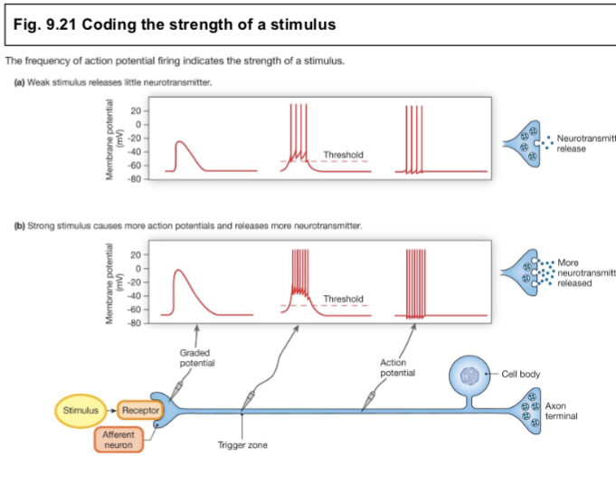
Stronger Stimuli Release More Neurotransmitter
• A single AP results in the
release of a constant
amount of
neurotransmitter
• AP’s code duration &
magnitude in the
frequency of AP’s
produced
• Tonic activity
• Burst activity
A patient has damage to the axon hillock of a motor
neuron. Which function would be most directly
impaired?
Integration of incoming signals and action potential
initiation
Loss of satellite cell function would most directly
affect:
B. Support of neurons within peripheral ganglia
A researcher selectively destroys growth cones in a
developing embryo. Which outcome is most likely?
Failure of many neurons to establish proper synaptic
targets
Which feature best distinguishes a nerve from a
neuron?
Nerves are bundles of axons wrapped in connective
tissue
Which scenario would most likely hyperpolarize a neuron?
Cl⁻ entry
A drug prevents opening of voltage-gated Na⁺
channels. Which phase of the action potential would be
most directly inhibited?
B. Depolarization (rising phase)
Why do graded potentials decrease in magnitude as they
travel?
C. Current leak and cytoplasmic resistance
Which characteristic is unique to action potentials but
not graded potentials?
Ability to travel long distances without decreasing
amplitude
Action potentials are considered examples of positive
feedback because:
Depolarization causes more Na⁺ channels to open
Which event prevents action potentials from traveling
backward?
C. Absolute refractory period
A mutation causes voltage-gated Na⁺ channels to fail
to inactivate. What is the most likely effect?
B. Excessive neuronal excitation
Compared with an unmyelinated axon of equal
diameter, a myelinated axon conducts faster because:
B. APs occur only at nodes of Ranvier
Which ion imbalance would make neurons easier to
excite?
B. Hyperkalemia
Which process would terminate neurotransmitter
activity most rapidly in the neuromuscular junction?
C. Acetylcholinesterase activity
Why can stronger stimuli produce larger responses
if each action potential releases the same amount of
neurotransmitter?
C. Stronger stimuli increase AP frequency
Why is regeneration after spinal cord injury usually poor
compared with peripheral nerve injury?
CNS glia fail to form regeneration tubes and release
growth-inhibiting factors
A toxin selectively destroys Schwann cells but leaves
axons intact. Which effect would occur first?
B. Slower action potential conduction in the PNS
A neuron receives several weak excitatory graded
potentials that individually fail to reach threshold. An
action potential occurs anyway. Which explanation is
most likely?
B. Graded potentials were integrated at the trigger zone
Which event would most likely reduce the speed of action
potential conduction without preventing action potentials
entirely?
C. Demyelination between nodes of Ranvier
During the falling phase of an action potential, the
membrane becomes less positive primarily because:
K⁺ exits through voltage-gated channels
Which feature would be expected in a neuron specialized
for very rapid communication over long distances?
Large-diameter myelinated axon
A researcher blocks acetylcholinesterase activity at a
synapse. Which outcome is most likely?
B. Neurotransmitter remains active longer in the cleft
Which CNS glial cell would be most directly involved in
removing pathogens after a brain infection?
D. Microglial cell
A patient with severe spinal cord injury receives a
treatment that neutralizes CNS growth-inhibiting
molecules. What is the primary goal?
B. Promote axonal regeneration
Which statement best explains why neurotrophic factor
therapy for CNS disorders is difficult?
Large molecules have difficulty crossing the blood-brain
barrier

Match each term with its description:
(a) axon- long process that transmits signals to the target cell
(b) dendrite-process of a neuron that receives incoming signals
(c) afferent-sensory neuron, transmits information to CNS
(d) efferent-neuron that transmits information from CNS to the rest of the body
(e) trigger zone- region of neuron where action potential begins
Axonal transport is best explained as which of the following?
movement of organelles and cytoplasm up and down the axon.

Match the numbers of the appropriate characteristics with the two types of potentials. Characteristics may apply to one or both types.
(a) Action potential
1. all-or-none (It fires completely once threshold is reached or not at all)
4. exhibits a refractory period (Features a recovery phase where another cannot easily fire)
(b) Graded potential
2. can be summed (Can undergo temporal or spatial summation)
3. amplitude decreases with distance (Signals decay as they travel along the membrane)
5. amplitude depends on strength of stimulus (Larger stimuli produce larger voltage changes)
6. has no threshold (Can be triggered by any level of stimulus)
Arrange the following events in the proper sequence:
(e) Sensory organ detects change in the environment.
A receptor first senses an internal or external stimulus.
(b) Afferent neuron reaches threshold and fires an action potential.
The sensory (afferent) neuron carries the electrical signal toward the central nervous system.
(d) Integrating center reaches decision about response.
The central nervous system (brain or spinal cord) processes the incoming information.
(a) Efferent neuron reaches threshold and fires an action potential.
The motor (efferent) neuron carries the command signal away from the integrating center.
(c) Effector organ responds by performing output.
A muscle or gland executes the final physical response.
Match the glial cell(s) on the right to the functions on the left. There may be more than one correct answer for each function.Match the glial cell(s) on the right to the functions on the left. There may be more than one correct answer for each function.Match the glial cell(s) on the right to the functions on the left. There may be more than one correct answer for each function.
a) modified immune cells: 3. microglia
(b) help form the blood-brain barrier: 1. astrocytes
(c) form myelin: 4. oligodendrocytes, 6. Schwann cells
(d) separate CNS fluid compartments: 2. ependymal cells
(e) found in peripheral nervous system: 5. satellite cells, 6. Schwann cells
(f) found in ganglia: 5. satellite cells
An action potential is (circle all correct answers)
the same size and shape at the beginning and end of the axon.
transmitted to the distal end of a neuron and causes release of neurotransmitter.
What causes the depolarization phase of an action potential? (Circle all that apply.)
Na+ entering the cell through voltage-gated channels.
Match the best term (hyperpolarize, depolarize, repolarize) to the following events. The cell in question has a resting membrane potential of -70mv.
a. -70 to -50 mv
b. -70 to -90 mv
c. +20 to -60 mv
d. -80 to -70mv
a. Depolarize
b. Hyperpolarize
c. repolarize
d. Depolarize
A neuron has a resting membrane potential of
. Will the neuron hyperpolarize or depolarize when each of the following events occurs? (More than one answer may apply; list all those that are correct.)
(a) Na+ enters the cell
(b) K+ leaves the cell
(c) Cl-enters the cell
(d) Ca2+ enters the cell
a. Depolarize
b. Hyperpolarize
c. Hyperpolarize
d. Depolarize
The presence of myelin allows an axon to (choose all correct answers):
conduct impulses more rapidly.