(A&P 1) Lab 8: Histology of Nervous Tissue
 Call Kai
 Learn
 Practice Test
 Spaced Repetition
 Match
 Flashcards
 Knowt Play
1/66

There's no tags or description
Looks like no tags are added yet.
67 Terms

How does the Endocrine system communicate/function?
By the means of chemical messengers (hormones) secreted into the blood

How does the Nervous System communicate/function?
Employs electrical and chemical means to send messages from cell to cell
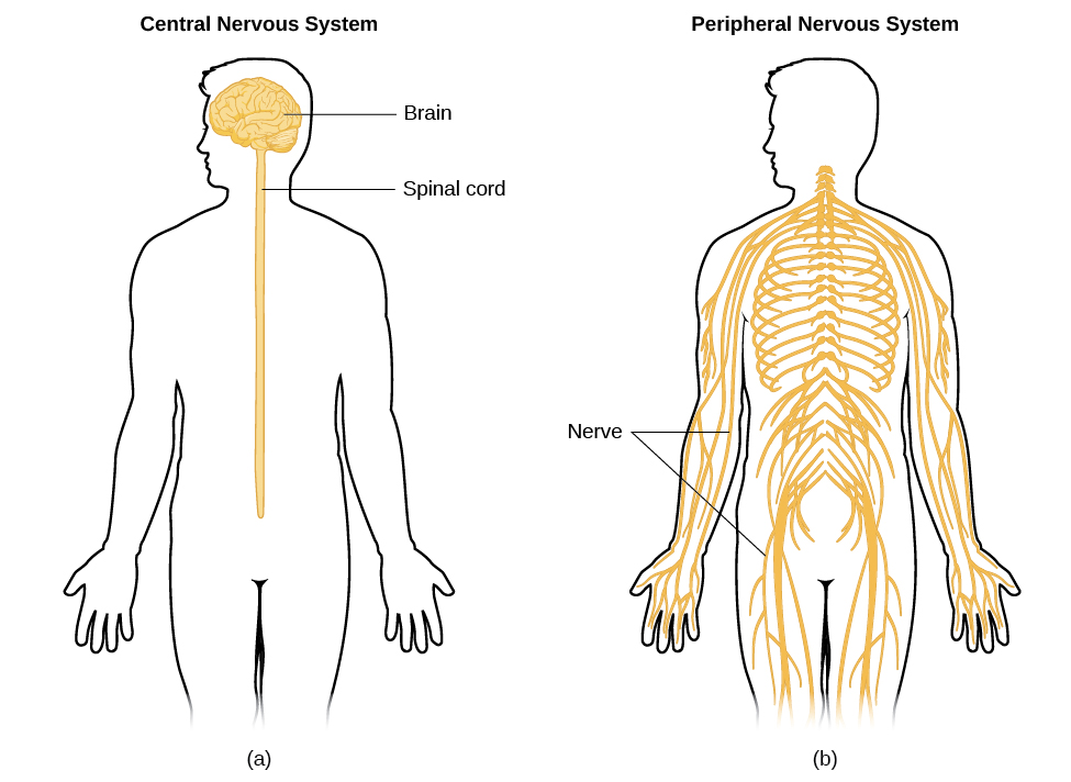
What do the two Nervous System Anatomical Subdivisions consist of?
CNS: Brain and spinal cord (enclosed and protected by the cranium and vertebral column)
PNS: Nerves and ganglia
Define a nerve and ganglion.
A nerve is a bundle of nerve fibers (axons) wrapped in fibrous connective tissue.
A ganglion is a knotlike swelling in a nerve where neuron cell bodies are concentrated.
What are the three steps of nervous system coordination?
1. Sensory input (receives information)
2. Integration (CNS processes information and determines response)
3. Motor output (issues commands to muscle/gland cells).
What is the difference between the somatic sensory division and visceral sensory division?
Somatic sensory division: Carries signals from receptors in the skin, muscles, bones, and joints.
Visceral sensory division: Carries signals from the viscera of the thoracic and abdominal cavities.
What is the difference between the somatic motor division and visceral motor division?
Somatic motor division: Carries signals to skeletal muscles for voluntary control.
Visceral motor division (Autonomic Nervous System): Carries unconscious signals to glands, cardiac muscle, and smooth muscle.
What are the two subdivisions of the visceral motor division (Autonomic Nervous System)?
Sympathetic- Arouses the body for action ("fight or flight").
Parasympathetic- Calms the body, slowing heart rate and aiding digestion ("rest and digest").
What are the two main types of cells in nervous tissue?
Neurons: Functional nerve cells that detect stimuli, respond, and transmit.
Neuroglia (Glial cells): Protective cells that support and assist neurons.
Define the neurosoma and dendrites.
Neurosoma: cell body containing the nucleus, cytoplasm, and standard organelles.
Dendrites: processes extending from soma that act as a site for receiving signals.
Define the axon and the axon hillock.
Axon: cylindrical process specialized for rapid conduction of nerve signals away from the soma.
Axon Hillock: specialized mound on one side of the neurosoma where the axon originates.
What are the specific terms for the cytoplasm and the cell membrane of an axon?
Cytoplasm: Axoplasm.
Cell Membrane: Axolemma.

What is found at the distal end of an axon?
A terminal arborization (fine branch network) ending in synaptic knobs
What are multipolar neurons?
Neurons with one axon and multiple dendrites.
(the most common structural type in brain and spinal cord)
What are bipolar neurons and where are they found?
Neurons with one axon and one dendrite. They are localized to specialized sensory regions like olfactory cells, retinal neurons, and inner ear sensory paths.
What are unipolar neurons
Neurons with only a single process leading away from the neurosoma that branches T-like into peripheral and central axons; they carry sensory signals directly to the spinal cord.
What are anaxonic neurons and where are they found?
Neurons with multiple dendrites but no axon that communicate locally and do not produce action potentials
found in the brain, retina, and adrenal medulla.

What are the primary functions of Astrocytes?
The most abundant CNS neuroglia,
form a supportive structural framework
assist in forming the blood-brain barrier
regulate chemical composition of interstitial fluid, and form scar tissue (astrocytosis) when damaged.
What are Microglia?
CNS macrophages that act as defensive cells to phagocytize debris, dead tissue, and pathogens.
What are Ependymal cells?
Cuboidal-like epithelial cells lining the internal ventricles of the CNS that produce cerebrospinal fluid (CSF) and use apical cilia to circulate it.
What is the function of Oligodendrocytes?
Glial cells that wrap around nerve fibers in the CNS to form insulating myelin sheaths.

What are Schwann cells?
Cells in the PNS that
envelop individual nerve fibers (assist in regeneration)
produce the peripheral myelin sheath

What are Satellite cells?
Support cells that surround neurosomas within PNS ganglia
providing electrical insulation
regulating microenvironment.
What is the composition of the myelin sheath and when is its production completed?
Composed of 20% protein and 80% lipid.
Myelination proceeds rapidly during infancy and is not fully completed until late adolescence.
What two factors determine the conduction speed of nerve fibers?
1. Fiber diameter: Larger, conduct signals faster
2. Myelination: The presence significantly increases conduction velocity.
What connects the two cerebral hemispheres, and how much of the brain's volume does the cerebrum make up?
The corpus callosum (a thick tract of nerve fibers) connects the right and left hemispheres.
The cerebrum constitutes 83% of total brain volume.
Define a gyrus, a sulcus, and a fissure.
Gyrus: A thick, raised fold on the surface of the brain.
Sulcus: A shallow groove separating adjacent gyri.
Fissure: A deep groove, (such as the longitudinal fissure dividing the hemispheres.)
What is the main function of the Frontal lobe?
Controls voluntary motor functions, planning, motivation, mood, and executive decision-making.
What is the main function of the Parietal lobe?
Primary sensory area responsible for receiving and interpreting general sensory input
What is the main function of the Occipital lobe?
The principal visual center of the brain, processing visual input from the optic pathways.
What is the main function of the Temporal lobe?
Responsible for hearing (audition) and smell (olfaction)
memory processing, learning, and aspects of emotional behavior.

What is the insula?
A deep mass of cortex concealed lateral to the insular region,
involved in taste, visceral sensation, and empathy.

What separates the frontal and parietal lobes, and which gyri sit on either side of it?
Separated by the central sulcus.
• Precentral gyrus (Primary motor cortex) lies anterior.
• Postcentral gyrus (Primary somatosensory cortex) lies posterior.
Name the key anatomical features of the cerebellum.
right/left hemispheres connected by the central vermis,
surface folds called folia
internal branching white matter pattern called the arbor vitae.
What are the most distinctive neurons in the cerebellum, and what is the cerebellum's main function?
(Purkinje Cells) Center for motor coordination, monitoring muscle contractions, and fine-tuning balance/posture.

Name the three cerebellar peduncles and what they connect to.
• Inferior peduncles: Connect to the medulla oblongata.
• Middle peduncles: Connect to the pons.
• Superior peduncles: Connect to the midbrain.

Contrast Gray matter and White matter in the brain.
Gray Matter: Outer surface cortex and deep nuclei; houses neurosomas, dendrites, and synapses.
White Matter: Deep to the cortex; composed of myelinated axon tracts connecting regions.
What are the three layers of the meninges from superficial to deep?
1. Dura mater (outermost, tough layer)
2. Arachnoid mater (middle spiderweb-like layer)
3. Pia mater (innermost delicate membrane)
Describe the structure of the Dura mater and dural sinuses.
Made of two layers: an outer periosteal layer and an inner meningeal layer.
Spaces between them form dural sinuses (e.g., superior sagittal sinus) that collect venous blood from the brain.

Describe the Arachnoid mater, Pia mater, and the space between them.
The Arachnoid mater and Pia mater are the two innermost layers of the meninges , and they are separated by the subarachnoid space.
What is meningitis, what age group is most vulnerable, and how is it diagnosed?
Inflammation of the meninges due to infection
Most critical in infants/children (3 months to 2 years).
Diagnosed via a spinal tap (lumbar puncture) examining CSF for pathogens and elevated white blood cells.
What is Cerebrospinal Fluid (CSF)?
A clear filtrate that fills the ventricles, canals, and subarachnoid space, providing physical protection, and chemical stability to the CNS.
What is the choroid plexus?
A network of blood capillaries situated in the brains ventricles (covered by ependymal cells) and filter blood plasma in CSF
What are the four ventricles and how are they connected?
Two lateral ventricles connect to the third ventricle via the interventricular foramina.
The third connects to the fourth ventricle via cerebral aqueduct, which drains into the central canal of the spinal cord.
How does interrupted blood flow affect the brain at 10 seconds, 1-2 minutes, and 4 minutes?
• 10 seconds: Causes loss of consciousness.
• 1–2 minutes: Significantly impairs neural function.
• 4 minutes: Results in irreversible, permanent brain damage.

What creates the blood-brain barrier (BBB)?
Strict tight junctions joining the endothelial cells that form the inner walls of brain capillaries
stimulated and maintained by astrocytic foot processes.
What substances is the brain barrier system (BBS) highly permeable to?
Highly permeable to water, glucose, and lipid-soluble substances
(e.g.,) oxygen, carbon dioxide, alcohol, caffeine, nicotine, and general anesthetics.
What are the three major components of the brainstem, from rostral to caudal?
1. Midbrain (rostral)2. Pons3. Medulla Oblongata (caudal, terminating at the foramen magnum)
What passes through the medulla oblongata?
All ascending sensory and descending motor nerve tracts connecting the brain to the spinal cord.
What is the reticular formation and what is habituation?
A loose network of gray matter nuclei running through the brainstem regulating somatic motor control, cardiovascular control, pain modulation, and sleep/consciousness.
Habituation: Process where the brain learns to ignore repetitive, non-significant stimuli.
Describe the roles of the three components of the Diencephalon
• Epithalamus: Houses the pineal gland (regulates circadian rhythms via melatonin)
• Thalamus: Major relay center for sensory information bound for the cerebral cortex.
• Hypothalamus: Core control center for autonomic function, endocrine regulation, body temperature, hunger, and thirst.
What are the gaps between the myelin sheaths (Schwann cells) along an axon called?
Nodes of Ranvier
What are branches off the main axon called?
Axon collaterals.
What are neurofibrils?
Bundles of actin filaments inside the neuron.
What is another term for the terminal branches of an axon?
Telodendria.
What does the lateral sulcus separate?
It separates the temporal lobe from the frontal and parietal lobes.
What does the lateral sulcus separate?
It separates the temporal lobe from the frontal and parietal lobes.
What fissure separates the cerebrum from the cerebellum?
Transverse cerebral fissure.
Name the fold of dura mater separating the right and left cerebral hemispheres.
Falx cerebri.

Name the fold of dura mater separating the cerebrum from the cerebellum
Tentorium cerebelli.

Name the fold of dura mater separating the right and left cerebellar hemispheres.
Falx cerebelli.
What is the function of arachnoid granulations?
They absorb cerebrospinal fluid (CSF) into the dural venous sinuses.

How does CSF flow from the fourth ventricle into the subarachnoid space?
Through the median and lateral apertures.

What thin membrane separates the left and right lateral ventricles?
Septum pellucidum.

What are the corpora quadrigemina?
The superior and inferior colliculi located on the midbrain. (The reflex center for sight and sound)

What is the optic chiasm?
The X-shaped structure where the optic nerves cross.
What are the three specific regions of the pituitary gland shown in the histology diagrams?
Pars distalis, Pars nervosa, and Pars intermedia.