Ch 19: Allosterism and Receptor-Ligand Interactions
 Call Kai
 Learn
 Practice Test
 Spaced Repetition
 Match
 Flashcards
 Knowt Play
1/36

There's no tags or description
Looks like no tags are added yet.
37 Terms
Allosteric regulation
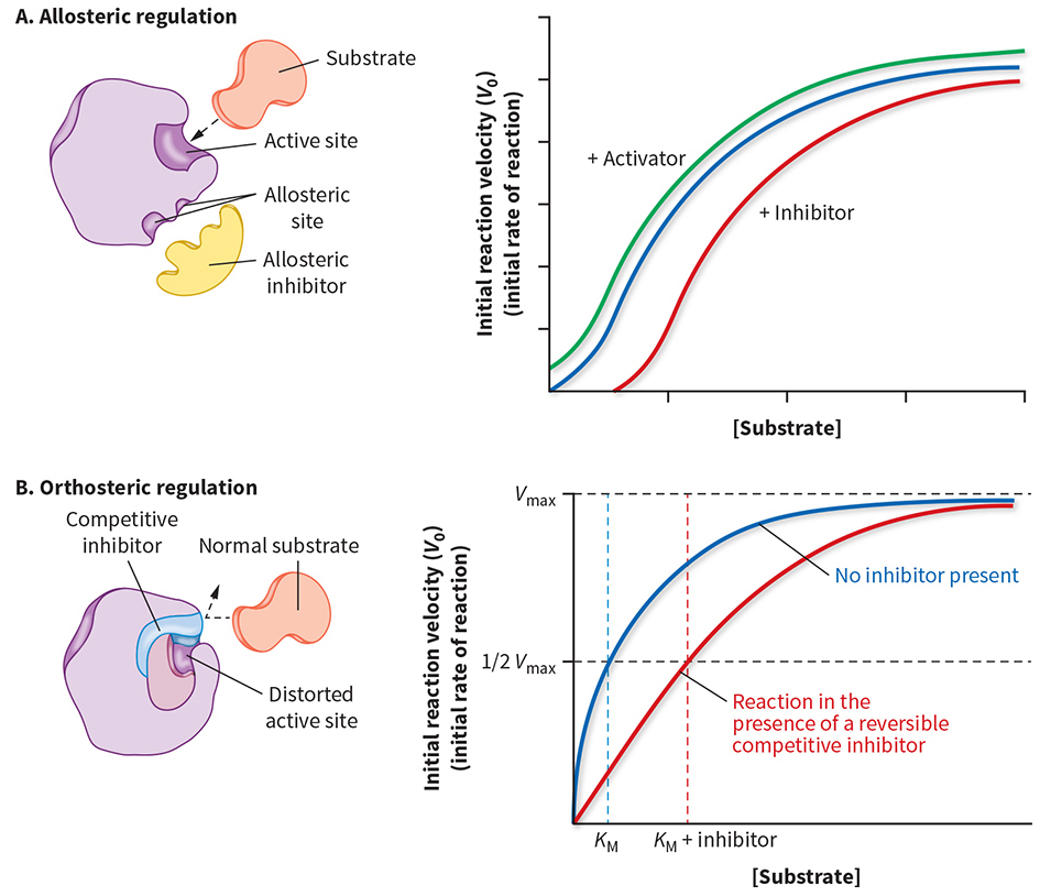
(how cells control when enzymes are active) The term allosterism literally means “other shape” or “other site.” It is used to describe the regulation of a molecule such as an enzyme, receptor, or transport protein through a conformational change induced by the binding of a regulator. Numerous proteins are regulated by allosteric mechanisms .
Allosteric regulation occurs when a regulator binds to a site other than the active site.
This binding changes the enzyme's conformation and can affect its activity.
Allosteric activation: Enzyme activity increases.
Allosteric inhibition: Enzyme activity decreases.
Allosteric binding is generally reversible, allowing activity to respond to changing cellular conditions.
The key idea is that binding at one site can influence the shape and function of another site.
Reversible covalent modification: Phosphorylation
Another way to regulate enzymes is by adding or removing a chemical group.
The lecture focuses on phosphorylation.
Kinase: Transfers a phosphate group, typically from ATP, to a target.
Phosphatase: Removes the phosphate group.
Common phosphorylation targets include the hydroxyl groups of:
Serine.
Threonine.
Tyrosine.
Phosphorylation can either activate or inhibit an enzyme, depending on the protein.
The professor uses Src as an example in which phosphorylation of a tyrosine residue produces a conformational change that makes the active site unavailable, decreasing activity.
The important distinction is that phosphorylation is a reversible covalent modification.
Proteolytic activation of zymogens
A zymogen is an inactive precursor of an enzyme.
Zymogens allow enzymes to be produced in one location and activated only where their activity is needed.
For example:
The pancreas produces chymotrypsinogen.
Chymotrypsinogen is secreted into the small intestine.
Proteolytic cleavage activates it to chymotrypsin.
Proteolytic activation is generally irreversible because peptide bonds are cleaved.
Why are digestive enzymes produced as zymogens?
The pancreas contains proteins and other cellular components that could be damaged by active digestive proteases.
Producing these enzymes in inactive forms helps prevent the pancreas from digesting itself.
Premature activation of digestive zymogens can contribute to pancreatic injury, as in pancreatitis.
Why are blood-clotting zymogens useful?
Blood-clotting proteins need to be activated quickly when an injury occurs. Having inactive precursors already present allows the body to activate the clotting pathway rapidly instead of waiting to synthesize the proteins.

Hemoglobin is a protein, not an enzyme
connecting hemoglobin to the previous discussion of allosteric regulation of ATPase.
ATPase is an enzyme that can be regulated allosterically.
Hemoglobin is not an enzyme, but it is also regulated allosterically.
Hemoglobin is a protein found inside red blood cells.
Its primary function is to transport molecular oxygen, O2, from the lungs to tissues throughout the body.
ATPase
a class of enzymes that break down adenosine triphosphate (ATP) into adenosine diphosphate (ADP) and an inorganic phosphate ion, releasing chemical energy that drives vital cellular processes.
Energy Transduction: They convert chemical energy stored in ATP bonds into mechanical, chemical, or transport work.
Active Transport: Many ATPases act as membrane pumps to move ions (such as Na⁺, K⁺, or Ca²⁺) or nutrients across cell membranes against their concentration gradients.
Muscle Contraction & Motor Movement: Proteins like myosin and kinesin use ATPase activity to generate force and move along cellular tracks.
Protein
A protein is a large, complex molecule made of long chains of amino acids, while an enzyme is a specific type of protein that acts as a biological catalyst to speed up chemical reactions.
• Building blocks: Proteins are made of smaller units called amino acids linked together by peptide bonds into chains called polypeptides.
• Structure: There are 20 different amino acids. They twist and fold into complex three-dimensional shapes. This shape decides what the protein does.
• Functions: Proteins are the "workhorses" of cells. They build structures (like keratin and collagen), transport oxygen (like hemoglobin), send chemical signals (like hormones), and protect against illness (like antibodies).
Enzyme
An enzyme is a specialized functional protein that speeds up chemical reactions inside and outside of cells.
• How they work: Enzymes lower the activation energy (the energy needed to start a reaction). They have a specific shape with an active site where target molecules—called substrates—fit like a lock and key.
• Not consumed: Enzymes are not used up or changed permanently by the reactions they speed up, meaning they can be used over and over again.
• Examples: Amylase (which breaks down starch in saliva) and lactase (which breaks down dairy sugar).
• The Rule of Thumb: All enzymes are proteins (with rare exceptions like RNA-based ribozymes), but not all proteins are enzymes.
The two essential parts of oxygen transport
Hemoglobin must perform two complementary functions:
Bind oxygen in the lungs, where oxygen availability is relatively high.
Release oxygen in peripheral tissues, where oxygen availability is lower and cells need oxygen for metabolism.
Think of hemoglobin as a delivery truck:
The lungs are the warehouse where the truck picks up its cargo.
Oxygen molecules are the packages.
Peripheral tissues are the delivery addresses.
Hemoglobin must both load and unload its cargo.
This analogy explains why simply binding oxygen strongly is not enough. If hemoglobin binds oxygen but cannot release it, oxygen cannot reach the tissues that need it.
Binding Component of hemoglobin's activity.
Oxygen binding can be measured using percent saturation.
Oxygen delivery is the other half of hemoglobin's physiological job.
A binding curve tells us how much oxygen is bound under different oxygen conditions; it does not, by itself, tell us everything about how effectively oxygen is delivered to tissues.
Always distinguish between oxygen binding and oxygen release. A high affinity for oxygen is not automatically an advantage in every part of the body.
(In chemistry, affinity is the tendency of a chemical species (like an atom, molecule, or drug) to react, combine, or bind with another species)

Myoglobin versus hemoglobin
Myoglobin is another oxygen-binding globin protein. It occurs inside muscle cells and also binds molecular oxygen, O2
(see image on front)
Essential terminology
Monomer: A protein consisting of one polypeptide chain.
Tetramer: A protein complex consisting of four subunits.
Homotetramer: Four identical subunits.
Heterotetramer: Four subunits that are not all identical.
Polypeptide: A chain of amino acids linked by peptide bonds.
Adult hemoglobin is a heterotetramer because its four subunits consist of two α-globin chains and two β-globin chains.
Its composition is written: (see image)
Each subunit can bind one oxygen molecule, so one adult hemoglobin molecule can bind up to four O2 molecules.

Globin genes and protein assembly
which genes must be expressed to produce hemoglobin. The relevant globin genes include:
Myoglobin gene: Encodes the myoglobin polypeptide.
α-globin gene: Encodes an α-globin chain.
β-globin gene: Encodes a β-globin chain.
γ-globin gene: Encodes a γ-globin chain, important in fetal hemoglobin.
The basic sequence of events is:
Gene Expression: The appropriate globin genes are transcribed and translated
Globin polypeptides are produced: Adult hemoglobin production requires alpha-globin and beta-globin chains.
Four subunits assemble: Two alpha-chains and two beta-chains form one a2b2 hemoglobin molecule
Functional oxygen-binding protein: Each globin subunit contains a heme group
The key distinction is that genes encode polypeptide chains, not the entire multimeric protein as a single chain. Different gene products assemble into one functional hemoglobin molecule.
Secondary and tertiary structure: α-helices
Both myoglobin and hemoglobin contain many α-helices.
Myoglobin contains approximately eight α-helices in the structure discussed in class; some descriptions count nine depending on how the helices are delineated.
The α- and β-globin subunits have similar sizes and overall three-dimensional folds.
The globin chains resemble myoglobin structurally despite differences in their amino acid sequences.
Why do α-helices matter?
An α-helix is a type of secondary protein structure. Its amino acid side chains, or R-groups, project outward from the helix axis.
This arrangement matters because some side chains can interact with other molecules. In globins, particular histidine residues are positioned so they can interact with the iron in the heme group.
Key distinction: A protein's amino acid sequence helps determine its three-dimensional structure, and that three-dimensional structure positions the chemical groups needed for oxygen binding.

The heme prosthetic group
A globin chain is not composed solely of amino acids. It also contains a non-protein component called heme.
What is a prosthetic group?
A prosthetic group is a non-polypeptide component that is tightly associated with a protein and is required for its function.
Heme is the prosthetic group that enables globins to bind oxygen.
Myoglobin contains one heme group.
Each hemoglobin subunit contains one heme group.
Adult hemoglobin therefore contains four heme groups.
Protein:
Myoglobin: 1 globin chain, 1 heme group, maximum O2 molecules bound: 1
Adult Hemoglobin: 4 globin chains, 4 heme groups, 4 maximum O2 molecules bound
(see image to see above view)
Heme contains a large, approximately planar ring system called protoporphyrin IX, with an iron ion at its center.
The ring contains four nitrogen atoms that coordinate the iron.
Important terms:
Porphyrin ring: The large ring system containing four nitrogen atoms.
Fe²⁺: Ferrous iron, the iron oxidation state emphasized in the lecture.
Coordination bond: An interaction between a central metal ion and a ligand that donates an electron pair.
Heme: The iron-containing prosthetic group within the globin protein.
The iron is crucial because it provides the site to which molecular oxygen binds.


How many bonds can the heme iron form?
(see image on front for bond summary)
the iron has six coordination positions:
Four coordinate bonds connect the iron to the four nitrogen atoms of the porphyrin ring.
A fifth coordinate bond connects the iron to a histidine residue from the globin protein.
The sixth position can bind molecular oxygen.
(see image)
The iron is therefore coordinated by the heme ring and the protein, while the remaining position provides the oxygen-binding site.
Scientific clarification: The professor describes Fe²⁺ as the usual iron state. In actual oxygenated heme, the electronic structure is more complex than a simple picture of an unchanged Fe²⁺ ion donating or losing a fixed quantity of electron density. For this course, the key structural concept is the reversible coordination of oxygen at the heme iron.

Proximal and distal histidine residues
Two histidine residues are particularly important for understanding globin function. (see image)
Proximal histidine: the mechanical connection
The proximal histidine directly forms the fifth coordination bond to the heme iron.
It is part of an α-helix in the globin protein. Therefore, when the iron moves, the proximal histidine can move, and that movement can be transmitted to the helix and the rest of the protein.
This is an important link between a small chemical event at the heme and a larger structural change in hemoglobin.
Distal histidine: controlling ligand geometry
The distal histidine does not directly coordinate the iron in the same way as the proximal histidine.
Instead, it sits near the ligand-binding site and influences the position and geometry of bound molecules.
The professor emphasizes that the distal histidine helps prevent ligands from binding in an unfavorable perpendicular orientation relative to the heme plane.
This matters because the geometry of ligand binding influences how strongly the ligand interacts with the iron and how readily it can dissociate.
Exam takeaway: Remember the distinction:
Proximal histidine = directly bonded to iron.
Distal histidine = influences the ligand-binding pocket and helps control ligand geometry.

Why oxygen binds at an angle
Mechanism
Oxygen approaches the iron at the heme center.
The distal histidine creates steric constraints within the binding pocket.
These constraints favor angled oxygen binding.
The angled geometry helps avoid the very strong, linear binding geometry that carbon monoxide favors.
The result is a binding pocket that supports reversible oxygen binding and release.
The professor's central point is that oxygen must not bind so strongly that hemoglobin cannot release it.
A useful comparison is:
Oxygen: Binds reversibly in a geometry influenced by the heme pocket and distal histidine.
Carbon monoxide (CO): Has a much stronger preference for linear binding to heme iron and can bind extremely tightly when the protein pocket does not sufficiently constrain its geometry.
Why is carbon monoxide dangerous?
Carbon monoxide binds heme iron with an affinity far greater than that of oxygen. The lecture gives an approximate comparison of 400 times stronger.
This is an approximate teaching figure; actual relative affinities depend on the heme environment and conditions.
When CO occupies a heme-binding site, oxygen cannot occupy that same site. This reduces the blood's capacity to transport oxygen and also makes the remaining oxygen bound to hemoglobin harder to release.
The second effect is especially important: CO binding can impair oxygen delivery even at heme sites that are not directly occupied by CO.
Why can hyperbaric oxygen help?
The professor explains the principle of hyperbaric oxygen treatment:
The patient breathes oxygen at a very high partial pressure.
This increases oxygen availability in the blood and tissues.
High oxygen availability favors oxygen binding when CO dissociates from heme.
Over time, CO can be displaced and eliminated as the patient breathes.
Hyperbaric oxygen is a clinical treatment used in selected cases of CO poisoning; it is not a guarantee of recovery, and suspected CO poisoning requires emergency medical evaluation.
Exam takeaway: CO poisoning is dangerous because CO occupies hemoglobin's oxygen-binding sites and interferes with the release of oxygen to tissues.

Oxygen availability in the lungs versus tissues
Hemoglobin loads oxygen in the lungs and releases it in peripheral tissues.
(see image)

How do we measure hemoglobin's oxygen binding?
For enzymes, we often plot reaction rate against substrate concentration. For oxygen-binding proteins, we instead plot fractional saturation with oxygen against oxygen partial pressure. These measurements are analogous, but they are not identical (see image)
What does percent saturation mean?
Percent saturation indicates what percentage of available oxygen-binding sites are occupied by oxygen.
For example:
0% saturation: no oxygen-binding sites are occupied.
50% saturation: half the available binding sites are occupied on average.
100% saturation: all available binding sites are occupied.
For hemoglobin, saturation reflects occupancy across its four heme sites. It does not mean that every hemoglobin molecule necessarily has exactly two oxygen molecules bound when the overall saturation is 50%.
What is oxygen partial pressure?
Oxygen partial pressure, PO2PO2, describes the contribution of oxygen to the total pressure exerted by a gas mixture.
It is commonly measured in mmHg in physiology.
For gases, partial pressure is related to the amount of that gas present. In the context of the lecture, it serves as a measure of oxygen availability.
Important distinction: Partial pressure is not literally the same quantity as molar concentration, although it is related to dissolved oxygen concentration under specified conditions.


Hyperbolic versus sigmoidal curves
Myoglobin's hyperbolic curve
Myoglobin has one polypeptide chain and one heme group.
Oxygen binds to its single heme site.
There are no other subunits within the same myoglobin molecule for that binding event to influence.
Its oxygen-binding behavior is therefore noncooperative.
Its curve is hyperbolic.
Myoglobin can still undergo a conformational change when oxygen binds. However, there is no second heme site within the same myoglobin molecule for the first oxygen-binding event to make more favorable.
Hemoglobin's sigmoidal curve
Hemoglobin contains four interacting subunits.
Oxygen binds to one heme site.
This binding changes the protein's conformation.
Because the subunits interact, that change influences the other subunits.
Subsequent oxygen binding becomes more favorable.
This behavior produces the characteristic S-shaped, or sigmoidal, curve.
Exam rule: Hyperbolic binding is characteristic of myoglobin; sigmoidal binding is characteristic of cooperative hemoglobin.
(check front and back images)

How does binding affinity relate to the curve?
Affinity describes how strongly a protein binds a ligand.
For oxygen-binding curves:
Higher oxygen affinity means the protein becomes substantially saturated at a lower oxygen partial pressure.
Lower oxygen affinity means a higher oxygen partial pressure is needed to reach the same saturation.
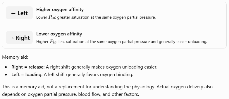
A curve shifted to the left generally indicates increased oxygen affinity. A curve shifted to the right generally indicates decreased oxygen affinity.
At the same oxygen partial pressure:
A left-shifted curve usually means greater oxygen saturation.
A right-shifted curve usually means lower oxygen saturation and a greater tendency to release oxygen.
This is why curve shifts matter physiologically: they indicate how readily hemoglobin loads or unloads oxygen under different conditions.
The P50 concept
P50 is the oxygen partial pressure at which a protein is 50% saturated with oxygen.
Lower P50 = higher oxygen affinity.
Higher P50 = lower oxygen affinity.
For a cooperative protein such as hemoglobin, P50 is a useful measure of the overall oxygen-binding behavior.

Why oxygen binding causes a conformational change
Step-by-step mechanism in myoglobin:
Oxygen approaches the heme iron: Before oxygen binds, the iron is positioned slightly out of the porphyrin plane toward the proximal histidine.
Oxygen coordinates the iron: Oxygen binds at the available coordination position on the heme iron. The electronic distribution around the iron changes as part of the coordination interaction.
The iron moves toward the porphyrin plane: The altered electronic structure favors a smaller effective iron radius and a position closer to the plane of the porphyrin ring.
The proximal histidine moves: Because the iron is coordinated to the proximal histidine, iron movement pulls or shifts the attached histidine.
The protein changes conformation: The movement is transmitted to the attached helix and changes the local structure of the globin protein.
What is a conformational change?
A conformational change is a change in a protein's three-dimensional arrangement without requiring the protein's amino acid sequence to change.
The key mechanism is:
O2 binding→heme iron movement→histidine movement→protein conformational change
a relatively small chemical event can produce a structural change in a protein.
Scientific clarification: The lecture's description of electron density being removed from iron is a simplified model. Oxygen coordination changes the electronic structure of the heme complex; the actual electronic behavior is more complex than simply subtracting electrons from an isolated Fe²⁺ ion.
Positive cooperativity in hemoglobin
The same initial conformational change occurs when oxygen binds to a hemoglobin subunit. The important difference is that hemoglobin has four subunits in contact with one another. A conformational change in one subunit can therefore influence the other subunits.
Step-by-step mechanism:
Hemoglobin begins in a relatively low-oxygen-affinity conformation.
An oxygen molecule binds to one heme group.
The iron moves toward the porphyrin plane.
The proximal histidine and attached protein structure shift.
This structural change is transmitted through interactions between the subunits.
The other subunits become more favorable for oxygen binding.
Additional oxygen molecules bind more readily.
The first oxygen-binding event therefore increases the likelihood of subsequent oxygen-binding events.
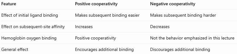
This is called positive cooperativity: Definition: Cooperativity occurs when ligand binding at one site of a multimeric protein affects ligand binding at other sites.
Positive cooperativity: Binding at one site increases the affinity of other sites for the ligand.
Negative cooperativity: Binding at one site decreases the affinity of other sites for the ligand.
The professor uses an analogy involving four children standing shoulder to shoulder. When one receives a favorite cookie, the others feel the excitement and become more eager for a cookie themselves. The conformational change in one hemoglobin subunit similarly influences the other subunits.
The analogy represents structural communication between the subunits (see image)
Important distinction: Positive cooperativity does not mean the first oxygen molecule itself becomes stronger. It means binding one oxygen molecule changes the protein so that the remaining sites bind oxygen more favorably.
Oxygen is a homotropic allosteric effector
An allosteric effector is a molecule that influences a protein's activity or ligand-binding behavior through an interaction that changes the protein's functional state.
Two important categories are:
Homotropic effector: The effector is the same molecule as the primary ligand.
Heterotropic effector: The effector is a different molecule from the primary ligand.
Oxygen binds hemoglobin and influences the binding of additional oxygen molecules.
Therefore: Oxygen is a positive homotropic effector of hemoglobin.
This terminology combines two ideas:
Positive: Oxygen binding increases the favorability of subsequent oxygen binding.
Homotropic: Oxygen is both the ligand and the effector.
Other molecules, such as protons and 2,3-BPG, also influence hemoglobin, but they are heterotropic effectors because they are not oxygen.
Why myoglobin does not show cooperativity
Myoglobin also undergoes a conformational change when oxygen binds. However, myoglobin is a monomer with only one oxygen-binding site. There are no other heme-binding sites within the same myoglobin molecule for the first binding event to influence. Therefore:
Myoglobin can undergo conformational changes.
Myoglobin can bind oxygen.
Myoglobin does not exhibit the inter-subunit positive cooperativity characteristic of hemoglobin.
Exam takeaway: A conformational change alone does not prove cooperativity. Cooperativity requires interactions between multiple binding sites within a protein complex.
Allosteric Regulators of Hemoglobin
Four main regulators:
Protons, H+
2,3-bisphosphoglycerate (2,3-BPG)
Carbon dioxide, CO2
The amino acid differences in fetal hemoglobin that alter 2,3-BPG binding
The first three are heterotropic regulators. Fetal hemoglobin is a different hemoglobin form whose structure changes how it responds to 2,3-BPG.

Protons and pH
A proton is a hydrogen ion, H+. Although it is very small, it can bind to sites on hemoglobin and influence the protein's conformation and oxygen affinity. Proton concentration changes hemoglobin's oxygen-binding curve. What happens when proton concentration changes Remember the relationship: pH = -log10[H+]
Consequently:
Higher H+ concentration means lower pH.
Lower H+ concentration means higher pH.
The direction of the oxygen-binding change is important. (see image on front) when there are fewer protons, the oxygen-binding curve shifts to the left, indicating increased affinity. When proton concentration increases, the curve shifts to the right, indicating reduced affinity.

What is the Bohr effect?
The Bohr effect is the decrease in hemoglobin's oxygen affinity caused by increased H+ concentration and increased CO2 under physiological conditions. The key idea is that acidity helps hemoglobin unload oxygen.
Step-by-step physiological mechanism
A tissue becomes metabolically active.
Cellular metabolism increases the production of acidic equivalents and often CO₂.
The local environment becomes more acidic, increasing H+H+ concentration.
Protons interact with hemoglobin and stabilize conformations with lower oxygen affinity.
Hemoglobin releases more oxygen at a given oxygen partial pressure.
The tissue receives additional oxygen to support metabolism.
Why is this useful during exercise?
Actively exercising muscle has a high demand for ATP and consumes oxygen rapidly.
Metabolic activity can increase proton and CO₂ concentrations in the local environment.
The resulting decrease in hemoglobin's oxygen affinity helps oxygen delivery match the tissue's needs.
The professor mentions lactic acid production. More precisely, lactate production is associated with high rates of glycolysis, and the relationship between lactate formation and pH is more complicated than saying that lactate itself is simply the cause of acidity. The exam-relevant point is that actively metabolizing tissues can develop conditions that promote oxygen unloading.
Bohr effect: cause and consequence:
Increased metabolism —> Increased local CO2 and/or H+ —> Lower hemoglobin oxygen affinity —> Increased oxygen unloading to tissues
Higher proton concentration lowers oxygen affinity. This is physiologically useful because active tissues need more oxygen.
2,3-Bisphosphoglycerate (2,3-BPG)
The full name is 2,3-bisphosphoglycerate, abbreviated 2,3-BPG.
Understanding the name
Glycerol is a three-carbon molecule with three hydroxyl groups. Glycerate is a related three-carbon molecule containing an oxidized carboxylate group. The name 2,3-bisphosphoglycerate indicates that phosphate groups are attached at positions 2 and 3 of the glycerate backbone. The lecture compares 2,3-BPG with 3-phosphoglycerate, which has a different phosphate arrangement. The structural difference matters because the two molecules do not have the same binding properties.
Where does 2,3-BPG bind?
2,3-BPG binds in a central cavity within hemoglobin, especially in the deoxygenated form. The binding pocket contains amino acid side chains that interact with the molecule's negatively charged phosphate groups. The professor specifically mentions:
Lysine residues
Histidine residues
These residues help stabilize 2,3-BPG binding through favorable electrostatic interactions and, where appropriate, hydrogen bonding. Lysine is especially important because its side chain is usually positively charged at physiological pH, allowing it to interact favorably with negatively charged phosphate groups. Histidine can also participate in electrostatic interactions depending on its protonation state.
Step-by-step mechanism of 2,3-BPG action
2,3-BPG approaches the central cavity of hemoglobin.
Its negatively charged phosphate groups interact with positively charged or otherwise complementary side chains.
These interactions stabilize the deoxygenated, lower-affinity conformation of hemoglobin.
Hemoglobin's oxygen affinity decreases.
At a given oxygen partial pressure, hemoglobin tends to be less saturated.
Oxygen release to tissues becomes easier.
The effect of 2,3-BPG:
More 2,3-BPG binding —> Stabilization of low-affinity hemoglobin —> Decreased oxygen affinity —> Increased oxygen unloading
What happens when 2,3-BPG is absent?
The professor compares a normal oxygen-binding curve with one in which 2,3-BPG has been removed. Without 2,3-BPG:
Hemoglobin's oxygen affinity increases.
The curve shifts to the left.
Hemoglobin binds oxygen more readily.
However, it becomes less effective at releasing oxygen at a given tissue oxygen partial pressure.
The professor notes that the curve can begin to resemble myoglobin's curve in its high-affinity behavior. This is a qualitative comparison, not a claim that hemoglobin becomes structurally identical to myoglobin or necessarily acquires a perfectly hyperbolic curve.
What happens when 2,3-BPG increases?
More 2,3-BPG generally stabilizes deoxygenated hemoglobin, reducing oxygen affinity and promoting oxygen unloading.
This makes 2,3-BPG a:
Negative heterotropic allosteric effector
Negative = it reduces oxygen affinity.
Heterotropic = it is different from oxygen itself.
Physiological significance: high altitude
At high altitude, oxygen availability is lower.
The body can adapt by increasing red blood cell 2,3-BPG concentrations.
This helps hemoglobin release oxygen more readily to tissues.
The professor describes endurance athletes training at high altitude and then returning to lower altitude for competition. The underlying principle is that physiological adaptations to altitude can improve oxygen delivery, although the magnitude and time course of changes in 2,3-BPG and athletic performance vary.
Blood doping: the lecture's example
The professor mentions substances used to stimulate red blood cell production and discusses the relationship to 2,3-BPG.
The essential concept to retain is that increasing the blood's oxygen-delivery capacity and adjusting hemoglobin's oxygen affinity are related but distinct processes.
More red blood cells can increase total oxygen-carrying capacity.
More 2,3-BPG can lower oxygen affinity and promote unloading.
Neither effect should be confused with the other.
Exam takeaway: 2,3-BPG binds in hemoglobin's central cavity and stabilizes the low-affinity form, promoting oxygen release.
Fetal hemoglobin versus adult hemoglobin
Why fetal hemoglobin binds oxygen more strongly than adult hemoglobin.
Subunit composition:
Adult hemoglobin, HbA: α2 β2
Fetal hemoglobin, HbF: α2 γ2
Both forms contain four subunits, but fetal hemoglobin uses two γ-globin chains instead of the two β-globin chains found in adult hemoglobin. The γ-globin gene is expressed during fetal development, whereas the pattern of globin gene expression changes as development proceeds.
How does γ-globin affect 2,3-BPG binding?
A histidine residue in the relevant β-globin position is replaced by a serine residue in γ-globin.
(see image). serine substitution makes fetal hemoglobin bind 2,3-BPG less strongly.
Step-by-step mechanism:
Adult hemoglobin has a central binding cavity that interacts with 2,3-BPG.
Positively charged residues help stabilize the negatively charged phosphate groups.
Fetal hemoglobin contains γ-globin chains with amino acid differences in this cavity.
The relevant histidine-to-serine substitution reduces favorable interactions with 2,3-BPG.
Fetal hemoglobin binds 2,3-BPG less strongly.
It is less strongly stabilized in the low-oxygen-affinity state.
Its oxygen affinity is higher than that of adult hemoglobin under comparable conditions.
Fetal hemoglobin still binds 2,3-BPG, but less strongly. It is not completely incapable of binding it.

Why does fetal hemoglobin need higher oxygen affinity?
A developing fetus receives oxygen from maternal blood across the placenta. The fetus does not directly breathe air through its lungs before birth. Instead, oxygen must be transferred from the mother's circulation into fetal blood. For efficient transfer, fetal hemoglobin must be able to bind oxygen at oxygen partial pressures where maternal hemoglobin is releasing it.
Because fetal hemoglobin has a higher oxygen affinity:
It can become more highly saturated at a lower oxygen partial pressure.
Maternal hemoglobin can release oxygen.
Fetal hemoglobin can capture that oxygen.
Oxygen transfer across the placenta becomes more effective.
Maternal-to-fetal oxygen transfer:
Maternal blood: Adult hemoglobin has relatively lower oxygen affinity (Oxygen then crosses the placenta)
Fetal blood: Fetal hemoglobin has higher oxygen affinity and captures oxygen more effectively
Oxygen crosses the placental barrier; maternal hemoglobin itself does not simply cross into fetal circulation as the oxygen carrier.
Exam takeaway: Fetal hemoglobin has an α2 γ2 composition, binds 2,3-BPG less strongly, and has higher oxygen affinity than adult hemoglobin
Carbon dioxide transport and hemoglobin
The bicarbonate transport pathway: Carbon dioxide produced in peripheral tissues can enter red blood cells, where carbonic anhydrase catalyzes the reversible reaction:
CO2 + H2O —> H2CO3
Carbonic acid can then dissociate:
H2CO3 —> H+ + HCO3-
The resulting bicarbonate ion, HCO3−, is a major form in which carbon dioxide is transported in blood.
Step-by-step in peripheral tissues
Cells produce CO₂ during metabolism.
CO₂ diffuses into the blood and red blood cells.
Carbonic anhydrase speeds up the reaction of CO₂ with water.
Carbonic acid forms and dissociates into H+ and bicarbonate.
Bicarbonate can move out of red blood cells and circulate in the blood.
The blood transports the carbon derived from CO₂ toward the lungs.
Step-by-step in the lungs
Bicarbonate is returned to red blood cells.
Bicarbonate and protons form carbonic acid.
Carbonic anhydrase catalyzes conversion of carbonic acid to CO₂ and water.
CO₂ diffuses into the alveoli.
CO₂ is exhaled.
The pathway is reversible, and the direction depends on the conditions in the tissues or lungs.
Hemoglobin can also bind CO₂ directly
CO₂ can bind reversibly to the N-termini of globin chains.
This forms a carbamate, often called carbaminohemoglobin.
The binding site is different from the heme iron used for oxygen binding.
Thus, hemoglobin contributes to CO₂ transport in two distinct ways:
It binds some CO₂ directly to globin amino groups.
It buffers protons produced in the bicarbonate pathway and participates in the transport of CO₂ indirectly through acid-base chemistry.
The bicarbonate pathway is the major form of CO₂ transport in blood; direct binding to hemoglobin is another important contribution.
CO₂ as a negative allosteric effector
Higher CO₂ conditions are associated with lower hemoglobin oxygen affinity. This is physiologically logical: tissues that produce large amounts of CO₂ during metabolism also need oxygen.
Step-by-step mechanism
A tissue increases its metabolic activity.
CO₂ production increases.
CO₂ can react with water to produce protons and bicarbonate.
Protons and CO₂-related interactions favor hemoglobin conformations with lower oxygen affinity.
Hemoglobin releases more oxygen.
The tissue receives oxygen to support its metabolism.
Direct CO₂ binding to globin can also contribute to the stabilization of deoxygenated hemoglobin. The effect of CO₂ overlaps with the Bohr effect because CO₂ can increase proton concentration, but direct CO₂ binding and proton-mediated effects are chemically distinct mechanisms. Therefore: CO2 is a negative heterotropic effector of oxygen binding.
Exam takeaway: High CO₂ is associated with reduced oxygen affinity and increased oxygen unloading in metabolically active tissues.
Comparison of allosteric regulators
(see image)
Remember that these are general physiological trends. The precise shape and position of the oxygen-binding curve depend on the combined conditions and on the form of hemoglobin being considered.
Negative versus positive allosteric effects
A negative effector of oxygen binding does not necessarily harm the body. In this case, it can improve oxygen delivery.
Negative effect on binding means lower oxygen affinity.
Lower affinity can make oxygen release easier.
The result can be beneficial when tissues require more oxygen.
This is one of the most important conceptual lessons of the lecture: the body regulates oxygen affinity to optimize delivery, not merely to maximize binding.

Review 1
see image

Review 2
see image

Review 3
see image