BIOL 252L: Lab 7 Cardiovascular Physiology
 Call Kai
 Learn
 Practice Test
 Spaced Repetition
 Match
 Flashcards
 Knowt Play
1/44

There's no tags or description
Looks like no tags are added yet.
45 Terms
What is the instrument used to listen for heart sounds?
Stethoscope
Closure of which two valves leads to the first heart sound ‘lub’?
Atrioventricular valves
Closure of which two valves leads to the second heart sound ‘dub’?
Semilunar valves
If extra heart sounds are heard, that is called a:
Murmurs

Label which valve you can hear at each auscultation point (orange colored dot). Pay close attention, it is not where you expect!!
Aortic valve

Label which valve you can hear at each auscultation point (blue colored dot). Pay close attention, it is not where you expect!!
Pulmonary valve
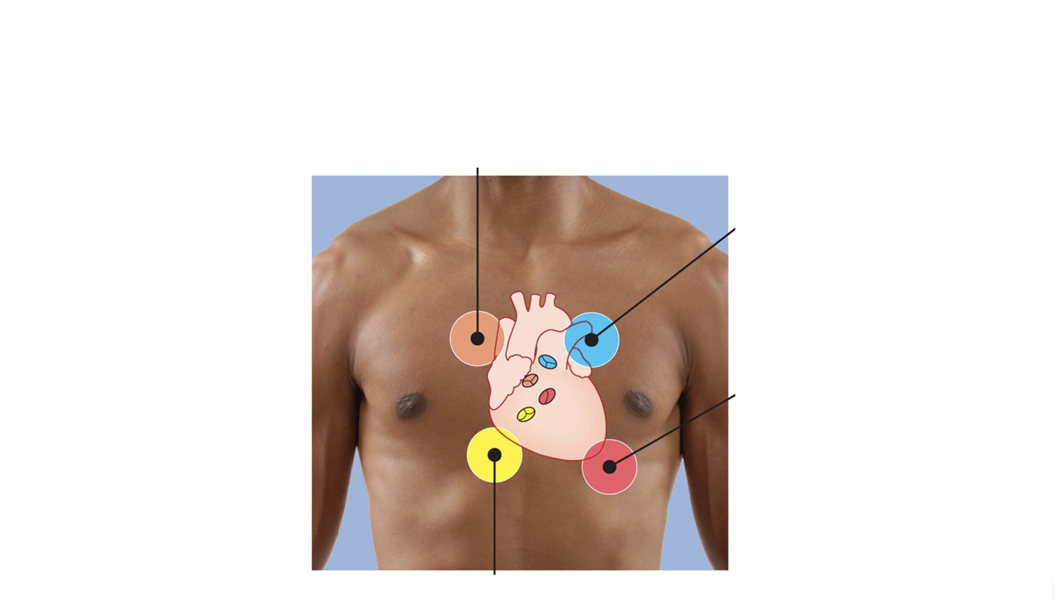
Label which valve you can hear at each auscultation point (red colored dot). Pay close attention, it is not where you expect!!
Bicuspid valve

Label which valve you can hear at each auscultation point (yellow colored dot). Pay close attention, it is not where you expect!!
Tricuspid valve

① Label the diagram below of the intrinsic conduction system.
Sinoatrial (SA) node (pacemaker)
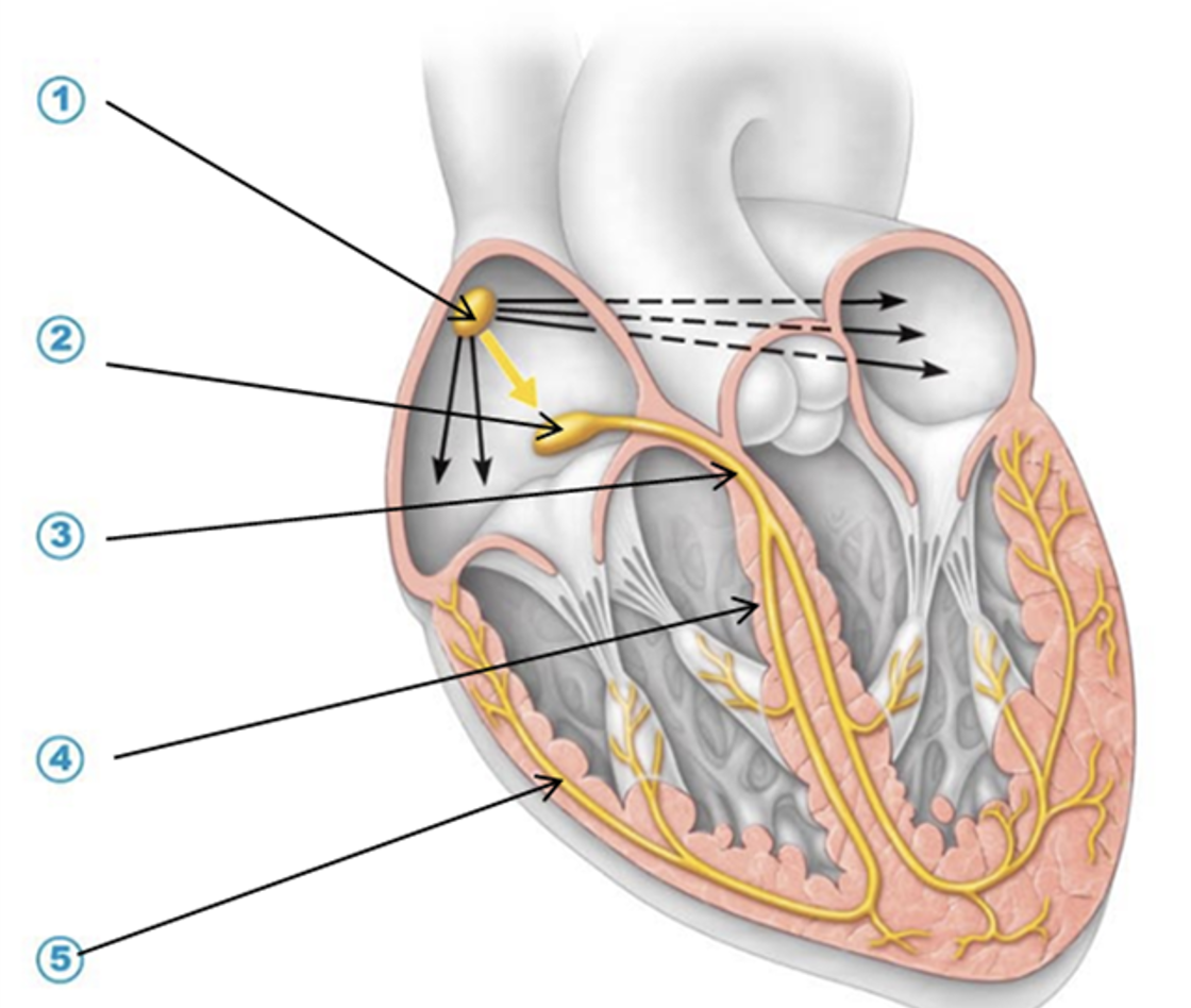
② Label the diagram below of the intrinsic conduction system.
Atrioventricular (AV) node

③ Label the diagram below of the intrinsic conduction system.
Interventricular bundle of His

④ Label the diagram below of the intrinsic conduction system.
Right bundle branch

⑤ Label the diagram below of the intrinsic conduction system.
Endocardial conducting (Purkinje) fibers
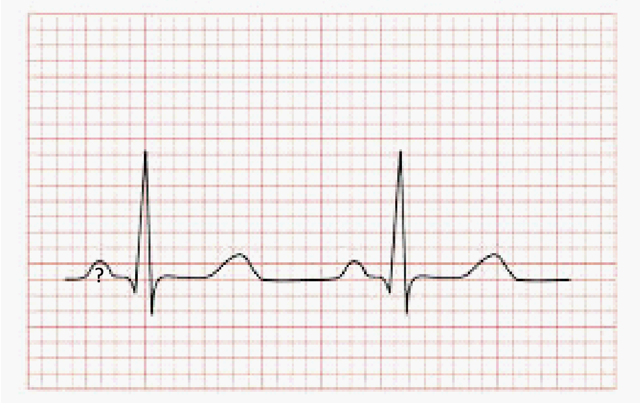
Label all the waves on this ECG.
P wave

Label all the waves on this ECG.
Q wave

Label all the waves on this ECG.
R wave

Label all the waves on this ECG.
S wave

Label all the waves on this ECG.
R wave

Fill up the table below with the electrical events and contractile events at each wave of the ECG.
Atria depolarize
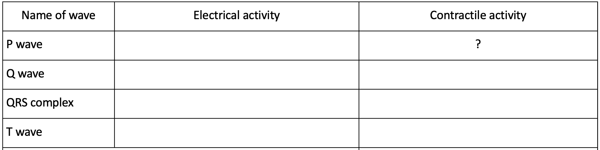
Fill up the table below with the electrical events and contractile events at each wave of the ECG.
Atria contract

Fill up the table below with the electrical events and contractile events at each wave of the ECG.
Atria repolarize

Fill up the table below with the electrical events and contractile events at each wave of the ECG.
Atria relax

Fill up the table below with the electrical events and contractile events at each wave of the ECG.
Ventricle depolarize

Fill up the table below with the electrical events and contractile events at each wave of the ECG.
Ventricle contract

Fill up the table below with the electrical events and contractile events at each wave of the ECG.
Ventricle repolarize

Fill up the table below with the electrical events and contractile events at each wave of the ECG.
Ventricle relax
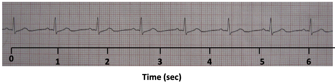
Patient 1 HR is : __________ bpm
72
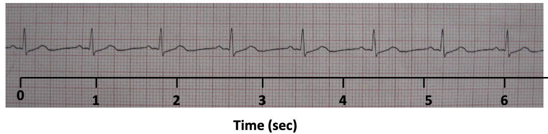
Describe the patient’s HR:
Normal (70-80 bpm)

Patient 2 HR is : __________ bpm
48

Describe the patient’s HR:
Bradycardia (too slow)
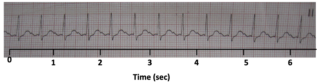
Patient 3 HR is : __________ bpm
120

Describe the patient’s HR:
Tachycardia (too fast)
What is the blood pressure cuff properly called?
Sphygmomanometer
Which blood vessel is being occluded?
Brachial artery
What are the sounds called?
Korotkoff sounds
When do you record the systolic pressure?
When taps appear
When do you record the diastolic pressure?
When taps disappear
Which generates a sound? Turbulent blood flow or steady blood flow?
Turbulent blood flow
What are the units of blood pressure?
120/80 mmHg
What happened to heart rate and blood pressure during exercise?
HR ↑ BP ↑
Why do heart rate and blood pressure NEED to increase?
Pump extra blood to working muscles
Signals in which arm of the autonomic nervous system will increase during exercise?
Sympathetic nervous system
How does the sympathetic nervous system affect the heart?
Heart rate ↑ x stroke volume ↑ → cardiac output ↑
How does the sympathetic nervous system affect skeletal muscle and skin blood vessels?
Dilate blood vessels
How does the sympathetic nervous system affect digestive and urinary blood vessels?
Constrict blood vessels