RAD 101 Unit 6 Anatomy of the Forearm, Elbow, and Distal Humerus
 Call Kai
 Learn
 Practice Test
 Spaced Repetition
 Match
 Flashcards
 Knowt Play
1/40

There's no tags or description
Looks like no tags are added yet.
41 Terms

Forearm in anatomical position
Radius is lateral, and ulna is medial.
Bones articulate at the proximal and distal radioulnar joints, which allow
rotation (pronation and supination) of the hand
Radius
shorter than ulna

Ulna

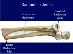
Two radioulnar joints
distal and proximal radioulnar joints
Ulnar notch

Head of the ulna fits into what joint
distal radioulnar joint

Radial notch
Head of the radius fits into
notch to form the proximal radioulnar joint.
Only the distal radius is directly involved
in the wrist joint.
When pronating the hand
it is the radius that rotates, the ulna remains stationary.
Proximal ulna primarily forms
the elbow joint
Head of the ulna is distal while
the head of the radius is proximal.
Olecranon process
posterior and easily palpated. It is the "point" on the posterior elbow

Coronoid process
anterior
Medial margin of coronoid process opposite radial notch

Coronoid tubercle (medial)

Head of radius

Neck of radius

Radial tuberosity

Body of the ulna

Head of ulna
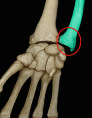
Ulna styloid process

Radius styloid process

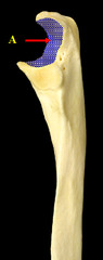
Trochlear notch of ulna
articulates with the distal humerus
AP Elbow
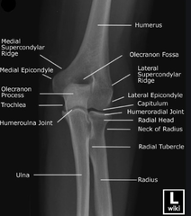
Humerus body

Radial fossa
The radial head fits into fossa during complete elbow flexion

Coronoid fossa
the coronoid process fits into fossa during complete elbow flexion

Lateral epicondyle
projection above capitulum

Medial epicondyle
larger and above the trochlea

Capitulum
articulates with the head of the radius

Humeral condyle
distal end of the humerus
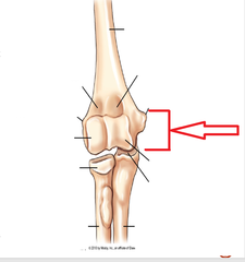
Trochlea
has 2 outer margins and a depressed center referred to as the trochlear sulcus
Articulates with the ulna

Trochlear Sulcus (Groove)

Olecranon fossa
olecranon process fits into fossa during complete flexion

Lateral elbow
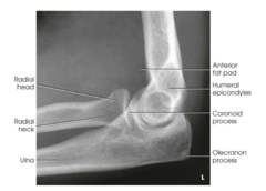
Three Concentric Arcs
Trochlear sulcus- smallest and will appear darker or more radiolucent.
Edges of capitulum and trochlea
Trochlear notch is largest


Anterior fat pad
shaped like a tear drop. Located anterior to distal humerus
Trauma will cause it to be elevated, more visible, and distorted in shape.

Posterior fat pad
D
located within the olecranon fossa, shaped like a sail
If visible suggests a joint effusion which displaced the fat pad

Supinator fat stripe
E
located anterior to proximal radius
If visible may indicate radial head or neck fractur

Monteggia fracture
fracture of the proximal half of the ulna, along with dislocation of the radial head, may result from defending against blows with a raised forearm