Anatomy Exam 3 Dr. Small Fall 26
 Call Kai
 Learn
 Practice Test
 Spaced Repetition
 Match
 Flashcards
 Knowt Play
1/142

There's no tags or description
Looks like no tags are added yet.
143 Terms
Neuron
a specialized cell transmitting nerve impulses; a nerve cell; when adequately stimulated te neuron fires an action potential
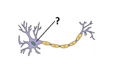
Soma (cell body)
the central, bulbous part of a neuron that houses the cell nucleus
Axon
the extension of a neuron, ending in branching terminal fibers, through which messages pass to other neurons or to muscles or glands

Axon Terminals (Terminal Buttons)
store and release chemical messengers called neurotransmitters to pass electrical signals across the synaptic cleft to neighboring neurons, muscles, or glands

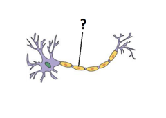
Myelin Sheath
a fatty, protective protein coating that wraps around the axons of nerve cells; Like the plastic insulation on an electrical wire, it insulates nerve fibers and allows electrical impulses to travel quickly and efficiently throughout the nervous system.
Action Potential
a neural impulse; a brief electrical charge that travels down an axon
Synapse
the specialized junction or tiny gap between two neurons—or between a neuron and another cell, like a muscle—where electrical or chemical signals pass from one cell to the next

Neurotransmitter
a chemical messenger produced and released by neurons to pass signals across a synapse (the small gap between cells) to neighboring neurons, muscles, or gland cells

Cells of the Central Nervous System (CNS):
- Astrocytes
- Oligodendrocytes
- Microglia
- Ependymal cells
Astrocytes (CNS)
maintain blood-brain barrier; provide structural support; regulate ion, nutrient, and dissolved gas concentrations; absorb and recycle neurotransmitters; form scar tissue after injury
Oligodendrocytes (CNS)
myelinate CNS axons; provide structural framework
Microglia (CNS)
immune cells of the nervous system; act as macrophages (scavengers) and phagocytize (engulf) damaged or infectious cells and debris. Are involved in inflammatory reaction in the brain
Ependymal Cells (CNS)
glial cells in the CNS that line the fluid-filled ventricles of the brain and the central canal of the spinal cord; assist in producing CSF and regulate chemical environment; features cilia and coordinated beating of their cilia helps circulate and move cerebrospinal fluid through the ventricular system
Cells of the Peripheral Nervous System (PNS)
- Schwann Cells
- Satellite Cells
Schwann Cells (PNS)
surround all axons in PNS; responsible for myelination of peripheral axons; participate in repair process after injury
Satellite Cells (PNS)
surround neuron cell bodies in ganglia; regulate O2, CO2, nutrient, and neurotransmitter levels around neurons in ganglia
Central Nervous System
brain and spinal cord
Divisions of the Brain:
- Forebrain (Prosencephalon)
- Midbrain (Mesencephalon)
- Hindbrain (Rhombencephalon)
Subdivisions of the Brain:
- Telencephalon
- Diencephalon
- Mesencephalon
- Metencephalon
- Myelencephalon
Structures within Telencephalon:
- Cerebral Cortex
- Basal Ganglia
- Limbic System
Structures within Diencephalon:
Thalamus and Hypothalamus
Structures within Mesencephalon:
Tectum and Tegmentum
Structures within Metencephalon:
Cerebellum and Pons
Structures within Myelencephalon:
Medulla Oblongata
Peripheral Nervous System
the sensory and motor neurons that connect the central nervous system to the rest of the body
Motor Neurons
send signals to activate muscles and glands
Sensory Neurons
send signals to our nervous system about our internal (body) and external worlds (environment)
Afferent (Sensory) Division
nerves transmit sensory signals from the periphery to brain; Sensory Neurons exclusively
Efferent (Motor) Division
carry information from the brain to periphery; Motor Neurons mostly
Somatic Nervous System
voluntary control of skeletal movement
Autonomic Nervous System
regulation of smooth muscle, cardiac muscle, and glands (involuntary)
Sympathetic Division (ANS)
fight or flight
Parasympathetic Division (ANS)
rest and digest
Meninges
covers the central nervous system
Layers of Meninges (superficial to deep)
1. Dura Mater
2. Arachnoid Membrane
3. Pia Mater
Dura Mater
"hard mother" the thick, tough outer layer that is flexible but cannot be stretched
Arachnoid Membrane
soft and spongy, lies beneath the dura mater
Pia Mater
"soft mother" is attached to the brain and spinal cord and contains smaller surface blood vessels
Subarachnoid Space
is a gap between the pia mater and the arachnoid membrane that is filled with cerebrospinal fluid (CSF)
Cerebrospinal Fluid (CSF)
circulates throughout the brain and spinal cord
Ventricular System
a system of fluid-filled cavities inside the brain
Ventricles of the Brain
- L Lateral Ventricle
- R Lateral Ventricle
- 3rd Ventricle
- 4th Ventricle

Primary Components of Grey Matter:
neuron cell bodies (soma), dendrites, unmyelinated axons, and neuroglia
Physical Appearance of Grey Matter:
gray-brown or pinkish-tan due to cell body density and lack of myelin
Anatomical Location (brain) of Grey Matter:
outer superficial layer (cerebral cortex, ~2-2.5 mm thick) and deep internal clusters (basal nuclei)
Primary Function of Grey Matter:
signal integration, sensory perception, decision-making, and motor planning
Major Brain Examples of Grey Matter:
cerebral cortex, cerebellar cortex, thalamus, and basal nuclei
Primary Components of White Matter:
bundles of myelinated axons
Physical Appearance of White Matter:
bright white due to the high lipid (fat) content of myelin sheaths
Anatomical Location (Brain) of White Matter:
deep inner region beneath the cerebral cortex, surrounding subcortical nuclei.
Primary Function of White Matter:
rapid signal transmission and communication between brain regions
Major Brain Examples of White Matter:
corpus callosum (interhemispheric connection) and internal capsule
Gyri (Gyrus)
bulges found on brain
Sulci (Sulcus)
small grooves found on brain
Fissures
large grooves found on brain
Epineurium (Nerve Structure)
surrounds the entire nerve
Perineurium (Nerve Structure)
surrounds the fascicle
Endoneurium (Nerve Structure)
surrounds the axon
Fascicle (Nerve Structure)
group of axons bundled together; many fascicles make up a nerve
Limbic System (HOME)
- Homeostasis (Hypothalamus)
- Olfaction (Olfactory Bulb)
- Memory (Hippocampus)
- Emotion (Amygdala)
Frontal Lobe (Brain Structure)

Primary Motor Cortex (Frontal Lobe)

Motor Homunculus (Frontal Lobe)
the body map on the motor cortex

Parietal Lobe (Brain Structure)

Primary Somatosensory Cortex (Parietal Lobe)
area of the parietal lobe where messages from the sense receptors are registered

Sensory Homunculus (Parietal Lobe)
a representation of the area of the somatosensory cortex that receives input from body regions

Temporal Lobe (Brain Structure)

Primary Auditory Cortex (Temporal Lobe)

Occipital Lobe (Brain Structure)

Insular Lobe (Brain Structure)

Subcortical Structures
thalamus, hypothalamus, hippocampus, amygdala, basal ganglia
Basal Ganglia (Subcortical Structure)
made up of nuclei responsible for motor control, learning, relating to routine behaviors or "habits", emotional processing, "action selection"
Hippocampus (Subcortical Structure)
"The Seahorse"; memory consolidation and storage; biological basis for learning (LTP) can be shown here; Adult neurogenesis; Sensitive to "stress" hormones (corticosteroids)

Amygdala (Subcortical Structure)
resp. for emotions / fear-induced responses

Thalamus (Diencephalon)
information relay station for filtering and projecting sensory and motor info, regulation of consciousness, sleep, and alertness.

Hypothalamus (Diencephalon)
it acts as the body's primary "smart control" center to maintain a stable internal state known as homeostasis. Links the nervous system to the endocrine system.

Tectum (Midbrain)
functions as a sensory reflex center; integrates visual, auditory, and somatosensory spatial maps for eye and head orienting movements and process sound localization; Tectum orients behavioral responses toward environmental stimuli
Tegmentum (Midbrain)
responsible for homeostatic, motor control, arousal, and reward-related functions; major source of dopamine for motor control and the reward pathways
Cerebellum (Hindbrain)
does not initiate movement, but helps; coordination, precision, and accurate timing of movement; role in attention and language

Pons (Hindbrain)
connects medulla, cortex, and cerebellum; regulates sleep, wakefulness, respiration, swallowing, bladder control, etc.

Medulla Oblongata (Hindbrain)
extension of the spinal cord; Autonomic (involuntary) functions of breathing, heart rate and blood pressure.

Spinal Gray Matter:

Neuron Morphological Types in the Spinal Cord:
Unipolar and Multipolar
Unipolar Neurons
a nerve cell that features a single process extending from the cell body (soma) which branches to function as both an axon and dendrites

Multipolar Neurons
a type of nerve cell that has a single axon and multiple dendrites extending from its cell body


Cranial Nerve I (CN I)
Olfactory Nerve (sensory)
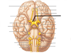
Cranial Nerve II (CN II)
Optic Nerve (sensory)
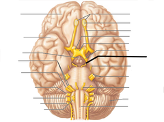
Cranial Nerve III (CN III)
Oculomotor Nerve (motor)

Cranial Nerve IV (CN IV)
Trochlear Nerve (motor)

Cranial Nerve V (CN V)
Trigeminal Nerve ("Dentist Nerve") (both)

Cranial Nerve VI (CN VI)
Abducens Nerve (motor)

Cranial Nerve VII (CN VII)
Facial Nerve (both)

Cranial Nerve VIII (CN VIII)
Vestibulocochlear / Auditory Nerve (sensory)

Cranial Nerve IX (CN IX)
Glossopharyngeal Nerve (both)

Cranial Nerve X (CN X)
Vagus Nerve (both)
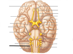
Cranial Nerve XI (CN XI)
Accessory / Spinal Accessory Nerve (motor)
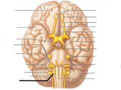
Cranial Nerve XII (CN XII)
Hypoglossal Nerve (motor)
Cervical Plexus (C1-C4)
Phrenic nerve, Intercostal nerves, and Subcostal nerves
Phrenic Nerve (C3-C5)
innervates the diaphragm
Intercostal Nerves
supply muscles of the ribs, anterolateral thorax, and abdominal wall