Anatomy and Physiology - Respiratory System
 Call Kai
 Learn
 Practice Test
 Spaced Repetition
 Match
 Flashcards
 Knowt Play
1/69

Earn XP
Description and Tags
LINK INCLUDED
70 Terms
Respiratory system
Respiration: movement of O2 and CO2 between air and systemic body cells
Cells need O2 for aerobic ATP production and need to dispose of CO2 that this process produces
The respiratory system provides the means for gas exchange
Consists of respiratory passageways in the head, neck, trunk, and lungs
Respiratory system functions
The respiratory tract is a passageway for air between atmosphere and alveoli as we breathe
Mucous membrane conditions inhaled air by warming and humidifying it and trapping most inhaled particles
Respiratory membrane is a site for gas exchange; oxygen diffuses from alveoli into blood & carbon dioxide diffuses from blood into alveoli
Olfaction: olfactory receptors in superior nasal cavity
Sound production: air moves across vocal cords of the larynx (voice box); vocal cords vibrate, producing sound
Label the respiratory system
https:// www.purposegames.com/game/respiratory-system-labeling-interactive-game

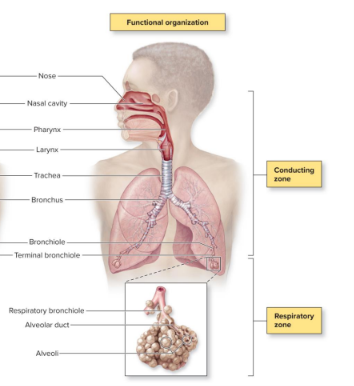
Structural organization
Upper respiratory tract: nose, nasal cavity, pharynx, larynx
Lower respiratory tract: trachea, bronchus, bronchiole, terminal bronchiole, lungs


Functional organization
Conducting zone: nose, nasal cavity, pharynx, larynx, trachea, bronchus, bronchiole, terminal bronchiole, lungs
Respiratory zone: respiratory bronchiole, alveolar duct, alveoli
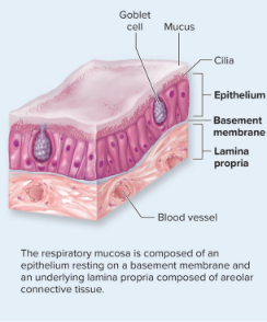
Respiratory mucosa
Lines the respiratory tract
Epithelium with cilia and goblet cells on a basement membrane
Underlying lamina propria composed of areolar connective tissue; MALT found here
Becomes thinner from the nasal cavity to the alveoli (pseudostratified ciliated columnar → simple ciliated columnar → simple cuboidal → simple squamous)
Exceptions are stratified squamous found in high abrasion areas: oropharynx, laryngopharynx, vocal cords, superior portion of larynx

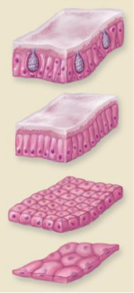
Progressively thinner epithelium layers
Pseudostratified ciliated columnar: nasal cavity, paranasal sinuses, nasopharynx, trachea, inferior portion of larynx, main bronchi, lobar bronchi
Simple ciliated columnar: segmental bronchi, smaller bronchi, large bronchioles
Simple cuboidal: terminal and respiratory bronchioles
Simple squamous: alveolar ducts and alveoli

Mucous secretions (respiratory mucosa)
Produced from secretions of goblet cells of epithelial lining and mucous and serous glands of the lamina propria
Contain mucin protein: increases mucus viscosity and serves to trap dust, dirt, pollen, etc.
Contains defenses against microbes: lysozyme (antibacterial enzyme), defensins (antibacterial proteins), immunoglobulin A (antibody)
Called sputum when coughed up with saliva and trapped substances
Rhinorrhea/runny nose
Occurs as a result of:
Increased production of mucus due to allergies, virus
Crying, due to increased secretions from lacrimal glands draining into the nasal cavity
Exposure to cold air due to water condensation mixing with mucus + less effective cilia unable to sweep mucus into nasopharynx

Paranasal sinuses
Air-filled cavities named for the bones in which they occur
Lined by pseudostratified ciliated columnar epithelium
Warms, humidifies, and cleanses inhaled air, like the nasal cavity does
Cilia sweep mucus; mucus drains through ducts into the nasal cavity → traps foreign materials of inhaled air → cilia sweep mucus into pharynx (throat) → mucus is swallowed

Sinusitis
Respiratory infection or allergy can cause inflammation of the ducts that drain from the paranasal sinuses
Drainage of mucus decreases and accumulates in the sinuses
Germs can grow in the accumulated mucus → sinus infection
Inflamed and blocked sinuses and pressure changes → sinus headaches
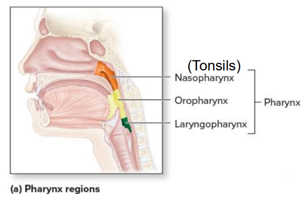
Pharynx
Funnel-shaped passageway posterior to nasal cavity, oral cavity, and larynx
Lateral walls composed of skeletal muscles
Partitioned into:
Nasopharynx: pharyngeal tonsils
Oropharynx: palatine + lingual tonsils
Laryngopharynx


Larynx
Pyramid-shaped airway structure between laryngopharynx and trachea
Produces sound (vocal folds vibrate during expiration)
Air passageway
Prevents ingested materials from entering respiratory tract
Epiglottis covers superior opening during swallowing


Vocal folds
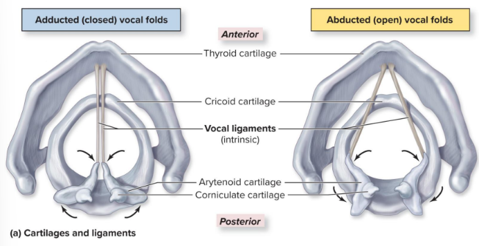
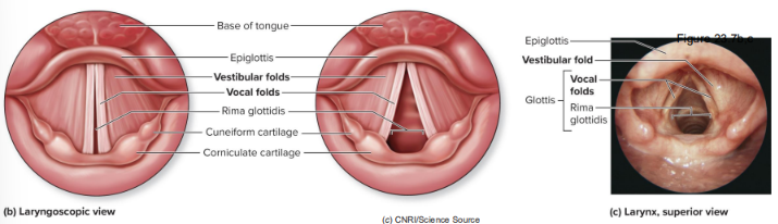
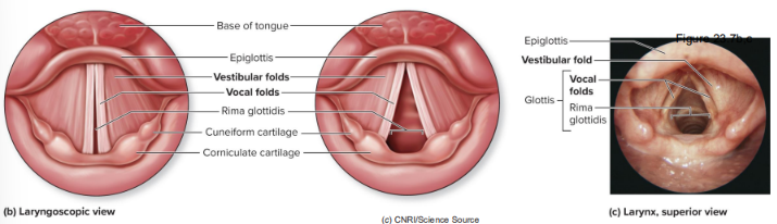
Consists of mucosa-covered vocal ligaments that can adduct (close) when instrinsic muscles contract and abduct (open) when instrinsic muscles relax; between the thyroid cartilage and the arytenoid cartilages
They adduct with speaking or singing, and abduct when breathing deeply
Sound production
Results from vocal cord vibration
Intrinsic laryngeal muscles narrow opening between vocal folds (called the rima glottidis)
Air is forced past vocal cords during expiration
Voice range (sound production)
Determined by length, thickness of vocal cords
Males have longer and thicker folds, and so deeper voices
Folds increase in length with growth, deepening range
Pitch/frequency (sound production)
Determined by tension on vocal cords
Increased tension = folds vibrate more = higher pitch
Regulated by intrinsic laryngeal muscles (cricothyroid)
Loudness (sound production)
Depends on force of air passing across cords; more air = louder sound
Laryngitis
Inflammation of the larynx
Symptoms of hoarse voice, sore throat, sometimes fever
Caused by bacterial or viral infection, or overuse (yelling)
Severe cases can extend to the epiglottis
May lead to sudden airway obstruction, especially in children
Trachea
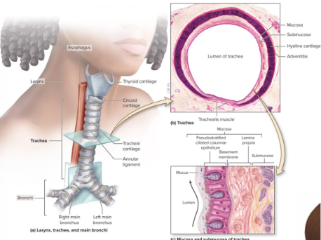
AKA the windpipe
Connects to the larynx superiorly (at its top) and to the main bronchi inferiorly (at its bottom); located anteriorly to the esophagus
Mucus moves upward towards the pharynx and larynx, where it may be swallowed or expelled

Tracheostomy (intubation)
Incision into trachea to facilitate breathing when:
Airway is blocked
Ventilation compromised by disease or injury
SARS COV-2 (COVID-19)
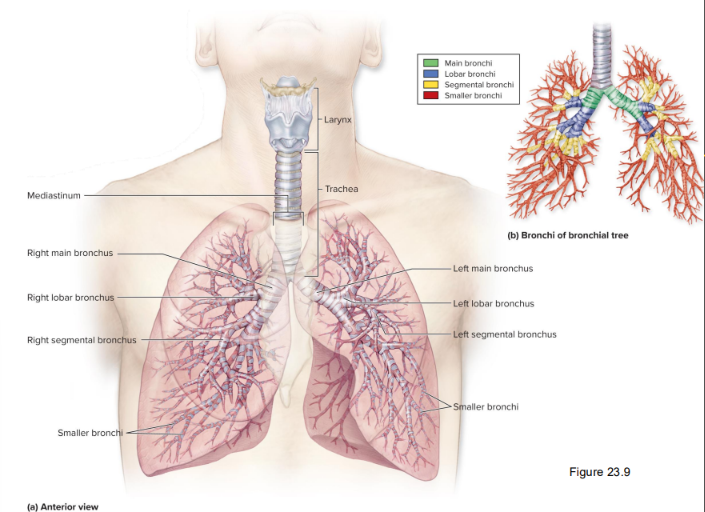
Bronchial tree
System of highly branched air passages, starting with main bronchi and ending at alveoli
Main bronchi → lobar bronchi → segmental bronchi → smaller bronchi
All covered in irregular cartilage plates of decreasing size
Then bronchiole → terminal bronchiole → respiratory bronchiole → alveolar duct → alveoli
No cartilage, but instead a proportionately thicker layer of smooth muscle that can bronchoconstrict or dilate to regulate amount of air reaching the alveoli

Acute bronchitis
Inflammation of the bronchi caused by bacterial or viral infection or inhaled irritants
Occurs during or after an infection
Coughing, sneezing, pain with inhalation, fever
Most cases resolve in 10 to 14 days
Chronic bronchitis
Inflammation of the bronchi caused by bacterial or viral infection or inhaled irritants
Occurs after long-term irritant exposure
Large amounts of mucus + cough lasting 3 months
Permanent changes to bronchi occur
Increases likelihood of future bacterial infections
Asthma
Episodes of bronchoconstriction, wheezing, coughing, shortness of breath, and excess mucus
Primary treatments: inhaled steroids and bronchodilators
Occupational asthma: asthmatic with sensitivity to airborne agent
Localized immune reaction occurs in bronchi & bronchioles → walls of the bronchi becoming permanently thickened
Exercise-induced asthma (bronchoconstriction) leads to excessive constriction of airways during exertion

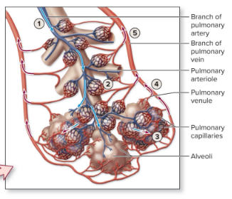
Bronchioles and alveoli of the lung
Each lung contains 300 to 400 million ALVEOLI
Terminal bronchioles → respiratory zone: respiratory bronchiole → alveolar ducts → alveoli
Pulmonary vessels (veins, arteries) are positioned alongside the bronchioles
Pulmonary capillaries form a vascular network around each alveolus for gas exchange
Alveoli have a honeycomb-like appearance, covered in elastic fibers


Alveoli
2 cell types form the alveolar wall
Alveolar type I cells (squamous alveolar cells)
Most common of the two cell types making up alveolar wall
95% of alveolar surface area; form the alveolar epithelium of the respiratory membrane
Alveolar type II cells (septal cells): not very numerous
Secrete oily pulmonary surfactant


Pulmonary surfactant
An oily fluid that prevents the collapse of alveoli by decreasing the surface tension of water and thus the attractive force of these molecules


Respiratory membrane
Comprised of:
Plasma membrane of the alveoli type 1 cell
Plasma membrane of the capillary endothelial cell
Fused basement membrane of the above 2 cells


Pulmonary circulation of lungs
Deoxygenated blood enters lungs thru branches of pulmonary artery
Moves into smaller pulmonary arterioles → further divide into capillary networks surrounding the alveoli
In the alveoli, CO2 diffuses out the blood into the air, while oxygen diffuses into the blood
Oxygenated blood enters pulmonary venules
Converges into branches of pulmonary vein, carrying O2-rich blood

Lung position
Within the thoracic cavity, lungs are bordered and protected by the thoracic ribcage
Lateral to the mediastinum
Base of each lung rests on the diaphragm
Apex is slightly superior and posterior to the clavicle

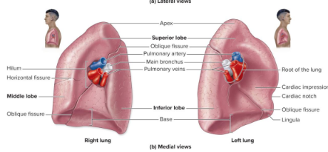
Surface indentations of the left lung
Accommodate heart and aorta
Cardiac impression on medial surface
Cardiac notch on anterior surface
Groovelike impression for aorta on medial surface


Lobes and bronchopulmonary segments of the lungs
Right lung has 3 lobes (superior, middle, inferior) → 10 segments
Left lung has 2 lobes (superior, inferior) → 8-10 segments
Autonomous units encapsulated with connective tissues
Each supplied with its own segmental bronchus
Supplied with its own branch of pulmonary artery and vein and lymph vessels
Can be removed individually in cases of disease

Lobules
Each bronchopulmonary segment organized into these
Marble-sized; each surrounded by connective tissue
Each supplied by a terminal bronchiole, arteriole, venule, and lymph vessel
Pneumonia
Infection of the lung, resulting in alveoli filling with fluid, exudate, or pus
Usually caused by bacterial or viral infection
Cough, fever, difficulty breathing, weakness, chills, increased heart rate, and chest pain while inhaling
Pneumonia with milder symptoms is called walking pneumonia (because the patient is not bedridden)
Older or immunocompromised individuals have a longer illness than younger adults
COVID-19 and mRNA vaccines
Coronavirus disease (COVID-19) is an illness caused by virus called SARS-CoV-2
Dry cough, fatigue, fever
May temporarily lose sense of smell or taste
Lingering symptoms (long COVID) in some individuals
Prevention through messenger ribonucleic acid (mRNA) vaccines
mRNA created in lab, induce body cells to make a viral protein (usually a spike protein)
Protein triggers immune response and memory, preventing severe illness in future encounter
Smoking
Smoking causes respiratory changes with increased risk of
Respiratory infections
Cellular or genetic changes to the lungs
Emphysema (damage to alveoli)
Cancer of the lungs, esophagus, stomach, and pancreas
Peptic ulcers
Atherosclerosis
Lower birth weight babies in pregnant women
Poor delivery of oxygen, nutrients to systemic tissues
Bronchitis, asthma, ear infections from secondhand smoke
Lung cancer
Highly aggressive and frequently fatal malignancy originating in respiratory epithelium
Smoking causes about 85% of all lung cancers
Symptoms include chronic cough, coughing up blood, excess pulmonary mucus, and increased likelihood of pulmonary infections
Respiration
Movement of gases between atmosphere and body’s cells; supports cellular respiration (glucose + oxygen → carbon dioxide and water)
Involves 4 processes:
Pulmonary ventilation: btwn atmosphere and alveoli
Pulmonary gas exchange: btwn alveoli and blood
Gas transport: (blood transport of respiratory gases) btwn lungs and systemic cells
Tissue gas exchange: btwn blood and systemic cells
Respiration steps
Inspiration: net movement of O2 from atmosphere → alveoli
O2 from alveoli → blood
O2 from lungs → systemic cells
O2 from blood → systemic cells
CO2 from systemic cells → blood
CO2 from systemic cells → lungs
CO2 from blood → alveoli
Expiration: net movement of CO2 from alveoli to atmosphere
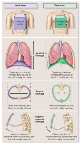
Skeletal muscles of (quiet) breathing
Includes the diaphragm and external intercostals
Inspiration: diaphragm contracts and moves down (V↑, P↓)
Vertical dimensions of thoracic cavity increases
Ribs elevated and thoracic cavity widens
Inferior portion of sternum moves anteriorly and thoracic cavity expands
Expiration: diaphragm relaxes and moves up (V↓, P↑); opposite of above

Boyle’s law
P1V1=P2V2
Inverse relationship between gas pressure and volume at constant temperatures
Air pressure gradient
An air pressure gradient exists when force per unit area is greater in one place than another
Air flows from high to low pressure until pressure is equal
Volumes and pressures with breathing
Respiratory rate for normal, quiet breathing is 12-15 breaths per minute (eupnea)
Diaphragm and external intercostals contract
Alveolar volume of lungs increases → 3. intrapulmonary pressure decreases
Air flows down pressure gradient into alveoli until intrapulmonary pressure = atmospheric pressure (760 mm Hg)

Respiratory center and skeletal muscles of quiet breathing
Autonomic nuclei within the brain coordinate breathing
Respiratory center of the brainstem
Medullary respiratory center: ventral respiratory group (VRG) and dorsal respiratory group (DRG)
Pontine respiratory center (pneumotaxic center) in pons
Brainstem neurons influence respiratory muscles
Medullary respiratory neurons synapse with lower motor neurons of skeletal muscles in spinal cord (cervical)
Lower motor neuron axons project to respiratory muscles
Axons innervating diaphragm travel in phrenic nerves from cervical plexus
Axons innervating external intercostals travel in intercostal nerves from spinal cord (thoracic)

Components of homeostatic system for breathing regulation
Hypothalamus increases breathing rate if body is warm (through respiratory center) - ↑ temp = ↑ metabolic rate = ↑ energy demand
Limbic system alters breathing rate in response to emotions (through respiratory center)
Frontal lobe of cerebral cortex controls voluntary changes in breathing patterns; bypasses respiratory center, stimulating lower motor neurons directly

Homeostatic regulation of breathing steps
Chemoreceptors detect stimuli (changes in blood gases)
Central chemoreceptors: in the medulla oblongata
Peripheral chemoreceptors: on aortic and carotid bodies
Aortic chemoreceptors send signals thru vagus nerves; carotid chemoreceptors send thru glossopharyngeal nerves; central chemoreceptors send directly to MRC
Respiratory center (both pontine and medullary) integrates input
Center alters signals relayed along somatic motor neurons to the skeletal muscles of breathing
Skeletal muscles are stimulated to contract and relax to ↑ respiration rate and depth → more CO2 expelled

Apnea
Absence of breathing
Can occur voluntarily (swallowing, holding your breath)
May be drug-induced
Can result from neurological disease or trauma
Sleep apnea = temporary cessation of breathing during sleep
May be treated with CPAP (continuous positive airway pressure) machine
Factors that influence airflow
F = ∆P ∕ R (flow = pressure gradient between atmospheric and intrapulmonary pressure/resistance)
Skeletal muscles of breathing establish pressure gradient
Resistance determined by:
Bronchiole diameter (bronchoconstriction/-dilation)
Compliance: the ease with which the chest walls and lungs expand + surface tension within alveoli (↓ with pulmonary surfactant secretion)
Stronger muscular force generated → steeper ∆P → F↑ and vice versa
R↓ (bronchodilation and/or increased compliance) → F↑ and vice versa
Tidal volume (TV)
The amount of air taken into or expelled out of lungs during a quiet breath
M/F: 500 mL
Inspiratory reserve volume (IRV)
The amount of air taken into lungs during a forced inspiration, following a quiet inspiration
Measure of lung compliance
M: 3100 mL
F: 1900 mL
Expiratory reserve volume (ERV)
The amount of air expelled from lungs during a forced expiration, following a quiet expiration
Measure of lung and chest wall elasticity
M: 1200 mL
F: 700 mL
Residual volume
The amount of air left (residual) in lungs following a forced expiration
M: 1200 mL
F: 1100 mL
Inspiratory capacity
TV + IRV
Total ability to inspire
M: 3600 mL
F: 2400 mL
Functional residual capacity
ERV + RV
Amount of air typically left (residual) in lungs after you expire quietly
M: 2400 mL
F: 1800 mL
Vital capacity
TV + IRV + ERV
Maximum amount of air that can be forcefully expired after a forced inspiration
M: 4800 mL
F: 3100 mL
Total lung capacity
TV + IRV + ERV + RV
Maximum amount of air the lungs can hold
M: 6000 mL, about 6 L
F: 4200 mL
Gas exchange
Oxygen diffuses across respiratory membrane from alveoli into the capillaries, moving down its partial pressure gradient
Carbon dioxide diffuses from blood to alveoli, moving down its partial pressure gradient
O2 and CO2 exchange until blood levels = alveoli levels
Levels in alveoli remain constant as fresh air continuously enters
(Levels in systemic cells remain constant as cellular respiration occurs)
Partial pressure
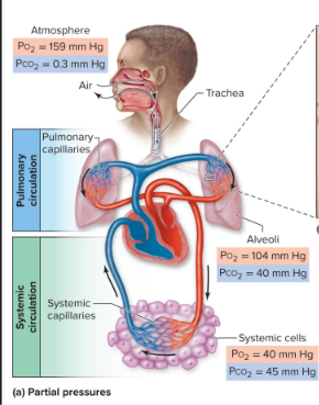
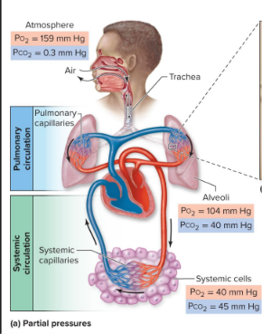
Pressure exerted by a single gas within a mixture of gases
A partial pressure gradient exists when partial pressure for a gas is higher in one region of the respiratory system than another
Gas moves from region of higher partial pressure to region of lower
Alveoli: PO2 = 104 mm Hg, PCO2 = 40 mm Hg
Systemic cells: PO2 = 40 mm Hg, PCO2 = 45 mm Hg
Pulmonary gas exchange
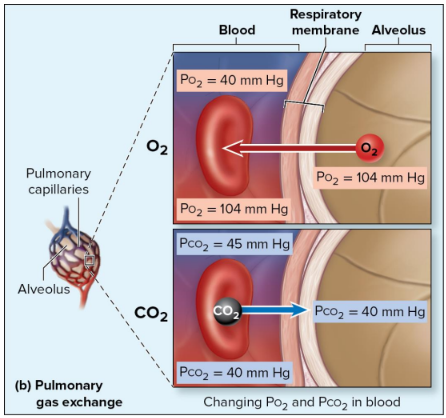
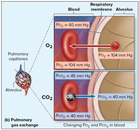
Oxygen moves from alveoli (PO2 = 104 mm Hg) → blood in the pulmonary capillaries (PO2 = 40 mm Hg) until blood PO2 increases to 104 mm Hg
Carbon dioxide moves from blood (PCO2 = 45 mm Hg) → alveoli (PCO2 = 40 mm Hg) until blood PCO2 decreases to 40 mm Hg

Tissue gas exchange
Oxygen moves from blood in systemic capillaries (PO2 = 100 mm Hg) → systemic cell (PO2 = 40 mm Hg) until blood PO2 decreases to 40 mm Hg
Carbon dioxide moves from cell (PCO2 = 45 mm Hg) → blood (PCO2 = 40 mm Hg) until blood PCO2 increases to 45 mm Hg

Pulse oximeter
Noninvasive and indirect way to measure oxygen; applied to finger or earlobe
Measure hemoglobin saturation by determining the ratio of oxyhemoglobin to deoxyhemoglobin
Healthy reading hemoglobin saturation >95%
Uses a light source emitting two wavelengths of light:
Red light (660 nm): Absorbed more by deoxygenated hemoglobin (Hb)
Infrared light (940 nm): Absorbed more by oxygenated hemoglobin (HbO₂)
Photodetector: measures the amount of light that passes through or is reflected by the tissue (e.g., fingertip, earlobe)
Hemoglobin
On the iron of heme groups: oxygen
On the globin portions: carbon dioxide, hydrogen ion
In tissue gas exchange, ↑ temperature in cells → ↑ hemoglobin flexibility → ↑ oxygen release and promoting the binding of carbon dioxide and hydrogen ions → ↓ the hemoglobin’s oxygen affinity
(2,3-BPG is a molecule that binds to hemoglobin to also cause release of additional oxygen)

Conversion of CO2 to HCO3- (systemic)
CO2 diffuses into an RBC
Carbonic anhydrase joins CO2 + H2O → HCO3- and H+
Chloride shift: HCO3- (negatively charged) exits from the erythrocyte; simultaneously, Cl- enters; meanwhile, H+ attaches to hemoglobin


Conversion of CO2 to HCO3- (pulmonary)
O2 enters the RBC, causing hemoglobin to release H+ and CO2 from within the cell
HCO3- moves into the RBC as Cl- moves out
Carbonic anhydrase joins HCO3- + H+ → CO2 and H2O (when joined, this is carbonic acid, or H2O3-)
CO2 diffuses out of the RBC into the plasma and into an alveolus

Percentages of carbon dioxide as different forms
As CO2 dissolved in plasma: 7%
As CO2 attached to amine group of globin portion of hemoglobin: 23% - HbCO2 is carbaminohemoglobin
As bicarbonate dissolved in plasma: 70%
From least to most: dissolved CO2 → on hemoglobin → dissolved bicarbonate
Variables that influence the % saturation of hemoglobin
More oxygen is released from hemoglobin (LOWERS % saturation) as…
Temperature ↑ (normal body temp is 37C)
pH ↓ (normal blood pH is 7.4)
Oxygen-hemoglobin saturation curve
Direct relationship between PO2 and % O2 saturation of hemoglobin
Pulmonary gas exchange (PO2 within the lungs): as altitude ↑, atmospheric oxygen pressure ↓ → alveolar PO2 and % saturation ↓
Tissue gas exchange (PO2 within systemic cells): as physical activity ↑, cells require more oxygen to make energy → tissue PO2 and % saturation ↓
Hyperventilation
Breathing rate or depth above body’s demand
Caused by anxiety, ascending to high altitude, or voluntarily
PO2 ↑ and PCO2 ↓ in the alveolar air
Additional oxygen does not enter blood because hemoglobin is already 98% saturated
There is greater loss of CO2 from blood, called hypocapnia
Hypoventilation
Breathing too slow (bradypnea) or too shallow (hypopnea)
Causes include: airway obstruction, pneumonia, brainstem injury, other respiratory conditions
O2 levels ↓, CO2 levels up in ↑
Blood PO2 decreases (hypoxemia) and can lead to low oxygen in tissues (hypoxia)
Blood PCO2 increases (hypercapnia)