radiographic lung patterns
 Call Kai
 Learn
 Practice Test
 Spaced Repetition
 Match
 Flashcards
 Knowt Play
1/4

There's no tags or description
Looks like no tags are added yet.
5 Terms
alveolar pattern
soft tissue opaque
border effacement of pulmonary vessels, other structures
poor visualization of heart margins
air bronchograms “trees in the fog”
red arrows
± lobar sign (blue arrow)
air in alveoli is replaced by fluid or not replaced (atelectasis)

unstructured interstitial pattern
regional, diffuse, hazy
increased opacity of interstitial tissue but pulmonary vessels still visible
cellular, fluid, or fibrotic change
mimicked by poor rad quality, expiration, obesity

structured interstitial pattern
nodules, masses, miliary
nodule must be >3 mm diameter to be resolved from surrounding lung tissue
can be solid or cavitated (gas-filled)

bronchial pattern
increased visibility of bronchial walls
thickening or mineralization of small airways
seen as “donuts” (cross-section) or “tram lines” (longitudinally)

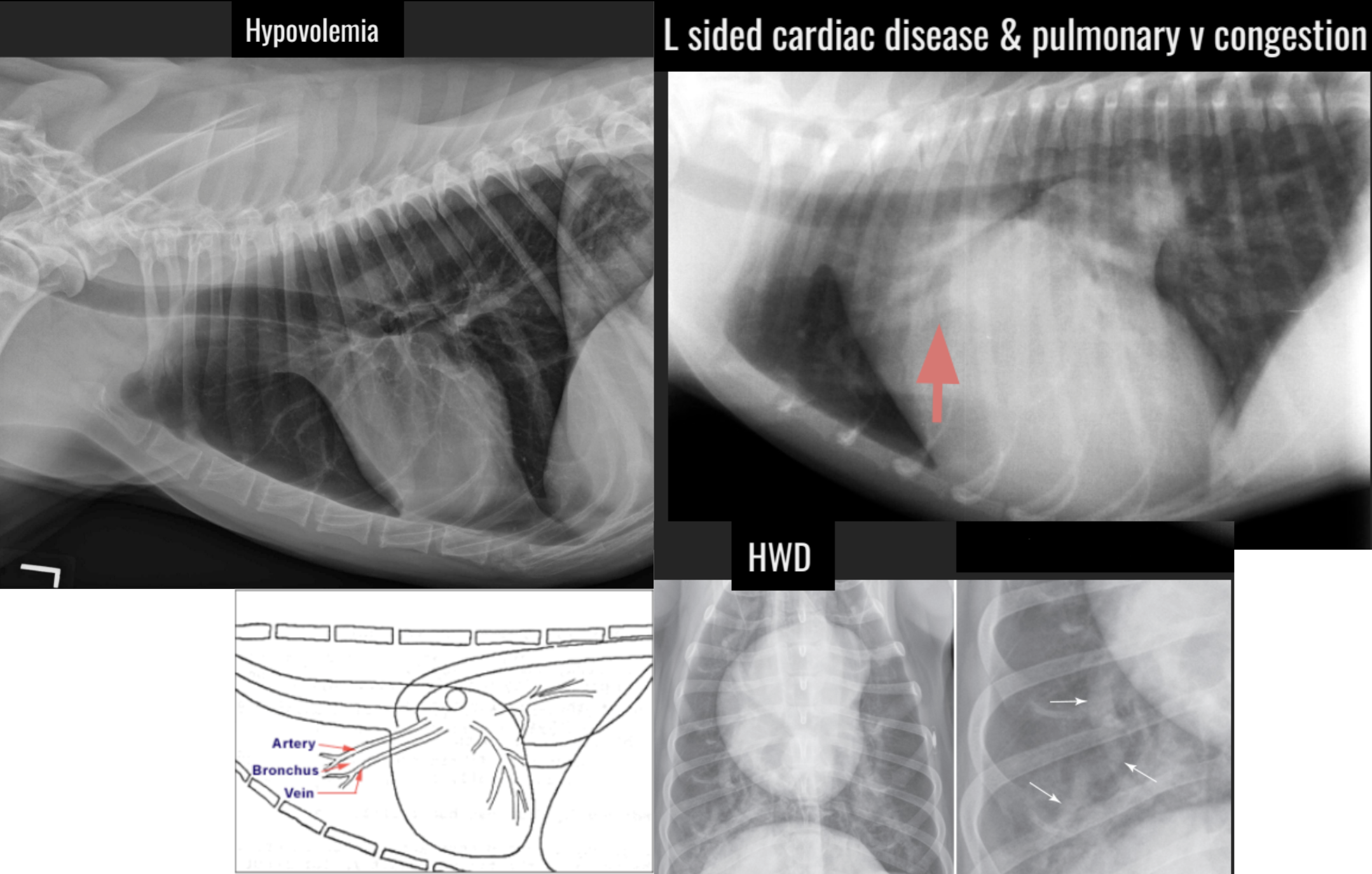
vascular pattern
dilation, narrowing, or altered course of pulmonary vessels
loss of normal tapering
most often associated with cardiac disease
reduced vascular pattern with hypovolemia or shock
may appear as hyperlucent lungs