Eyes - Packet and PowerPoint 4
 Call Kai
 Learn
 Practice Test
 Spaced Repetition
 Match
 Flashcards
 Knowt Play
1/84

There's no tags or description
Looks like no tags are added yet.
85 Terms
Where is bleeding for subconjunctiva hemorrhage?
Between the conjunctiva and the sclera
Where is bleeding with hyphema?
In the anterior chamber between the cornea and the iris of the eye
How are hyphemas characterized?
Pooling of blood in the anterior chamber that may be visible to the naked eye
Why do hyphemas happen?
Blood vessels in the iris bleed and leak into the clear aqueous fluid
What are the typical causes of hyphema?
Blunt, compressive force acting on the globe creates tears in the ciliary body, iris, and other anterior segment structures —> These tears cause shearing of blood vessels and blood leaks into the clear aqueous fluid
Where does blood come from in a hyphema?
Torn or injured blood vessels in the iris
When you are hit with a blunt, compressive force acting on the globe, where does it create tears when dealing with hyphema?
Creates tears in the ciliary body, iris, and other anterior segment structures
What do the tears in the ciliary body, iris, and other anterior segment structures cause when dealing with hyphema?
Shearing of blood vessels and blood leaks into the clear aqueous fluid
How do we manage hyphema?
Restrict movement
Shield to injured eye
Immediate referral to ophthalmology
Where does blood supply to the retina originate?
Carotid artery —> Ophthalmic artery —> Central retinal artery and ciliary arteries —> Retina
What artery is occluded in central retinal artery occlusion?
Central retinal artery
How does the eye get blood in central retinal artery occlusion?
Ciliary arteries (they continue to profuse because they are a different vessel)
What are the typical fundoscopic findings of central retinal artery occlusion?
Pale retina with a cherry red macula (the cherry red spot)
Why do people with central retinal artery occlusion have pale retinas with cherry red maculae?
It’s the result of obstruction of blood flow to the retinal from the central retinal artery, causing pallor, and continued supply of blood to the choroid from the ciliary artery, resulting in a bright red coloration at the thinnest part of the retina (the macula)
What are the symptoms of central retinal artery occlusion?
Painless
Sudden, unilateral blindness
What may be the cause of the occlusion in central retinal artery occlusion?
Embolism (disseminated atherosclerotic plaques, endocarditis, atrial fibrillation)
Thrombosis in a sclerotic central artery
Is thrombosis stationary or moving?
Stationary
Is embolism a piece of a thrombosis getting stuck in an artery?
Yes
What are the physical exam findings of central retinal artery occlusion?
Pale
Opaque fundus with red fovea (cherry-red spot)
What is the treatment for central retinal artery occlusion?
Reperfusion of the eye
Why is it important to treat central retinal artery occlusion?
If not relieved quickly, retinal infarction occurs and blindness is permanent
CRAO stands for what when dealing with eyes?
Central retinal artery occlusion
What happens in retinal detachment?
Retina peels away from underlying layer of support tissue becoming lifted or pulled from its normal position
What is the retina?
A thin disc-shaped layer of light-sensitive tissue on the back wall of the eye
What does the retina do?
Translates what we see in neural impulses and sends them to the brain via the optic nerve
What is a typical cause of retinal detachment?
Posterior vitreous separation (Decrease of fluid pressure buildup in older people (less fluid pressure = retina not being pushed against the wall))
How does retinal detachment progress without intervention?
Initial detachment (may be localized) —> Retina continues to detach (do to no treatment )—> Vision loss and blindness
Is retinal detachment a medical emergency?
Yes it can cause blindness and vision loss without rapid treatment
What is a rhegmatogenous retinal detachment?
A tear that leads to fluid getting behind the retina, leading to it peeling away from the eye
What does rhegma mean?
Greek word for tear
What is the disorder that involves a tear, hole, or break in the retinal that allows fluid to pass into the subretinal space (between the sensory retina and retinal pigment epithelium) leading to the peeling away of the retina from the back wall of the eye?
Rhegmatogenous retinal detachment
In rhegmatogenous retinal detachment what happens when the continuity between the sensory retina and the retinal pigment epithelium is disrupted?
Neurosensory retina becomes ischemic (photoreceptors degenerate, resulting in vision loss)
What are most case of rhegmatogenous retinal detachment associated with?
Posterior vitreous separation (a natural part of aging)
Why does posterior vitreous separation happen (the cause of most cases of rhegmatogenous retinal detachment)?
Natural part of aging - Due to the natural contraction, or shrinkage, of vitreous humor, which creates traction on the retina producing a retinal break
What is vitreous humor?
Gel-like substance that makes up 80% of the ocular globe volume
What age is it normal for the vitreous humor to begin to liquefy and shrink?
45-55 years of age
What may patients experience that are going through posterior vitreous separation?
Briefly see floaters and even flashing lights in their vision (usually subside and may leave small residual floater in the vision)
How often does PVD (posterior vitreous separation) cause a retinal tear or hole?
Rarely, but still is the cause of most cases of rhegmatogenous retinal detachment
What are the risk factors for retinal detachment?
Severe myopia
Cataract surgery
Trauma
Aging
What are the symptoms of retinal detachment?
The 3 f’s
Light flashes in the eye (Photopsia)
Sudden/Gradual increase of floaters
Field loss (vision loss)
What are floaters?
Shadows cast from particulate matter suspended in the vitreous over the retina (float in a person’s vision)
What is the medical term for light flashes in the eye?
Photospia
Why do we have photopsia (light flashes in the eye) with retinal detachment)?
Flashes represent retinal stimulation from vitreous traction
Do you suffer from peripheral or central vision loss with retinal detachment?
It depends on what part of the retina is off or torn
What kind of vision loss has the worst prognosis when dealing with retinal detachment?
Central vision because it represents detachment at the area of the macula
What is the treatment for retinal detachment?
Refer to ophthalmologist
Pneumatic retinopexy
Laser surgery
Cryopexy
If a patient comes in and reports sudden flashes, floaters, and field loss what should you do?
Refer emergently to an ophthalmologist because retinal detachment requires emergency surgery to prevent its spread into the fovea region
Why is early diagnosis and treatment essential for retinal detachment?
Longer duration of retinal detachment the worse the prognosis
What does pneumatic retinopexy involve?
Putting an air bubble in the eye to push the retina back and having the patient have his face down for most of the day for 3 weeks
What is the problem with pneumatic retinopexy?
The patient needs to keep their head down all day to make sure the air stays in for a few weeks
And it might not even work
How do we treat the small holes and tears of retinal detachment?
Laser therapy
Cryopexy
What does laser therapy involve with retinal detachment?
Tiny burns are made around the hole to weld the retina back into place
What does cryopexy involve with retinal detachment?
Freezing the area around the hole to help reattach the retina
What is a retinal hemorrhage?
Bleeding or hemorrhage in the retina
What is bleeding in a retinal hemorrhage?
Blood vessels in the retina leak or rupture
What is it when their is bleeding in the eye?
Intraocular bleeding
What are the common causes of a retinal hemorrhage?
Diabetes
Rupture of neovascularization vessels
High blood pressure
Blood disorders
Hemophilia
von Willebrand disease
Cardiovascular problems
Trauma
Aging
What can make a retinal hemorrhage harder to control?
Less controlled diabetes and hypertension
What are the symptoms of retinal hemorrhage?
Mild
Floaters or dark lines in vision
Advanced
Sudden, painless vision loss
Ability to have only perception of light due to obscuring by the blood
What is found in a physical exam of retinal hemorrhage?
Blood in the vitreous humor
What is the treatment for retinal hemorrhage?
Nothing (eye will suck up the blood)
Our challenge is to trying to find out why and how to stop it from reoccurring

What disease is this?
Subconjunctival hemorrhage

What disease is this?
Hyphema

Where is the blood?
In the anterior chamber between the cornea and the iris

Where is the blood?
Between the conjunctiva and the sclera

What disease is this?
Hyphema

What disease is this?
Hyphema

What disease is this?
It normal

Which eye is this?
Left eye

What eye is this
Right eye

What disease is this?
It’s normal

What eye is this?
Left eye

What disorder causes this?
Central retinal artery occlusion

What can be visibly seen (describe the characteristics that make you think that it is the disorder that it is)?
Pale fundus
Cherry-red spot

What disorder is this?
Central retinal artery occlusion

What disorder is this?
Central retinal artery occlusion
An 84-year-old man with recent onset of headache and fatigue presents to the emergency department with sudden vision loss. Examination reveals a visual acuity of "light perception only" in the right eye and 20/20 in the left eye. An afferent pupillary defect (Marcus Gunn pupil) is seen in the right eye. Examination by direct ophthalmoscope reveals a diffusely appearing pale retina except for a bright red spot in the fovea (Figure 1). What is the most likely diagnosis?
Central retinal artery occlusion
A 65-year-old Asian woman awakens in the middle of the night with severe left eye pain associated with nausea and vomiting. On presentation to the emergency department, visual acuity is noted to be "counting fingers at 1 foot" in the left eye and 20/40 in the right eye. The left eye is very injected and the cornea appears hazy. The left pupil shows minimal reaction to light and appears slightly dilated. The right eye is completely normal. What is the most likely diagnosis?
Closed-angle glaucoma

What disorder is this?
Retinal detachment
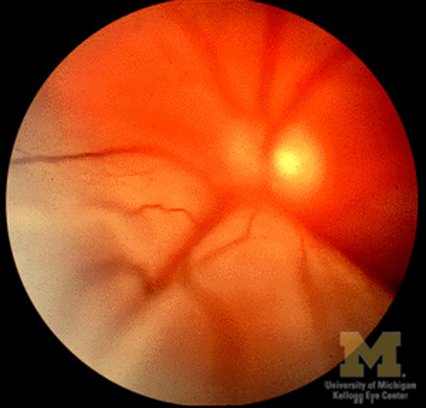
What disorder is this and where is it?
Retinal detachment above

What treatment do we use these for?
Pneumatic retinopexy
What are 2 disorder that can significantly increase your chance of bleeding in the eye?
Diabetes and hypertension (1+1=4)

What risk factors could have caused this?
Diabetes
High blood pressure
Blood disorders (hemophilia and von Willebrand disease)
Trauma
Aging
Anticoagulants can also be a cause
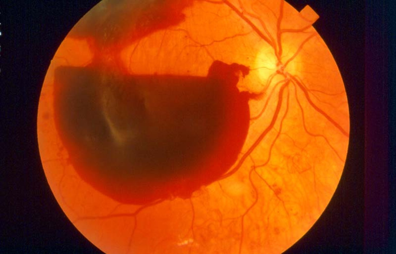
What disorder is this?
Retinal hemorrhage
What can diabetes cause that leads to retinal hemorrhage?
Rupture of neovascularization vessels