Liver
 Call Kai
 Learn
Learn Practice Test
 Spaced Repetition
 Match
Match Flashcards
 Knowt Play
1/109

There's no tags or description
Looks like no tags are added yet.
Name | Mastery | Learn | Test | Matching | Spaced | Call with Kai | Chat |
|---|
No analytics yet
Send a link to your students to track their progress
110 Terms
Provide definitions for the following terms
Term | Definition |
Liver disease | |
Liver failure (aka. hepatic insufficiency) | |
Acute liver failure/disease
| |
Chronic liver failure/disease
| |
Cirrhosis (end-stage) liver
| |
Cholestasis | |
Hepatitis | |
Cholangitis | |
Cholangiohepatitis | |
Cholecystitis |
Term | Definition |
Liver disease | Very BROAD term describing ANY disease process affecting the liver
|
Liver failure (aka. hepatic insufficiency) | Severe liver disease where >70 - 80% of functional liver is lost USUALLY associated with clinical signs and changes in bloods |
Acute liver failure/disease
| Hepatic dysfunction develops RAPIDLY (usually without pre-existing liver disease) Gross appearance: Liver looks NORMAL (no time for remodelling and changes) |
Chronic liver failure/disease
| Hepatic dysfunction develops SLOWLY over months/years due to progression of liver disease or damage Gross appearance: Small, fibrotic and firm |
Cirrhosis (end-stage) liver
| Chronic and irreversible result of progressive liver disease → Diffuse hepatic fibrosis Gross appearance: Bands of small, pale, firm liver (fibrosis) with interspersed nodules of regenerating liver parenchyma |
Cholestasis | Reduced bile secretion and flow → Accumulation of bilirubin and bile acids in blood
|
Hepatitis | Inflammation of the liver parenchyma (acute OR chronic) |
Cholangitis | Inflammation of the biliary tract |
Cholangiohepatitis | Inflammation centred on the biliary tract with extends to involve the adjacent hepatic parenchyma |
Cholecystitis | Inflammation of the gallbladder |
Which sheep has acute liver disease? Which has chronic liver disease?

Flossie = Acute liver disease
Bronzer and swollen
± More friable
Jezebel = Chronic liver disease
Small, pale and friable
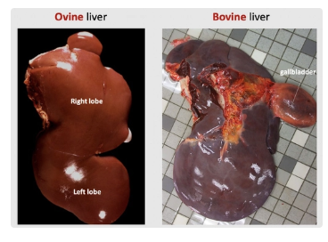
4 Macroscopic features of a NORMAL liver
Smooth outer capsule
Dark red-brown parenchyma divided into lobes (varies between species)
Ruminant = Left (ventral) and right (dorsal)
Gallbladder protrudes from RIGHT lobe
Camelid = Lobe fimbriation (frilly edges)
Sharp (tapering) lobe edges
Round margins = Swelling
Firm-ish texture (can place finger through)

4 Species that lack a gallbladder
“HARD”
Horse
Alpaca → Continuous secretion of bile
Deer
Rats (not mice)
2 Gallbladder features to assess in PM
Ensure patency
Oedema around gallbladder = Shock
Microscopic Structure of the Hepatic Lobule
Hepatocytes
Sinusoids
Kupffer cells
Bile flow
Hepatocytes: Long-lived epithelial cells making up most of the parenchyma
Arranged in plates (1 cell thick) separated by sinusoids
Metabolically active (eg. bile production)
Good regeneration
Sinusoids: Fenestrated liver capillaries through which blood flows and separate hepatocyte plates
= Blood from portal vein and hepatic artery (infarction difficult due to duel blood supply)
Kupffer Cells: Liver-resident macrophages within sinusoids which phagocytose aged/damaged RBCs and pathogens
Bile: Produced by hepatocytes → Bile canaliculi → Larger bile ducts → Gallbladder OR directly into the duodenum via the bile duct
Gallbladder = Concentrates and stored bile
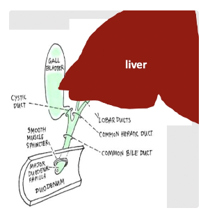
Hepatic Lobule
Structure
Portal area
Central vein
3 Zones of hepatocytes (+ hypoxia)
Toxin exposure
Structure: Each composed of thousands of hepatocytes arranged into a rough hexagon and separated by other lobules with connective tissue
Grossly observable in pigs
Portal Area: Each corner of the hexagonal hepatic lobule makes up a portal area which contains branches of:
Hepatic artery*
Portal vein*
Bile ducts*
Lymphatic vessels
Central Vein: Branch of the hepatic vein located at the centre of each hexagonal liver lobule which drains blood
Zones:
Periportal hepatocytes
Midzonal hepatocytes
Centrilobular hepatocytes (around central vein)
Contain enzymes involved in drug metabolism
Most susceptible to hypoxia as they receive blood (O2) last (periportal zones receive oxygenated blood first)
Toxins: DIFFERENT regions susceptible depending on the specific toxin
Although periportal area receives toxin first (eg. from GIT), different parts of the lobule have different enzymes for toxin metabolism
Blood flow vs. bile flow in the hepatic lobule
Blood flow = Within hepatic sinusoids from the outer portal areas INWARDS towards the central vein
Bile flow = Moves from the centrilobular regions OUTWARDS towards the portal areas

Describe FIVE functions of the liver
Bile and bilirubin formation, secretion and metabolism
Bile = Water + cholesterol + bile acids + degraded waste products (eg. bilirubin)
Functions:
Lipid digestion in intestine (bile acids) → Resorption and recycling
Waste and toxin excretion (bilirubin)
Carbohydrate, lipid and vitamin metabolism
Synthesis of glucose, cholesterol, vitA, B12, D and K
Protein synthesis and secretion
Albumin
Coagulation factors for haemostasis (FII, VII, IX and X)
Ammonia released from protein breakdown → Urea
Detoxification and "safe" body storage = Liver's position and bloody supply allow it to detoxify/modify substances absorbed from the GIT before they enter systemic circulation
Excretion of waste products, drugs and toxins in bile OR urine (liver makes insoluble toxins soluble → allows elimination from circulation by kidneys in urine)
Storage of substances (eg. iron and copper)
Immunity
Production of acute phase proteins
Hepatic location of cells of adaptive/innate immune systems (eg. Kupffer cells and lymphocytes)
Supply and drainage of liver blood supply
Liver receives blood from TWO main sources:
Portal vein = Drains GIT and provides 75% of the blood that the liver receives (nutrient-rich and O2-poor)
Hepatic artery = Provides remainder of blood to liver (O2-rich) Hepatic vein = Vessel which drains the liver of blood → Caudal vena cava → Heart

List 3 long-term (chronic) responses of the liver to injury
Regeneration
Fibrosis
Biliary hyperplasia
What is this?
Growth response
Indication
Gross appearance
Outcome


Regeneration
Growth Response: Hyperplasia (increased number of cells)
Cause: Mild/short-lived hepatic injury will resolve quickly through regeneration assuming sufficient ECM is intact
Gross Appearance: Nodules of new liver as surviving hepatocyte form islands to produce more (mistaken for neoplastic masses)
Outcome: Up to 70% of liver can be surgically removed without serious consequences → Return to normal size and function within a few weeks
What is this?
Cause
Gross appearance
Outcome

Liver Fibrosis
Cause: Chronic/severe injury → Hepatic necrosis
Gross appearance: Affected parts of liver are firmer, paler and smaller than normal (distribution affects nature of causative injury)
Outcome: Fibrosis (collagen) replaces functional hepatobiliary function
Biliary Hyperplasia
Definition
3 Causes
Gross appearance
Definition: Injury to bile ducts → Proliferation of bile ductules in portal triads ± fibrosis
Causes:
Obstruction
Severe hepatocyte injury
Portal inflammation
Gross appearance: NONE! (only microscopic)
3 Potential outcomes of liver injury + causes
Resolution = With regeneration of damage liver and return to normal function
Irreversible progression (end-stage liver/cirrhosis) = Loss of normal lobular architecture due to replacement by extensive fibrosis ± nodular regenerations
In-between = Part of liver is severely damaged → Fibrosis not regeneration BUT other parts unaffected → Regeneration


When are clinical signs of hepatobiliary damage evident?
Liver has large functional reserve → Clinical signs only apparent when damage is severe and advanced (heading towards liver failure) OR there is cholestasis
5 Clinical signs of mild liver disease
Clinically silent OR mild, intermittent or non-specific signs
Anorexia
Weight loss
Lethargy
Vomiting
Diarrhoea Similar clinical signs as other body systems (eg. renal and GIT)
4 Additional tests to confirm suspicion of hepatobiliary disease due to mild/non-specific clinical signs)
Bloods (CBC and biochemistry ± urinalysis)
Imaging of liver and associated structures (eg. U/S for liver margins, cannot see parenchyma well)
Sampling liver (FNA for cytology or biopsy for histology)
± Exploratory laparotomy
Describe FOUR clinical signs of advanced liver failure (>70 -80% loss of liver function)
Pathogenesis
Clinical signs
Oedema
MoA: Failing liver cannot produce enough albumin to maintain COP → Hypoalbuminaemia → Fluid leakage from vessels Ascites #1
Clinical Signs: Ascites #1
Neurological Signs = Hepatic Encephalopathy
MoA: Ammonia produced from protein breakdown → Failing liver cannot convert ammonia and other toxins from the blood into non-toxic metabolites (eg. urea) → Toxins remain in blood and cross BBB → Neurotoxic
Clinical Signs: Altered behaviour, depression, head-pressing, pacing, aimless wandering, ataxia ± seizure
Increased Bleeding
MoA: Failing liver cannot produce enough coagulation factors required to achieve haemostasis → Haemorrhage
Clinical Signs: Pale MM, haemothorax, haemothorax, joint haematoma, prolonged bleeding from wounds
Photosensitisation in Production Animals
Ascites caused by FIP vs. liver disease
Although liver disease is associated with FIP, there is not liver failure
Ascites is due to increased vascular permeability (inflammation) NOT liver failure
Leakage vs. Induction Liver Enzymes
3 Examples
Function
Duration of formation
Leakage Enzymes = Leak into blood when hepatocytes are damaged/die
ALT = Alanine aminotransferase
AST = Aspartate aminotransferase
GLDH = Glutamate dehydrogenase
Function: Indicate hepatocellular damage
Duration: RAPID increase after event (hours)
Induction Enzymes = Synthesis increases (induced) in response to damage/stimuli due to bile retention (or various drugs)
ALP = Alkaline phosphatase
GGT = Gamma glutamyltransferase
Function: Indicate cholestasis
Duration: Longer to increase after cholestatic event (>1d) as enzymes must be induced/synthesised
HALT HaMSTers with SCALPelS
HALT = Hepatocellular ALT
HaMSTers = Hepatocellular and Muscle (A)ST (also used in equine and ruminants)
SCALPelS = Steroids, Cholestasis, ALP elevates (+ Skeletal)
GLDH = L and H for liver/hepatocellular
GGT = CCC
4 Disadvantages of serum liver enzyme tests
NOT specific for liver damage/cholestasis (may increase when there is nothing to do with the liver i.e 2˚)
Species variation in liver specificity of enzyme increase and serum half-lives
Do NOT indicate prognosis (i.e. if liver damage is reversible or not)
Do NOT indicate how much functional liver tissue is present still (i.e. do NOT test for liver function)
Leakage Enzymes = ALT, AST, GLDH
Function
Lifespan
Disadvantage
DDx for elevation
ALT
Function: Liver-specific leakage enzyme reflecting acute hepatocellular injury/death in cats and dogs
NOT useful in large animals
Lifespan: Increases within 12hr of SINGLE liver injury → Peaks 1 - 2d post-injury → Returns to RR within 1 - 3w
Ongoing injury → Persistent marked ALT increase
-ve: None (different DDx for liver injury)
DDx:
Marked increase (>10x RR) → 1˚ Liver disease
Variable increase → Endocrinopathy, pancreatitis, anaemia/dehydration/cardiac disease, drugs, hepatic neoplasia (extensive)
No change/mild increase → Chronic liver failure OR incidental in older animals
AST
Function: NON-liver specific leakage enzyme used as an indicator of hepatocellular OR muscle injury/necrosis in large animals (also dogs and cats)
Lifespan: Half-life shorter than ALT and GLDH (returns to normal faster after liver injury)
-ve: Requires CK and PCV to exclude non-hepatic causes of AST increase (i.e. muscle damage or haemolysis respectively)
DDx:
Liver damage
Cardiac/skeletal muscle damage
Interpret with clinical signs as AST has longer half-life than CK (may have normal CK but elevated AST with muscle injury → Days since injury)
Haemolysis
GLDH
Function: Liver-specific leakage enzyme used in large animals as an indicator of acute hepatocellular injury/death
Large animal version of ALT in small animals
SDH in USA
Creatinine vs. creatine kinase (CK)
Creatinine
Creatinine produced at constant rate by muscles and excreted by kidneys
Less muscle mass = Less creatinine
Increases as part of azotaemia
Does NOT indicate muscle necrosis
Creatine Kinase
Large enzymes in muscle cells → Requires damage to cell membrane for leakage into serum
Does NOT change with weight loss/ill-thrift as muscle breakdown for energy is orderly process without cell membrane damage
Induction Enzymes = ALP and GGT
Function
Lifespan
Disadvantage
DDx for elevation
ALP
Function: NON-liver specific induction enzyme used in dogs and cats to indicate cholestasis
NOT useful in large animals
Lifespan: Takes 1 - 2d to increase post-damage
Dogs = Long half-life ~3d → Peaks 3 - 4w after and returns to normal within months
Cats = Less sensitive and shorter half-life ~6hr
Any increase is usually significant in cats
-ve: Complex interpretation (esp. dogs) due to many different causes of increased ALP which is typically not specific to the isoform on routine biochemistry
Tests to separate the different isoforms are not routinely available in NZ
DDx:
Hepatic (cholestasis) isoform
Cholestasis (eg. cholangitis and cholangiohepatitis)
Feline hyperthyroidism (hepatic and bone ALP isoform + ALT)
Diabetes mellitus
Liver disease causing hepatocellular swelling → Increased ALT and ALP (due to 2˚ biliary obstruction)
Bone isoform
Normal young and rapidly growing animals → Increased osteoblast activity associated with bone growth) + mild increase in calcium and phosphorus
Bone diseases → Increased osteoblastic/osteoclastic activity eg. osteosarcoma, healing fractures and fibrous osteodystrophy
Corticosteroid and other drug-induced ALP isoforms
Cushing's disease
Chronic stress
Iatrogenic
Various anti-seizure and anaesthetic drugs → Marked ALP elevations in dogs 12 - 24hr after
GGT
Function: Liver-specific induction enzyme previously used in large animals over ALP
Increasing use in cats and dogs in addition to ALP
Cats = GGT used in combination with ALP to give more information about likely disease process
Increased GGT in cats more specific for cholangitis and ALP more specific for hepatic lipidosis
DDx: 3 C's
Cholestasis
SA: Cholangitis/cholangiohepatitis
LA: Recent FE, fascioliasis, ragwort toxicity
Corticosteroids
Colostrum intake in neonates (except horses and cats)
Hepatic ALP vs. bilirubin in dogs vs. cats
Dogs: Hepatic ALP increases BEFORE increased bilirubin
Cats: Hepatic ALP increases AFTER increased bilirubin
Hepatic ALP has much shorter half-life in cats (6hr vs. 3 days in dogs)
Any hepatic ALP increase in cats is significant
Liver Function Tests
Function
2 Types (+ examples)
Function: Evaluate liver's capacity to perform functions unique to liver (vs. enzyme tests)
Liver function can be abnormal despite maintenance of hepatocyte membranes (i.e. normal liver enzyme activity)
Types:
Substances in Serum REMOVED by Liver
Bilirubin
Bile acids
Ammonia → Increased with liver dysfunction
Substances in Serum MADE by Liver
Albumin
Urea
Cholesterol*
Glucose*
Coagulation factors
*Unreliable decrease
Are all hyperbilirubinaemia cases jaundiced?
NO >50µmol/L required for clinical signs
Describe FIVE Mechanisms of Hyperbilirubinaemia
Signalment
Pathogenesis
Magnitude
DDx
Fasting
Signalment: Horses
Smaller increases in cattle, pigs and cats (not dogs)
Pathogenesis: Fat mobilisation interferes with bilirubin uptake by hepatocytes → Increase in unconjugated bilirubin
Magnitude: Mild
DDx: Anorexia
Pre-hepatic (Haemolytic) Hyperbilirubinaemia
Signalment: ALL species (#1 in ruminants)
Pathogenesis: Acute haemolytic disease → Rapid Hb breakdown → Rate of bilirubin formation > hepatocyte capacity to conjugate and excrete bilirubin
Magnitude: Marked
DDx: IMHA, Leptospirosis in calves/lambs, Theileriosis, Cu/Zn toxicity
Hepatic Hyperbilirubinaemia
Signalment: ALL species
Pathogenesis: Extensive acute/chronic liver damage → Decreased functional hepatic mass → Impaired uptake and conjunction of bilirubin
Magnitude: Mild
DDx: FIP, liver failure
Post-hepatic (Cholestatic) Hyperbilirubinaemia
Signalment: ALL species
Pathogenesis: Cholestasis → Regurgitation of bilirubin into circulation
Magnitude: Marked
DDx:
Extra-hepatic: Obstruction in large bile ducts
Intra-hepatic: Obstruction in bile canaliculi between hepatocytes (hepatocellular swelling)
Sepsis-Associated
Signalment: Dog and cat
Pathogenesis: Increased inflammatory mediators (esp. endotoxin) → Fewer transport proteins for bilirubin → Decreased bilirubin excretion
DDx: E. coli pneumonia, peritonitis, endocarditis, FIP
Bilirubinuria
Conjugated bilirubin is water-soluble → Freely filtered by glomeruli into urine (detected grossly or with dipstick)
Fill out the following table
RBCs | WBCs | Leakage enzymes | Induction enzymes | Other signs | |
|---|---|---|---|---|---|
Fasting | |||||
Haemolytic | |||||
Hepatic | |||||
Cholestasis | |||||
Sepsis |
RBCs | WBCs | Leakage enzymes | Induction enzymes | Other signs | |
|---|---|---|---|---|---|
Fasting | WNL | WNL | WNL | WNL | Follows anorexia and variable jaundice |
Haemolytic | Regenerative/pre-regenerative anaemia | Variable (WNL/stress/inflammation) | WNL (or mild increase if hypoxia) | WNL (or mild increase with hypoxia) | Jaundice ± Other signs of haemolysis |
Hepatic | WNL (or non-regenerative if chronic) | Variable (WNL/stress/inflammation) | Often increased (can be WNL if chronic) | WNL (or increased if also cholestasis) | Variable |
Cholestasis | WNL (or non-regenerative anaemia if chronic) | Variable (WNL/stress/inflammation) | WNL (or increased if hepatocyte damage too) | Increased | ± Bilirubinuria |
Sepsis | WNL or relative erythrocytosis (dehydration) | Inflammatory leukogram | WNL (unless liver involved) | WNL unless liver involved | Very sick animal ± pyrexia |
Bile Acids
Normal production and recycling
2 Mechanisms of increased serum bile acids (+ DDx)
Normal:
Bile acid synthesised and conjugated in liver from cholesterol
→ Excreted in bile
→ 95% resorbed in intestine
→ Enterohepatic recirculation into portal vein
→ Liver for removal from blood and recycling to make more bile
Increased BA:
Decreased clearance of BA from portal blood
Moderate/severe reduction in functional liver mass
Portosystemic shunts (blood and bile acid bypass liver and end up in peripheral blood)
Decreased excretion of bile acids = Cholestasis → Returns bile acids to serum due to regurgitation into peripheral blood

Measuring Bile Acids
Function
3 Disadvantages
Sampling requirements
Interpretation of results
Function: Increased serum bile acids indicate liver disease and decreased hepatobiliary function
Most useful when liver disease is suspected but routine biochemistry is equivocal eg.
Increased ALP but normal bilirubin
Increased bilirubin but normal ALP
Normal liver enzymes but other liver signs (eg. hepatic encephalopathy)
-ve:
NOT useful with jaundiced animals if have already ruled out non-hepatobiliary causes (i.e. haemolytic and sepsis-associated hyperbilirubinaemia ruled out)
Does NOT indicate cause or severity of disease (requires further testing eg. U/S or biopsy)
NOT routine and must be specifically requested (red top tube)
Sampling: TWO serum samples → Higher Sn and Sp
1st sample (baseline pre-prandial) collected 12hr post-fasting
2nd sample (post-prandial) collected 2hr after protein- and fat-containing meal
Interpretation:
Normal liver = Pre-prandial → LOW bile acids and post-prandial → MILD increase in bile acids
Damaged liver/PSS/cholestasis = Pre-prandial → Increased bile acids and post-prandial → MARKED increase in bile acids
Hypoalbuminaemia with Liver Failure
Pathogenesis
Chronicity
Exception
2 Other laboratory features
Pathogenesis: Liver failure/hepatic insufficiency → Liver cannot make enough albumin → Hypoalbuminaemia
Chronicity: Because albumin has a long half-life (7 - 10d) hypoalbuminaemia ONLY occurs with chronic or chronic-active liver failure as must turnover existing albumin in blood
Exception: Horses (may be seen with acute hepatic disease instead)
Other Features:
Decreased A:G ratio (hyperglobulinaemia)
Portal blood from gut to liver brings in Ag from intestine → Not processed by liver → More Ag in systemic circulation → More Ag presentation in systemic circulation → Hyperglobulinaemia
Hypocalcaemia 2˚ to hypoalbuminaemia
No clinical signs (does not affect ionised calcium which controls membrane potentials)
Urea in Liver Failure
Normal production
Pathogenesis
Other laboratory feature
Normal:
Toxic NH3 produced by breakdown of dietary protein/amino acids by GIT bacteria →
Absorbed into portal circulation →
Removed by liver through conversion to less toxic urea →
Urea excreted into systemic circulation →
Kidney removes urea in urine
Pathogenesis: Impaired hepatic function → Liver cannot convert ammonia to urea → Increased blood ammonia + decreased blood urea
Ammonia difficult to measure
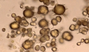
Other Feature: Ammonium biurate crystals = Brown-yellow spherical bodies with irregular protrusions (aka. thorn-apples) in urine
Esp. with portosystemic shunts
Normal in Dalmatians and bulldogs
Cholesterol in Liver Failure
Normal production
Pathogenesis
5 DDx for hypercholesterolaemia
Disadvantage
Normal: Synthesised in liver → Converted to bile acids → Excreted in bile
Pathogenesis: Impaired hepatic function → Decreased cholesterol synthesis → Hypocholesterolaemia
DDx:
Cholestasis
Post-prandial (use fasting sample)
Altered fat metabolism (eg. pancreatitis)
Nephrotic syndrome
Endocrine diseases (hypothyroidism, diabetes and Cushing's)
-ve: VARIABLE hypocholesterolaemia
Biliary excretion is ALSO reduced with liver failure → Normal cholesterol may be seen (± hypercholesterolaemia) depending on balance between synthesis and output of cholesterol
Hypocholesterolaemia is supportive evidence of liver failure but does NOT change diagnostic process if normal
Main disadvantage of glucose testing for liver failure
VARIABLE glucose levels (hypoglycaemic/hyperglycaemic/normoglycaemic)
Hypoglycaemia due to decreased hepatic gluconeogenesis and glycogen breakdown
Hyperglycaemia due to decreased glucose uptake by liver post-eating → Interpret in context of case
Effect of liver failure on coagulation factors
Significant hepatic dysfunction → Decreased production of clotting factors → Defective 2˚ haemostasis pathway → ± Unexpected bleeding
Clinical pathology findings of liver failure are similar to what haemostasis defect? How to distinguish?
VitK antagonism (i.e. rat bait toxicity)
Distinguish with history as liver failure = CHRONIC non-specific findings vs. rat bait = 3d onset post-toxin ingestion
EXAMPLE: “FITZ”
Signalment: 7yr Flat Coat Retriever dog
History: Weight loss over several months and occasional “weird episodes” with disorientation and restlessness
PE: BCS 2/9

Interpretation:
RBCs: Mild normocytic (normal MCV) and normochromic (normal MCHC) anaemia (low Hb, HCT and RBC). Given that MCV and MCHC are normal (and reticulocytes have not increased), the bone marrow has yet to respond to the anaemia with regeneration. This may be a pre-regenerative OR non-regenerative response. Given the chronic history of weight loss over several months, I am leaning more towards a non-regenerative response due to chronic illness. Furthermore, there is no evidence of haemolysis or haemorrhage in the history to indicate a pre-regenerative response. We can definitively rule out a pre-regenerative anaemia with blood sample in 3 - 5 days.
WBCs: Mild eosinopenia. Given the mild nature, this is likely an insignificant finding.
Calcium: Moderate hypocalcaemia likely due to the moderate hypoalbuminaemia. Because ~40% of calcium is bound to albumin, any decrease in albumin results in a decrease in serum calcium.
Mild increase in ALP: Hepatic isoform increase most likely but significance not certain (cholestasis vs. mild and insignificant increase)
DDx:
Insignificant → May be insignificant as close to RR
Cholestasis
Steroid isoform → No history of steroid administration and no stress leukogram
Bone isoform → Ruled out on history as not a young and growing animal AND no evidence of lameness indicating bone pathology
Mild decrease in urea: Given the non-specific clinical signs, low urea may be due to liver disease and reduced liver functional mass → Reduction in production in liver. The liver cannot take up NH3 for conversion into urea, resulting in mild reduced urea. NOT impaired renal function as typically urea is high.
MAY indicate starvation, but not consistent with history
Proteins: Moderate hypoalbuminaemia with mild hyperglobulinaemia (with low A:G ratio). This evidence supports liver disease and dysfunction. The liver cannot synthesise albumin as normal resulting in a hypoalbuminaemia and concurrent hyperglobulinaemia (may be due to reduced Ag processing in the liver resulting in increased Ag presentation in systemic circulation).
Unlikely blood loss or decreased protein intake, protein-losing nephropathy or enteropathy as not indicated in history and would expect concurrent hypoglobulinaemia
Hyperglobulinaemia due to inflammation/antigenic stimulation
Neoplasia → Spinal pain associated with myeloma
Dehydration → Would expect concurrent increase in creatinine (pre-renal azotaemia)
NORMAL leakage enzymes → Chronic liver damage = Fewer hepatocytes secreting enzymes
DDx: Evidence of significant liver disease (likely failure) and dysfunction
Canine chronic hepatitis
Diagnostic Steps:
Bile acids due to equivocal results → Confirms liver disease and reduced hepatobiliary function
High pre- and post-prandial bile acids → Consistent with liver disease
Abdominal U/S ± liver sampling
3 Disadvantages of liver biopsy
Cost and time (~3 days for results)
Risk to patient (GA required in sick animals)
Haemorrhage (esp. with depleted coagulation factors as with liver failure)
What is this?
Signalment
Gross appearance
Histology
Significance


Nodular Hyperplasia (5)
Signalment: Older animals (esp. dogs)
Appearance: Single/multiple firm, spherical nodules projecting from liver surface and extending into the parenchyma
Often slightly paler than rest of liver (accumulate fat and glycogen) → Mistaken for neoplasia
Histology: Normal
Significance: NONE
What is this?
Signalment
Gross appearance
Significance
2 DDx (+ how to differentiate)


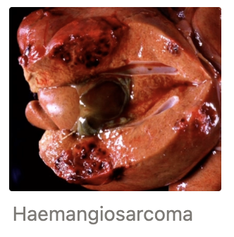
Telangiectasis (3)
Signalment: Cattle and older cats
Appearance: Multiple dark red circumscribed spots = blood-filled cavities (dilated sinusoids)
Significance: NONE
DDx:
Haemangiosarcoma metastasis = Raised with compressive mass effect (assess 1˚ masses in spleen)
Petechiae = Smaller than telangiectasis
2 Reasons why the liver undergoes rapid PM decomposition
Abundant nutrient content
Close proximity to GIT bacteria
Label the following images
 |  |  |
 |  |  |
 |  |
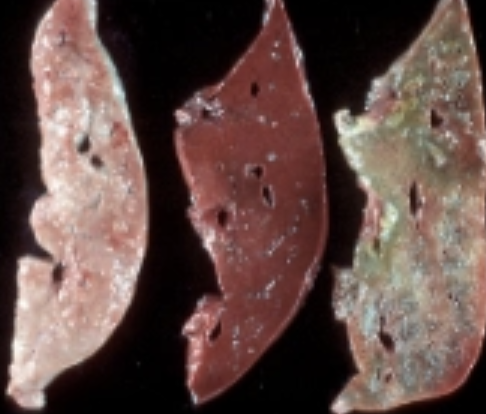
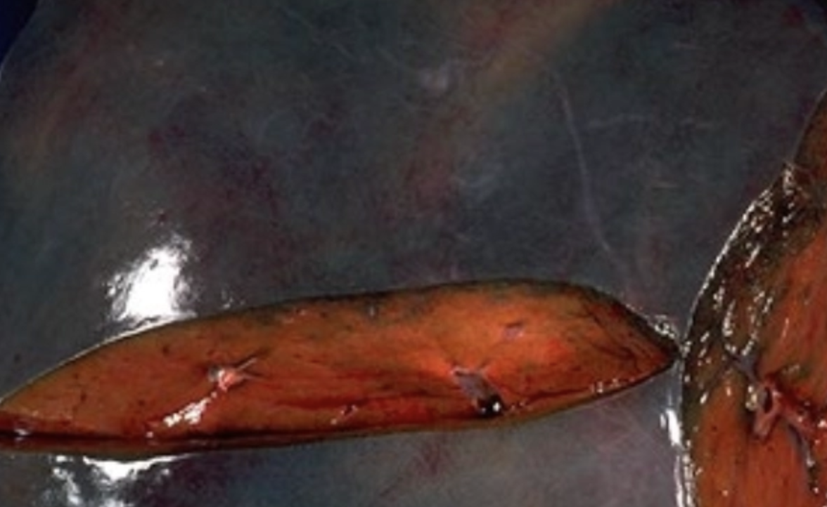
Putrefaction = Irregular pale, soft and stinky foci ± gas bubbles on capsular surface (may extend into parenchyma)
Autolysis = Diffuse softening of liver → Putty (partly due to bacterial action)
Pseudomelanosis = Green-black pigmentation of tissues in contact with GIT (typically only confined to outside and caudal aspects of liver)
Bile imbibition = Yellow-brown staining of tissues in contact with gallbladder (or bile ducts)
Putrefaction (tight liver full of gas)
Pseudomelanosis
Bile imbibition
Haemoglobin imbibition = Red-staining of blood vessel endothelium and adjacent liver
2 Causes of passive hepatic congestion (+ which is most common)
Right-sided heart disease #1 (CHF, right AV endocardiosis or endocarditis etc.)
Partial blockage of hepatic vein/caudal vena cava due to thromboembolism, abscess (eg. caval syndrome) or neoplasia
What is this?
Appearance

Acute Passive Hepatic Congestion
Appearance:
Diffusely enlarged and dark red colouration
Abundant blood ooze on incision
Central veins and centrilobular sinusoids distended by dark blood ± Enhanced lobular pattern when cut (mottled) but NOT nutmeg liver
What is this?
Appearance

Chronic Passive Hepatic Congestion eg. RCHF → Nutmeg liver = Enhanced lobular pattern with dark red centrilobular areas contrasting starkly with pale periportal areas
Rounded lobe margins
Centrilobular regions (veins and sinusoids) remain congested and dark red (filled with poorly oxygenated blood)
Persistently hypoxic hepatocytes in centrilobular region degenerate and die
Periportal hepatocytes undergo swelling and degeneration → Pale (less affected by hypoxia)
What is this?
Appearance
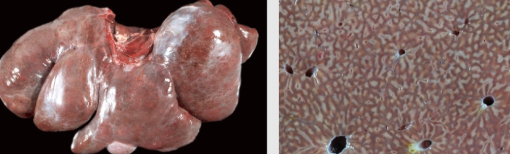
Very Chronic Passive Hepatic Congestion
Increased fibrosis = White and firm areas replacing dead hepatocytes
Cardiac fibrosis = Thickened capsule
Purple tinge to liver (deoxygenated blood is more purple than normal due to cardiac fibrosis)
Effects of ACUTE/SEVERE anaemia on the liver
Degeneration and death of centrilobular hepatocytes (centrilobular regions receive oxygenated blood last)
MARKED PCV decrease (chronic anaemia can be compensated) and mild increase in leakage liver enzymes as 2˚ liver disease
eg. Severe haemorrhage, IMHA, Theileriosis
Portosystemic Shunt (3)
Definition
2 Effects
Gross appearance
2 Anatomic types (+ signalment)
2 Aetiologies
Diagnosis
Definition: Abnormal vascular connection between portal vein and systemic circulation allowing blood from GIT to bypass the liver → Shunted back to systemic circulation via the caudal vein (occasionally azygous vein or hepatic vein)
Effects:
Liver does not receive nutrients and factors necessary for growth and function → Atrophy
Decreased liver function (eg. hepatic encephalopathy and oedema) and cannot recycle bile
Anatomic Types:
Extrahepatic = Most abnormal vascular connection sits OUTSIDE liver (cats and small breed dogs eg. Yorkies)
Intrahepatic = Most abnormal vascular connection running WITHIN liver (large breed dogs eg. ductus venosus)
Aetiologies:
Congenital PSS = Juvenile dogs (also cats esp. Birmans)
Acquired PSS = ALL species but older dogs #1
Diagnosis:
Bile acid serum concentration pre- and post-prandial to assess liver function (equivocal liver enzyme results) → PSS = Marked pre- AND post-prandial bile acid concentration
Definitive diagnosis with imaging (eg. portography, scintigraphy or CT) to ID abnormal vessels between portal and systemic circulation
Congenital PSS
Appearance
Aetiopathogenesis
3 Clinical signs
Appearance:
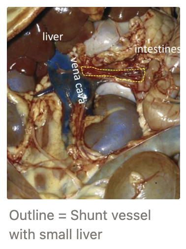
Single abnormal vascular connection (difficult to locate at PM)
Microhepatica (otherwise normal shape and consistency)
Aetiopathogenesis: Failure of ductus venosus to close after birth OR microvascular dysplasia
Clinical Signs:
Poor growth (thin and small for age)
± Neurological signs due to hepatic encephalopathy
± Late stage ascites (uncommon) due to end-stage hypoalbuminaemia (NOT portal hypertension)
Acquired PSS
Appearance
Aetiopathogenesis
3 Clinical signs
Appearance:
Number of tortuous abnormal vascular connections in mesentery (easier to locate at PM)
Portal vein distension
Small, firm and fibrotic liver
Aetiopathogenesis:
Chronic severe liver disease
Remodelling and fibrosis compresses blood vessels (eg. canine chronic hepatitis)
2˚ to portal hypertension due to chronic liver disease/vascular obstruction
Portal blood cannot pass through liver
→ Creates escape valve to allow itself back into systemic circulation
Clinical Signs:
Ascites common and EARLY in clinical course (due to portal hypertension NOT liver failure and hypoalbuminaemia)
± Neurological signs

Clinical Pathology Findings of PSS
Liver enzymes
Bile acids
Bilirubin
Liver function markers
Urinalysis
RBCs
Creatinine
Liver enzymes (ALT, AST and ALP) mildly increased/normal as chronic process and fewer hepatocytes in a small liver
Bile acids markedly increased (BOTH pre- and post-prandial)
Increased bilirubin (acquired PSS)
± Signs of liver failure depending on severity and stage (decreased albumin, urea, glucose, cholesterol + increased PT/APTT)
± Ammonium biurate crystalluria (thorn apples)
± Mild microcytic non-regenerative anaemia (not well-understood)
Low creatinine due to low muscle mass
What is this?
Gross appearance
Clinical signs
4 Risk factors
4 Diseases

Hepatic Lipidosis (3)
Appearance: Excess lipids in liver due to altered fat metabolism
Enlarged (rounded lobe margins)
Diffusely pale (red-pink → orange-yellow)
Soft, greasy and friable (may float in water due to high fat content)
Clinical Signs: Liver disease/failure when severe
Risks:
Obesity
Starvation
Pregnancy
Lactation
DDx:
Ketosis
Diabetes mellitus
Feline fatty liver syndrome
Equine hyperlipaemia
Ketosis
2 Signalments
Clinical signs
Pathogenesis
2 Diagnostic tests
Signalment:
Multiple-bearing ewes in late gestation (aka. pregnancy toxaemia)
Dairy cattle post-partum and early lactation
Clinical Signs: Weak + recumbent → Death ± ketone (onion) smell on PM
Pathogenesis:
Insufficient glucose precursors in diet to meet high metabolic demand
Negative energy balance
Markedly increased fat mobilisation from adipose tissue to compensate
Hepatic lipidosis and increased ketone production
Diagnosis:
Elevated ketones = β-hydroxybutyrate (BOH) measured on serum biochemistry or urine dipstick or milk
Hypoglycaemia
Feline Fatty Liver Syndrome
Signalment and pathogenesis
3 Clinical signs
Signalment: Obese cats that become anorexic for a short period of time (eg. illness or go missing for several days)
Clinical signs: Hepatic failure with encephalopathy and jaundice ± death

Equine Hyperlipidaemia
Signalment
Gross appearance
Signalment: Overweight OR lactating mare ponies (esp. Shetland), miniature horses and donkeys following period of anorexia
Appearance: Markedly fatty and friable liver that often ruptures while alive
What is this?
Aetiology
2 Example DDx
Gross appearance

Glycogen Accumulation (3)
Aetiology: Excessive glycogen accumulation in liver due to metabolic diseases altering glucose metabolism Insulin and corticosteroids help convert glucose → glycogen
DDx:
Diabetes mellitus in cats and dogs (lipids AND glycogen present in liver)
Hyperadrenocorticism in dogs (endogenous OR exogenous) as steroids induce glycogen synthase activity
Appearance: Diffuse/patchy swelling and pallor (difficult to distinguish from mild hepatic lipidosis)
EXAMPLE: “LOKI”
Signalment: 9m castrated Cairn Terrier
Clinical Signs: Poor growth, lethargy, progressive anorexia and occasional head pressing against wall AND vomiting
BCS 2/9 with pale pink gums
Interpretation:
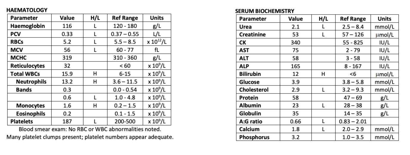
There is a mild microcytic (low MCV) and normochromic (normal MCHC) non-regenerative or pre-regenerative (normal retics., MCV and MHCH) anaemia (low Hb, PCV and RBC). This may be a pre-regenerative OR non-regenerative response. Given the chronic history of poor growth, I am leaning more towards a non-regenerative response due to chronic disease. Low MCV is highly indicative of abnormal Fe metabolism potentially due to congenital PSS. Potentially also young age is a contributing factor (young animals have lower RBC values). Furthermore, there is no evidence of haemolysis or haemorrhage in the history to indicate a pre-regenerative response. We can definitively rule out a pre-regenerative anaemia with blood sample in 3 - 5 days.
Bone marrow disease = Expect other WBC changes
There is a mild leukocytosis (high WBCs) characterised by a mild mature neutrophilia (high neuts. and normal bands), lymphopenia (low lymphocytes) and monocytosis (high monocytes). This is most consistent with a stress leukogram and elevated corticosteroids due to being ill.
Given that there are many platelet clumps present on blood smear and numbers appear adequate, the mild thrombocytopenia (low platelets) is most likely an artefactual change (eg. traumatic venipuncture activating platelets and causing them to clump).
There is a mild decrease in urea. Given the non-specific clinical signs, low urea may be due to liver disease and reduced liver functional mass → Reduction in production in liver. The liver cannot take up NH3 for conversion into urea, resulting in mild reduced urea.
There is a mild decrease in creatinine. Normal production of creatinine occurs at a fairly constant rate and depends on the patient’s muscle mass. Given that Loki’s BCS is 2/9, he likely has poor muscle mass which is causing the reduction in creatinine.
Liver enzymes (AST, ALT and ALP) are normal, but the high end of the reference range. This makes an acute liver pathology unlikely, however, we cannot rule out a chronic disease process. This is because a cirrhotic or smaller liver has very few functioning hepatocytes to produce an expected elevation in liver enzymes. Not only is this consistent with the chronic history, but the changes in liver function tests (i.e. bilirubin, cholesterol and albumin) too.
A mild hyperbilirubinaemia (high bilirubin) could be due to one of five mechanisms:
Fasting = Unlikely given this is a dog (more common in horses)
Haemolysis = Unlikely given the mild nature of the hyperbilirubinaemia and normal RBCs on blood smear examination
Hepatic disease = Most likely given the other biochemistry changes indicating reduced liver function
Cholestasis = Unlikely as induction enzyme (ALP) is normal
Sepsis = Lack of inflammatory leukogram rules out cause of hyperbilirubinaemia
There is a mild hypocholesterolaemia (low cholesterol) and mild hypoalbuminaemia (low albumin) with a moderately low A:G ratio. These changes are most likely due to impaired hepatic function and loss of significant liver mass (liver normally synthesises cholesterol and albumin) resulting in reduced circulation of these substances.
The mild hypocalcaemia (low calcium) is likely due to the hypoalbuminaemia. Because ~40% of calcium is bound to albumin, any decrease in albumin results in a decrease in serum calcium.
DDx: Congenital portosystemic shunt (most likely extrahepatic given small breed)
Pathogenesis of head pressing:
Ammonia produced from protein breakdown and microbial digestion in the large intestine
Ammonia absorbed into bloodstream through GIT → Portal vein
Congenital microvascular dysplasia → Blood vessels from portal vein bypass the liver straight into systemic circulation (eg. into caudal vena cava)
Ammonia and other toxins enter systemic circulation (cannot be converted to non-toxic urea for elimination)
Toxins travel to brain and cross BBB causing hepatic encephalopathy
Clinical signs of CNS dysfunction result (eg. head pressing, depression, seizures, altered behaviour and mentation)
Diagnostic tests:
Bile acid serum concentration pre- and post-prandial to assess liver function (equivocal liver enzyme results) → PSS = Marked pre- AND post-prandial bile acid concentration
Imaging for definitive diagnosis of PSS to ID the abnormal vessels between portal and systemic circulation (eg. scintigraphy)
4 DDx for pale liver on PM
Glycogen = Tan and large
Autolysis = Normal sized liver
Putrefaction = Rotten with gas bubbles
Lipid = Orange/yellow and large
What are the following causes of the liver FNAs?

A = Normal liver FNA (lipofuscin granules = Normal aging change)
B = Neutrophilic hepatitis
C = Hepatic lipidosis (lipid packed within cytoplasmic vacuole)
D = Autolysis (vacuolation OUTSIDE hepatocytes)
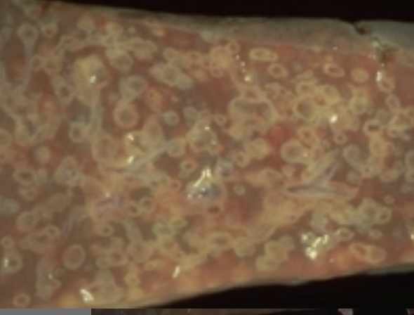
3 Portals of bacterial entry into the liver (+ examples)
Blood (eg. portal vein, hepatic artery, umbilical veins in neonatal calves or lambs) #1 most common
Biliary system = Biliary stasis → Ascending infection from bile ducts (eg. due to inflammation or obstruction as in suppurative cholangiohepatitis in dogs and cats)
Direct extension of inflammatory process from tissues adjacent to liver (eg. reticulum in hardware disease in cattle)
Gross appearance of hepatitis due to septicaemia
Highly variable manifestation of bacterial infection which depends on:
Bacterial species
Host species
→ Cannot ID specific bacteria from gross appearance
3 Different presentations of bacterial liver infections due to septicaemia (+ bacterial agent examples)
Multifocal tiny white spots randomly scattered throughout the parenchyma = Tiny multifocal areas of hepatic inflammation and necrosis (eg. Yersinia in lambs)
No gross features (eg. Salmonella spp. or Actinobacillus equuli in neonatal foals)
Large, single/multiple, well-demarcated lesions = Often incidental due to walling off of infection (eg. Abscess = Trueperella pyogenes, granuloma = Mycobacteria spp., coagulative necrosis = Fusobacterium necrophorum)


Liver Abscesses in the Neonatal Period (3)
Signalment
Pathogenesis
3 Agents
Prognosis
3 Methods of diagnosis
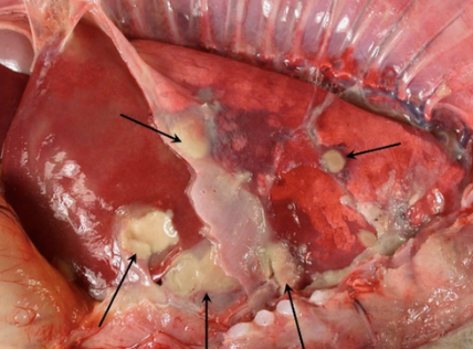
Signalment: Calves, lambs ± foals in first 2 - 6 weeks of life
Pathogenesis:
Inadequate colostrum intake in first 24hr → Lack of maternal antibody → Immunosuppression
Predisposes to umbilical infection (or GIT)
Septicaemia
Liver abscessation (also lungs, spine, brain and joints)
Agents: Mixed environmental contaminants
Fusobacterium necrophorum → Multifocal areas of green-yellow coagulative necrosis
Trueperella pyogenes → Classic abscessation
E. coli
Prognosis: Fatal
Diagnosis: Culture is NOT useful as it only shows a mixed environmental bacterial infection
Signalment (neonatal lamb/calf with history of dubious colostrum intake)
Gross lesions at PM
High GGT → Indicates adequate colostrum intake (GGT activity in mammary glands)
Signalment of liver abscesses in older animals (+ significance)
Cattle #1 → Incidental findings at PM as infection is wall off and large functional reserve
Pathogenesis of pulmonary embolic aneurysm in cattle

Invasion and rupture of hepatic abscess into hepatic vein or caudal vena cava
→ Dissemination of bacterial embolic around the body in the blood
Septic emboli cause 2˚ lesions in various organs (eg. vegetative valvular endocarditis and lung abscesses)
Bacterial proliferations within lung abscesses → Invasions of pulmonary vessels = Pulmonary embolic aneurysm
Pneumonia, vessel rupture, haemoptysis and epistasis
→ Sudden death
Overview of Leptospirosis
Source
Portal of entry
TWO syndromes
Agent
Signalment
Clinical signs
Source: Infected animals intermittently shed leptospires in urine → Infection from contaminated water sources (eg. rat urine after floods)
Portal of entry: MM and damage skin
Syndromes:
Acute intravascular haemolytic anaemia
Agent: Pomona
Signalment: Young calves, lambs and fawns
Clinical Signs: Weak, jaundice (pre-hepatic), anaemia, redwater
No haemolytic anaemia in adults
Acute concurrent severe renal and liver injury
Agent: Copenhageni (also Pomona, Hardjo, Tarassovi)
Signalment: Dog of ANY age
Clinical Signs: Lethargy, anorexia, fever, vomiting, abdominal pain, jaundice → High mortality or liver failure later
What is the main DDx for dog presenting with severe renal and/or liver disease?
Leptospirosis
Fill out the following table for Leptospirosis syndromes
Young calf/lamb | Dog/puppy | |
|---|---|---|
Syndrome | ||
RBC changes | ||
WBC changes | ||
Urine | ||
Jaundice (+ bilirubinaemia) | ||
Azotaemia | ||
Leakage enzymes | ||
Induction enzymes | ||
Other |
Young calf/lamb | Dog/puppy | |
|---|---|---|
Syndrome | Haemolysis | Renal and liver damage (either OR both) |
RBC changes | Regenerative anaemia with normal TPP | No |
WBC changes | Variable - MAY seen inflammatory leukogram | Often inflammatory leukogram (neutrophilia with left shift and toxic changes) |
Urine | Red due to haemoglobinuria | Variable, but not red |
Jaundice (+ bilirubinaemia) | Yes = Haemolytic jaundice (pre-hepatic) | Yes = Hepatic/cholestatic ± sepsis associated jaundice |
Azotaemia | None | Significant azotaemia ± Increased phosphorus (AKI) |
Leakage enzymes | No/mild increase in GLDH 2˚ to anaemia | Marked increase in ALT and AST |
Induction enzymes | No/mild increase in GGT 2˚ to anaemia | Marked increase in ALP |
Other | Multiple animals usually affected with weakness, lethargy ± death | Very sick with lethargy, vomiting, pyrexia, abdominal pain (kidneys) + death common |
THREE methods of leptospirosis diagnosis in dogs
Sample
Disadvantages
Serology MAT Ab
Sample: Paired samples (red top) taken 2 - 4w apart → Significant rising titre of Ab
-ve:
Slow turnaround = Animal dead or recovered by the time 2nd titre is obtained (treat empirically while awaiting results)
Must select specific serovars
Vaccination → False positives
PCR
Sample: Urine, kidney, liver ± blood (purple top) → Submit blood AND urine for PCR at the same time (Lepto goes through leptospiraemic phase in blood BEFORE being shed in urine → Test blood and urine simultaneously to increase chances of detecting it in either phase)
Intermittent bacterial shedding in urine and seldom in blood
Fresh kidney (most reliable) sample difficult to obtain AM
Does NOT ID serovar involved
Long turnaround time (treat while awaiting results)
PM findings and histology of liver and kidney = Definitive diagnosis
Culture is difficult
Prevention of leptospirosis in dogs + cross-protection
Vaccination containing Leptospira interrogans serovar Icterohaemorrhagiae
Protects against Leptospira interrogans serovar Copenhageni
Unknown cross protection against other serovars (including those that are livestock-associated)
Why must care be taken with diagnosis and treatment of leptospirosis?
ZOONOTIC! With lifelong effects → PPE: Eye protection, masks and gloves for PM or treatment
What is this? (pig liver)
Pathogenesis
Gross appearance
Significance

Nematode Migration (3) = Ascaris suum → Milk spotted liver
Pathogenesis:
Larvae migrate through liver → Tracts of necrosis and inflammation
Tracts eventually replaced by small linear areas of fibrosis
± Abscess and granuloma
Appearance: Multifocal pale white/pink areas of fibrosis over the liver
Significance: Clinically insignificant in many domestic species
What is this? (sheep liver)
Pathogenesis
Example
Appearance
Clinical significance

Cysticercus spp.
Pathogenesis: Larval cestodes of Taenia genus develop encysted forms within the liver of the IH
Adults are clinically insignificant in the DH
Example: Cysticercus tenuicollis (Taenia hydatigena)
Appearance: Single small fluid-filled cysts in ruminants → Firm fibrotic nodular foci
Significance: Clinically insignificant BUT result in carcass downgrade at slaughter and indicates dog (DH) exposure to offal and defaecation on pasture
What is this? (sheep liver)
Significance
Hosts
Gross appearance
Echinococcus granulosus
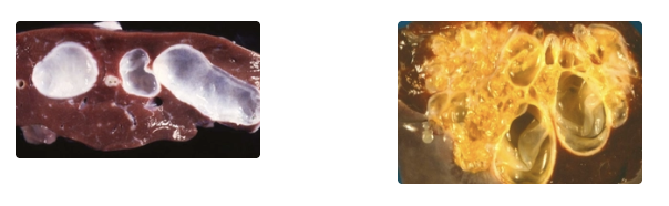
Significance: Zoonotic hydatid disease that has been eradicated from NZ (notify MPI if suspected)
Hosts:
DH = Dogs
IH = Most species (eg. sheep and people) → Cysts
Appearance: Multiple large, thick-walled fluid-filled cysts in liver ± lungs
What is this?
Hosts
Signalment
Pathogenesis
3 Clinical pathology findings
GGT
Bilirubin
GLDH
3 Clinical signs
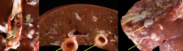
Fasciola hepatica
Hosts: DH = Adult sheep (less commonly cattle and goats)
IH = Lymnaea spp. of aquatic snail (more common disease in swampy areas)
Pathogenesis:
Ruminants ingest infective larvae
Larval migration to liver → Extensive hepatic damage
Enter bile ducts to mature to adults
Early = Adults cause cholangitis and bile duct obstruction → Cholestasis
Chronic infection = Dilation and fibrosis of bile ducts
Appearance: Hosepipes of enlarged, thickened and firm, white fibrotic bile ducts
Clinical Pathology Findings:
Mild GGT increase (more mild than facial eczema)
± Hyperbilirubinaemia
Normal GLDH
Clinical Signs: Variable severity
Ill-thrift
Anorexia
± Anaemia (fluke ingest blood)
What is this? (rabbit liver)
Pathogenesis
Pathogenesis
PPP
Signalment
7 Clinical signs
Gross appearance
3 Methods of diagnosis

Hepatic Coccidiosis in Rabbits = Eimeria steidae
Pathogenesis:
Naive rabbits ingest sporulated oocysts which survive for months in environment
Sporozoites released from the oocysts in the duodenum
Sporozoites invade intestinal wall to migrate to the liver via the portal vein and lymphatic vessels
Coccidial organisms live and reproduce in the bile duct epithelium
PPP: 15 - 18d
Signalment: Weanlings (adults = resistant)
Clinical Signs:
Anorexia
Weight loss
Diarrhoea/faecal staining
± Hepatomegaly
± Ascites
± Jaundice
± Sudden death
Appearance: Small multifocal white/pale yellow nodules in liver
Diagnosis:
Faecal flotation for oocysts (requires differentiation from intestinal infections with other types of Eimeria)
Impression smears of liver for oocysts = Refractile oval structures
Histology at PM for oocysts

2 Ways toxins cause liver damage
Direct hepatotoxic damage
Biotransformation = Liver convert substances into excreted metabolites via intermediate reactive compounds which are more hepatotoxic than the original ingested compound (eg. cytochrome P450 enzyme)
What is this?
Seasonality
Pathogenesis
Signalment
Clinical signs
Gross appearance
Clinical pathology findings


Chronic Facial Eczema
Seasonality: Late summer and autumn = Warm and humid conditions favour growth of Pithomyces chartarum on dead pasture litter
Pathogenesis:
Animal ingests fungal spores which release sporidesmin mycotoxin
Sporidesmin absorbed into blood, removed by liver and excreted in bile
→ Direct bile duct damage results in cholestasis and jaundice + damage into adjacent liver parenchyma
Damaged liver cannot effectively excrete phytoporphyrin (phylloerythrin) from ingested green plant breakdown in GIT
→ Retention of phytoporphyrin in blood
Sunlight passes through skin to activate phytoporphyrins in blood → Photosensitisation
Signalment: Older animals with sporidesmin exposure over multiple seasons
Appearance: OBVIOUS boxing-glove liver
Atrophy and fibrosis of left lobe → Small flap of fibrotic tissue (blood from GIT hits left lobe first)
Hypertrophy and fibrosis of right lobe → Firm and round (compensatory hypertrophy)
Clinical Pathology Findings:
Normal GLDH and GGT
Rare serum biochemical changes of liver failure (only when >70 - 80% loss)
What is this?
Onset
Signalment
3 Clinical signs
PM findings
3 Clinical pathology findings

Acute/Subacute Facial Eczema
Onset: ≥10d from ingestion
Signalment: Young animals with no prior FE damage → Recent sporidesmin ingestion
Clinical Signs:
± Jaundice
Red, wet, sloughing and painful photosensitisation (equivalent to 3rd degree burns)
Acute death
PM: NORMAL! ± Slightly bronze and enlarge liver (difficult to see)
Clinical Pathology Findings:
Markedly increased GGT (≤20 - 30x)
Mild/moderate increased GLDH
Hyperbilirubinaemia
What is this?
Source
Pathogenesis
Signalment
4 Clinical signs (+ onset)
PM findings
2 Clinical pathology findings

Source: Ragwort = #1 NZ plant containing PAs
Acute intoxication = Rare (most plants containing PAs are unpalatable) unless animals facing complete starvation and forced to eat unpalatable toxic plants
Chronic intoxication = Common due to repeated and seasonal intoxication
Pathogenesis: Indirect hepatotoxicity = Ingested PAs converted to hepatotoxic metabolites by CYP450 (pyrollic esters = alkalising agents that destroy cell components)
Signalment: Cattle and horses #1 (Sheep and goats more resistant to intoxication and pigs seldom exposed to ragwort)
Clinical Signs: 1 week to several months post-ingestion
Weight loss
Diarrhoea
Jaundice
Neurological signs due to hepatic encephalopathy (hyperexcitability, aimless wandering and ataxia)
PM: Cirrhosis = Small, pale and firm liver with variable interspersed nodules of regenerating liver parenchyma separated by thick fibrous bands
Clinical Pathology Findings:
Normal liver enzymes (GLDH and GGT)
± Serum biochemical changes of liver failure (low urea, glucose, cholesterol etc.)

What is the following diagnoses?

Chronic FE = Markedly abnormal size, colour and shape → Left lobe atrophy and right lobe hypertrophy → Boxing glove appearance
Chronic ragwort = Markedly abnormal size, colour and shape → Classic cirrhosis: Smaller, paler and firmer liver with variable interspersed nodules of regenerating liver parenchyma separated by thick fibrous bands
Chronic liver fluke = Normal size, colour and shape → Dilations and fibrosis of bile ducts (hosepipes)
Copper Toxicity (3)
Pathogenesis
Signalment
Risk factors
3 Causes (+ example DDx)
Pathogenesis: Hepatotoxic at high concentrations = Free radical production and oxidative damage of hepatocytes and other cells
Signalment:
Sheep #1 (tendency to store copper in liver and reduced ability to excrete Cu in bile)
Dogs (eg. Bedlington Terriers) = Hepatotoxic at high concentrations = Potential cause of canine chronic hepatitis
Risks: VitE or Mo deficiency = Cu antagonists
Causes:
Primary metabolic defect in copper metabolism
eg. Autosomal recessive mutation in Cu transport proteins (COMMD1) → Accumulation of excess Cu in hepatocytes
Signalment: Bedlington Terriers (also WHWT and Dalmatians = Familial Cu accumulation in many other dog breeds)
Altered hepatic biliary excretion = Sheep less able to excrete copper in bile
Dietary copper excess = Copper over-supplementation in sheep and cattle
Acute Copper toxicity
Aetiology
5 Clinical signs (+ onset)
PM findings
Aetiology: Ruminants after accidental administration of single large dose of oral or parenteral copper → Hepatic necrosis and acute gastroenteritis ± Concurrent Se of vitE deficiency
Supplementation NOT voluntary intake
Clinical Signs: ≤48hr post-ingestion
Abdominal pain
Diarrhoea
Paralysis
Collapse
Death
PM: Grossly dark red and swollen liver (often mottled)
What is this?
Signalment
Pathogenesis
3 Clinical signs (+ onset)
4 PM findings
Diagnosis
5 Clinical pathology findings
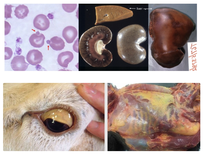
Chronic Copper Toxicity
Signalment: Sheep (also calves)
Pathogenesis: Acute presentation but chronic 2-step process
Copper accumulates in hepatocytes over weeks to months due to dietary excess (esp. low Mo or other Cu antagonists)
Crisis initiates sudden release of stored copper from hepatocytes (eg. hepatic damage due to ragwort ingestion, stress, transport, starvation)
Copper release initiates hepatic necrosis and oxidative damage of erythrocytes
→ Acute severe haemolytic Heinz body anaemia and severe IV haemolysis
Clinical Signs: 12hr post-stressful event
Jaundice (haemolytic, hepatic AND post-hepatic)
Haemoglobinuria
High mortality
PM:
Soft, enlarged and pale orange liver
Dark red-black kidneys (gun metal) due to Hb oxidation to methaemoglobin
Jaundiced carcass
Red urine
Diagnosis: Fresh liver and/or kidney samples for toxicology (Cu levels)
Clinical Pathology Findings:
Severe regenerative and haemolytic anaemia (often still in pre-regenerative phase) = Haemolysis
Many Heinz bodies in RBCs = Haemolysis
Haemoglobinuria = Haemolysis
Hyperbilirubinaemia + bilirubinuria = Haemolysis AND hepatocyte damage ± post-hepatic 2˚ to hepatocellular swelling
Marked increase in ALL liver enzymes (GLDH and GGT in large animals) = Hepatocyte damage
Species most likely to have toxicosis from therapeutic drugs
Cats more susceptible to intoxication due to low activity of key hepatic detoxification enzyme
List 4 therapeutic drugs that can cause hepatotoxicity (+ signalment)
Paracetamol (aka. Panadol, acetaminophen) = ALL cats
Heinz body anaemia
Acute liver necrosis with increased ALT and ALP
Idiosyncratic drugs = SMALL number of animals due to unknown mechanisms
Carprofen (aka. rimadyl, vetprofen) = Occasional acute hepatic necrosis in dogs (esp. Labradors)
Diazepam (aka. valium) = Occasional acute hepatic necrosis in cats
Anticonvulsants (eg. phenobarbital, phenytoin, primadone) = Chronic hepatotoxicity, cirrhosis and chronic liver failure in some dogs on long-term therapy
List 6 other toxins causing hepatic damage
Xylitol
Metaldehyde
Aflatoxins (eg. Aspergillus spp.)
Brassicas
Mushrooms
Blue-green algae
List 2 other (orphan) hepatic diseases
Canine chronic hepatitis (aka. "chronic-active hepatitis") (3)
Liver haemorrhage and/or rupture (3)
Define canine chronic hepatitis
Non-specific diagnosis describing appearance of liver due to persistent inflammation ≥ 6 months in duration
Canine Chronic Hepatitis (3)
Signalment
4 Causes
Clinical pathology findings
ALT/AST
ALP and bilirubin
Liver function enzymes
RBCs
Prognosis
PM findings
Diagnosis
Signalment: Middle-aged to older dogs
Causes: Original initiating cause often unknown an irrelevant → Unresolved inflammation
Inherited/familial defects of copper metabolism in Bedlington Terriers, WHWT and Dalmatians
Infectious canine hepatitis
Leptospirosis
Hepatotoxic drug damage
Clinical Pathology Findings:
Variable increase in ALT and AST → Normal with increasing chronicity
Increased ALP and bilirubin
Decreased urea, glucose, cholesterol and albumin
Increased coagulation times (eg. PT and APTT)
Increased bile acids
Non-regenerative anaemia of chronic disease
Prognosis: Poor due to complete liver failure within weeks or months of diagnosis
PM: Mildly small, firm nodular and fibrotic liver → Complete cirrhosis
Diagnosis: Biopsy and histology
Cannot determine inciting cause
Suspected copper storage abnormalities → Test liver copper levels in fresh biopsy samples

What is this?
2 Mechanisms (+ DDx)
Prognosis
PM findings
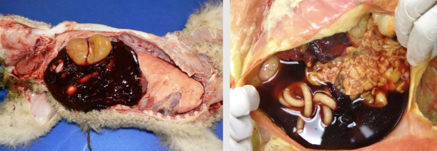
Liver Haemorrhage/Rupture
Mechanisms:
Normal OR foetal liver with MAJOR trauma
Traumatic HBC in dogs and cats
Traumatic dystocia in lambs and calves
Diseases causing diffuse hepatic enlargement and friability + MINOR trauma
Acute hepatitis
Severe congestion
Hepatic lipidosis
Neoplasia
PM: Torn liver capsule and underlying liver parenchyma with free abdominal fluid

Lymphocytic Cholangitis (3)
Signalment
Aetiology
Pathogenesis
Clinical signs
PM findings
Diagnois
Clinical pathology findings
Signalment: Common CHRONIC disease in middle-aged to older cats
Aetiology: Unknown
Pathogenesis: Inflammatory cells (mainly lymphocytes) surround and destroy bile ducts and adjacent hepatocytes
Clinical Signs: Mild, non-specific CHRONIC clinical signs (vomiting and lethargy) and cyclical
Jaundice UNCOMMON
PM: NORMAL liver
Diagnosis: Liver biopsy for definitive diagnosis and exclusion of other DDx with hepatocytes and lymphocytes
May miss lesions with cytology AND cannot differentiate from small cell lymphoma
Clinical Pathology Findings:
Normal/mild increase in liver enzymes (ALT, AST and ALP)
Normal/mild increase in bilirubin Only more significant when disease progression becomes more severe
Acute (Suppurative) Cholangitis (4)
Signalment
Cause
6 Clinical signs
PM findings
2 Methods of diagnosis
3 Clinical pathology findings
Signalment: ALL species and ages (most common in dogs and cats)
Cause: Ascending bacterial infection of biliary tree from GIT → Cholangitis ± cholangiohepatitis
Triaditis = Pancreatitis, IBD and cholangiohepatitis seen together in cats as pancreatic duct and bile duct enter the duodenum at the same major duodenal papilla
Clinical Signs: SEVERE and ACUTE (vs. lymphocytic cholangitis)
Pyrexia
Lethargy
Anorexia
Vomiting
Jaundice
Abdominal pain
PM: Normal
Diagnosis:
Presumptive diagnosis with antibiotic treatment
FNA for cytology or biopsy (definitive diagnosis) = Large numbers of neutrophils and intralesional bacteria
Clinical Pathology Findings:
Increased liver enzymes (ALT, AST and ALP) but variable magnitude
Inflammatory leukogram
Hyperbilirubinaemia
Malignant Primary Neoplasms of the Liver
4 Defining features
2 DDx
Features:
Variable single, large and extensive mass (± effacement of organ)
OR multiple masses
Poorly demarcated
± Masses in other organs if has metastasised
DDx:
Hepatocellular adenocarcinoma
Cholangiocellular adenocarcinoma


Primary Benign Neoplasms of the Liver
3 Defining features
3 DDx
Features:
Single (focal)
Well-demarcated
Small(ish) mass
DDx:
Hepatocellular adenoma
Cholangiocellular adenoma
Nodular hyperplasia (often well within liver parenchyma but difficult to distinguish on histology)


Secondary Malignant Neoplasms of the Liver
3 Defining features
2 DDx
Features:
Multiple masses OR
Diffuse organ enlargement
Masses in other organs
DDx:
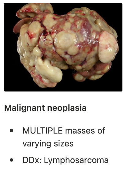
HSA (spleen) = Bloody and dark red
LSA (creamy or diffusely pale)


CASE 1: “PRINCESS”
Signalment: 3m Yorkshire Terrier puppy
History: Intermittent seizures becoming more frequent. Smaller than littermates

Interpretation:
There is a mild normocytic (normal MCV), normochromic (normal MCHC) non-regenerative or pre-regenerative (normal retics.) anaemia (low Hb and HCT). Given the chronic history of poor growth (smaller than littermates) and the lack of clinical signs suggestive of haemorrhage or haemolysis, a non-regenerative anaemia is more likely. This is most likely due to chronic disease. Young animals ALSO have physiological anaemia (RR may be taken for adult). MCV is lower end of RR which is most consistent with a portosystemic shunt (congenital aetiology fits with the age of the patient).
There is a mild leukocytosis (high WBC) characterised by a mild, mature (normal bands) neutrophilia (high neutrophils) and a mild lymphocytosis (high lymphocytes). The mature neutrophilia is most likely physiological due to fear (fight-flight) response. This is because it is very mild in nature and the patient is very young. Increased catecholamines results in migration of mature neutrophils from the marginal to circulating pool in blood vessels. The mild lymphocytosis is most likely due to a physiological fear/excitement response as we see with the neutrophilia. It may also be due to chronic inflammation due to Ag stimulation of T cells. This may be due to congenital portosystemic shunt which is causing Ag from the gut to bypass the liver for processing. Because more Ag enter systemic circulation, there is more Ag presentation which is causing the lymphocytosis.
NOT a stress leukogram as we would expect a lymphopenia
NOT inflammation as we would expect a left shift
Mild increase in ALT and ALP: Likely due to hepatocellular damage. Although the parameters are only mildly increased, we cannot rule out liver disease as a potential DDx as a dysfunctional liver (eg. cirrhosis or microhepatica) would not produce enough leakage enzymes or induction enzymes to cause expected elevation. Increased ALP may also be the bone isoform that is due to a growing patient
Low urea: Most consistent with reduced functional liver mass. Because the liver is responsible for producing urea from NH3 and protein breakdown products, when there are fewer functional hepatocytes, less urea (and more ammonia) will accumulate in circulation.
Low creatinine: Because creatinine is proportional to muscle mass, the low creatinine is most likely due to the low muscle mass of the puppy as indicated in the history of poor growth (smaller than littermates).
Mild hypocholesterolaemia and mild hypoalbuminaemia with a low A:G ratio: This is most likely due to reduced functional liver mass which reduces production of cholesterol and albumin.
Albumin has long half-life and so hypoalbuminaemia indicates chronic disease process
Mild hypocalcaemia (low calcium) is most likely due to concurrent hypoalbuminaemia. Because ~40% of calcium is bound to albumin, we often see a decrease in calcium due to reduced albumin.
Mild hypoglycaemia: Reduced functional liver mass is the most likely cause due to reduced glycogenolysis and gluconeogenesis capabilities of the liver.
DDx: Congenital portosystemic shunt (most likely extrahepatic given the signalment = Small breed)
Urinalysis:
Isosthenuria due to low urea causing medullary solute washout (lack of an osmotic gradient in the medulla which is required to concentrate urine)
Ammonium biurate crystalluria due to elevated levels of ammonia in blood (reduced transformation to urea by the dysfunctional liver) → Thorn-apple appearance
PM: Microhepatica but otherwise normal shape and appearance
Pathogenesis of seizures:
Congenital portosystemic shunt allows portal blood from the GIT to bypass the liver (including NH3 produced from bacterial breakdown in the colon)
Reduced functional liver mass (lack of nutrients delivered)
Reduced urea synthesis from NH3 (product of protein breakdown)
NH3 accumulates in systemic circulation as it can bypass the liver for transformation to urea
NH3 crosses the BBB and causes neurotoxicity to the neurons
Seizures due to hepatic encephalopathy
Diagnosis:
Bile acid serum concentration pre- and post-prandial to assess liver function (equivocal liver enzyme results) → PSS = Marked pre- AND post-prandial bile acid concentration
Imaging for definitive diagnosis of PSS to ID the abnormal vessels between portal and systemic circulation (eg. scintigraphy) and plan surgical treatment (ligation)