BIOL Week 5
 Call Kai
 Learn
 Practice Test
 Spaced Repetition
 Match
 Flashcards
 Knowt Play
1/113

There's no tags or description
Looks like no tags are added yet.
114 Terms
What is the general definition of Quorum Sensing in Bacteria
A method of communication that allows bacteria to detect population density (bacteria communicate based on how many bacteria are around.)
When the population gets large enough, they change their behaviour together.
How does quorum sensing work? (changing behaviour)
Bacteria release signals → more bacteria = more signal → signal reaches a high concentration → receptors detect it → signalling pathway activates → bacteria change behaviour together.

Why doesn't quorum sensing occur at low cell density?
Few bacteria → few signal molecules → signal concentration is too low → receptors aren't sufficiently activated → no group response.

Why is the correct receptor important?
Signal must bind its specific receptor → receptor activates → signalling begins. No correct receptor = no response.

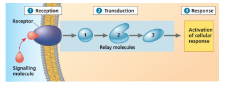
What is the general overall cell communication process? What are the 3 stages of cell signalling?
Produce signal → detect with receptor → transduce signal inside cell → cellular response with behavioural change
Reception → Transduction → Response.
Reception = detect signal;
transduction = relay it;
response = cell changes.

What is local signalling and the four types?
Cell-cell signalling occurs at local-distance scales in multicellular organisms. (occurs between nearby cells)
direct contact
Gap junctions
Paracrine signalling
Synaptic signalling
How does direct-contact signalling work?
cells communicate by touching each other through surface proteins.
Two cells touch → receptor/surface proteins on one cell recognize molecules on the other → communication occurs.

How do gap junctions (animals) / plasmodesmata (gap) work?
They form direct connections between neighbouring cells → signals can pass directly from one cell to another.

How does paracrine signalling work?
secretion of signalling molecules that are detected by nearby cells. One cell releases signals that affect multiple nearby target cells
Cell releases signalling molecules → signals travel a short distance → nearby target cells detect them → response.

How does synaptic signalling work?
Nerve cell releases a signal → signal crosses the synapse → one specific target cell detects it → response.

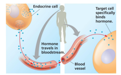
How does long-distance/endocrine signalling work?
communication between cells far apart, located in different parts of the organism (two organs)
Based on the usage of hormones that are secreted from the cell → signalling molecules that travel through the body.
Cell releases hormone → hormone travels through body → reaches distant cells → cells with correct receptor respond.
Animals: hormones travel mainly through the bloodstream → called endocrine signalling. • Plants: hormones can travel through vessels, cells, or even through the air as gases.
What happens during reception? general process
Ligand (any molecule that binds to another changing its shape) binds specific receptor → receptor changes shape → receptor becomes activated.

2 main kind of receptors
Cell-surface receptors → located in the plasma membrane (embedded proteins)
Intracellular receptors → located inside the cell (cytoplasm or nucleus)
Cell-Surface Receptors (eukaryotic cells) - What are G-Protein Coupled Receptors (GPCRs)?
A large family of cell-surface receptors that detect molecules outside the cell and activate internal signal transduction pathways.
(G-protein coupled receptors activate only one signal transduction pathway)
The receptor and G protein are specific → they only work with certain signals and partners.
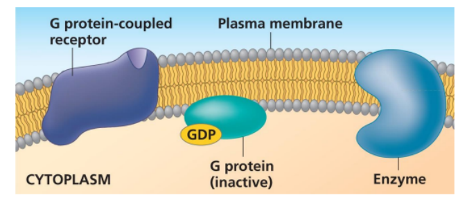
What is a GPCR like when inactive? What is inactive state?
Inactive state: G-protein coupled receptor is not associated with a signal molecule, and the G protein is associated with the GDP molecule
No signal bound → GPCR inactive → G protein is bound to GDP → G protein OFF.
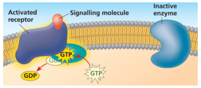
How is a G protein activated?
Ligand binds GPCR → GPCR changes shape → interacts with G protein → GDP leaves → GTP binds → G protein ON.
What does the activated G protein do?
G protein-GTP moves along membrane → binds a target enzyme → enzyme changes shape → enzyme activated or inhibited.

How is the G protein turned OFF?
G protein hydrolyzes GTP → GDP + phosphate → G protein becomes inactive again. - returns to neutral state

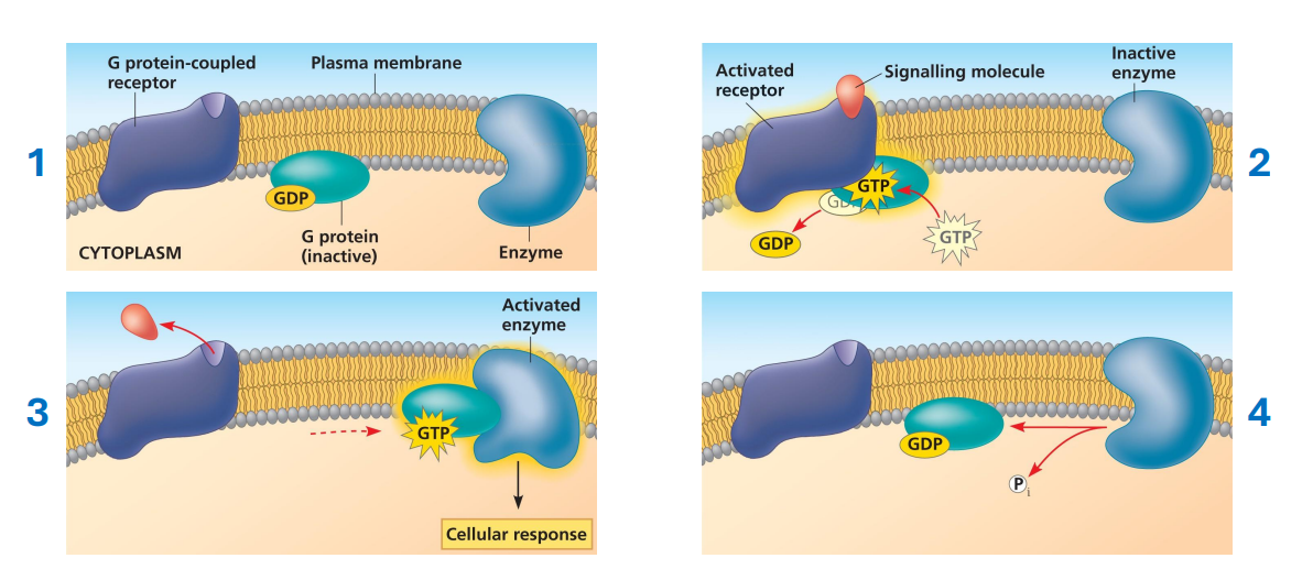
Explain the Reception process using G-Protein Coupled Receptors - cell surface receptors
1. Inactive: No signal → GPCR inactive → G protein has GDP.
2. Reception: Ligand binds GPCR → receptor changes shape.
3. G protein activation: GPCR interacts with G protein → GDP replaced by GTP → G protein activated.
4. Transduction: G protein-GTP moves along membrane → binds target enzyme.
5. Enzyme: Target enzyme changes shape → becomes activated or inhibited → leads to cellular response.
6. Shutoff: G protein hydrolyzes GTP → GDP + phosphate → G protein becomes inactive again.
Cell-Surface Receptors (eukaryotic cells) - What is receptor tyrosine kinase (RTK)(monomer) general
RTKs are another type of cell-surface receptor.
One RTK can activate multiple signalling pathways (up to 10).
• The part of the RTK inside the cell acts as a tyrosine kinase (monomer). (outside binds signalling molecule)

How does an RTK become activated?
Ligand binds (2 of them) → 2 RTK monomers come together → dimerization forms dimers → kinase activity activates.

what is Dimerization?
When the signal molecule binds to the receptor tyrosine kynases, two RTKs (monomers) come together through a process called dimerization. (The resulting quaternary structure is known as a dimer, meaning it contains two protein subunits.)

What happens after dimerization?
RTKs use ATP → add phosphate groups to their own tyrosine amino acids → receptor becomes phosphorylated and fully active.

What happens after phosphorylation?
Relay proteins bind phosphorylated RTK → relay proteins change shape → signal transduction pathways activate → cellular responses.

Why can RTKs cause multiple responses?
One activated RTK can activate multiple relay proteins/pathways, producing different responses.
How is RTK signalling turned OFF?
Phosphates are removed → RTKs separate → return to inactive monomers.
Receptor Tyrosine Kinase (RTK) signalling process
1. Inactive: RTKs exist as separate monomers.
2. Reception: Ligand binds → two RTKs come together (dimerization).
3. Activation: Dimerization activates their tyrosine kinase activity.
4. Phosphorylation: RTKs use ATP to add phosphate groups to their own tyrosine amino acids → fully active phosphorylated RTK.
5. Transduction: Relay proteins bind to the phosphorylated RTK → change shape → activate signalling pathways.
6. Response: Pathways produce one or more cellular responses.
7. Shutoff: Phosphates are removed → RTKs separate → return to inactive monomers.**

Cell-Surface Receptors (eukaryotic cells)- What are Lingand-gated ion Channel Receptors general & what is it like in inactive state
Ligand-gated ion channels are the third type of cell-surface receptor.
• They act as ion channels/transporters, allowing ions like Na⁺ and Ca²⁺ to move across the membrane
Inactive state: the channel is closed, so ions cannot pass through
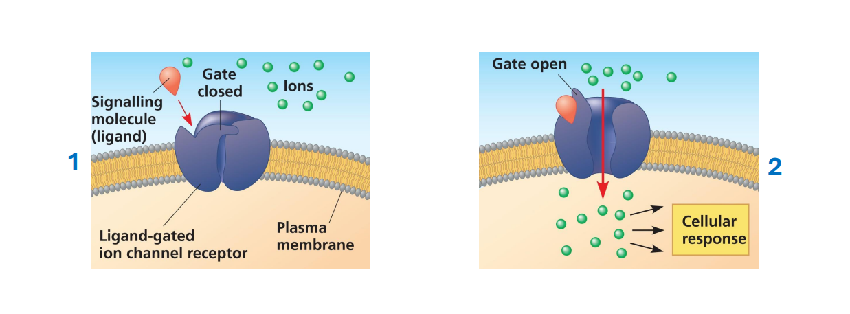
How does a ligand-gated ion channel work?
Ligand binds → receptor changes shape → gate opens → ions move through by facilitated diffusion → cellular response.

How does the channel turn OFF?
Ligand dissociates → gate closes → ions can no longer cross.
Ligand-gated vs. voltage-gated channel receptors?
Ligand-gated: opens when ligand binds.
Voltage-gated: opens/closes when electrical charge across membrane changes.
What is a Voltage-Gated Channel Receptor?
A type of ion channel that opens or closes in response to changes in membrane potential, allowing ions to flow across the membrane. Particles move with their concentration gradient.

Ligand-gated ion channel receptor process.
1. Inactive: No ligand → channel is closed → ions cannot cross.
2. Reception: Ligand binds receptor → receptor changes shape.
3. Channel opens: Structural change opens the gate.
4. Ion movement: Ions such as Na⁺ or Ca²⁺ move through the channel by facilitated diffusion.
5. Response: Ion movement causes a cellular response.

Cell-Surface Receptors (Bacteria cells) - What are Histidine kinases (HKs)
HKs are the main type of cell-surface receptor in bacteria.
• Like RTKs, they can detect signals and have kinase activity.
• Instead of adding phosphate to tyrosine, HKs add phosphate from ATP to a histidine.
How does a histidine kinase pathway work?
Signal activates HK → ATP provides phosphate → histidine phosphorylated → phosphate transferred to aspartate on response regulator → regulator activates → cellular response often inducing gene expression
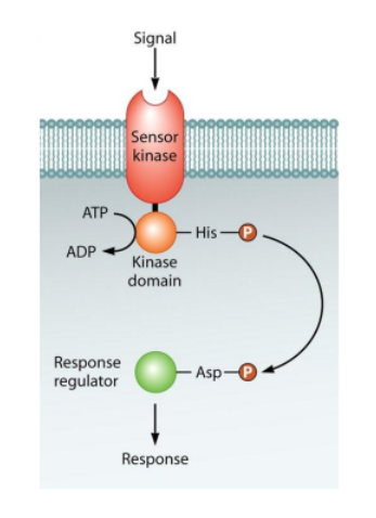
What does the activated response regulator often do? in Histidine kinase
It often changes gene expression, producing the cellular response.
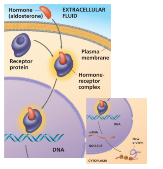
What signals can use intracellular receptors?
Signals must be able to cross membrane by themselves → usually hydrophobic (steroid hormones) or small (nitric oxide).

How does intracellular signalling work? (How does this produce a cellular response?)
Signal crosses plasma membrane → binds intracellular receptor → activated receptor moves into nucleus and affects DNA → transcription changes → protein production changes.
How does this produce a cellular response?
Transcription increases/decreases (This changes how much of a specific protein the cell makes.) → amount of specific protein changes → cell behaviour changes.
What is signal Transduction and what happens? general
Signal transduction = converting the information from signal reception into a form that can cause a specific cellular response.
A signal is detected by a receptor → the information from that signal is passed through the cell → relay molecules carry/convert the information → a specific cellular response occurs.
Does the original signalling molecule travel through the transduction pathway? and what are the two versions of transduction?
No. The signalling molecule activates the receptor, but then the information from the signal is passed along
Transduction can be:
○ One step for transduction → common in some bacterial pathways, such as histidine kinase pathways.
○ Multiple steps for transduction → called a signal transduction pathway. (more common) information passed through a series of relay molecules.
What are relay molecules?
Molecules that pass signal information through the cell. They are usually proteins, but can also be ions.
Why do signalling pathways often have multiple steps? and describe these factors
Multiple steps allow signal amplification + regulation. The signal can become stronger and the cell gets more places to control the response.
How does signal amplification work?
One activated molecule activates many molecules → each of those activates many more → a small original signal produces a large cellular response
Photo example: 1 receptor → 100 G proteins → 100 adenylyl cyclase → eventually 1,000,000 active glycogen phosphorylase molecules - - More steps = much greater signal amplification.
How do multiple steps improve regulation?
More steps = more places where the pathway can be controlled, allowing cells to fine-tune signal strength and produce different responses.

Why are scaffold proteins needed? & How do scaffold proteins improve signalling?
Without them, relay molecules would randomly diffuse through the cytoplasm trying to find each other → signalling would be slower and less efficient.
Scaffold protein holds relay molecules together → keeps them close → information passes faster and more efficiently.
Scaffold gathers relay proteins → proteins stay close → faster signal transfer → more efficient pathway.
How does phosphorylation regulate a protein?
adding a phosphate group (P) to a protein.
Protein kinase adds phosphate → protein's activity changes → usually becomes active (although phosphorylation can sometimes inhibit it).
How does dephosphorylation regulate a protein?
Protein phosphatase removes phosphate → protein usually returns to inactive state.
What is kinase? Enzymes: What does a protein kinase do? & What does a protein phosphatase do?
Kinase is an enzyme that sticks phosphates onto something else
Protein Kinase: Adds a phosphate group to a protein. Think: Kinase = P ON.
Protein Phosphatase: Removes a phosphate group from a protein. Think: Phosphatase = P OFF.
Explain the complete phosphorylation cascade.
A phosphorylation cascade = a signal transduction pathway made up of a series of phosphorylation events.
1. First activated relay molecule activates a protein kinase. (2 nd relay molecule)
2. Protein kinase 1 phosphorylates → activates protein kinase 2.
3. Protein kinase 2 phosphorylates → activates protein kinase 3.
4. Protein kinase 3 phosphorylates → activates a target protein.
5. Target protein causes a cellular response.
Signal → Relay molecule 1 → Kinase 1 → Kinase 2 → Kinase 3 → Target protein → Cellular response

How is a phosphorylation cascade turned OFF?
Can’t stay active forever!!
Signal leaves receptor → protein phosphatases remove phosphates from pathway proteins → proteins return to inactive states → pathway stops
Each kinase and phosphatase is specific, meaning it only works on certain proteins.
Kinase → adds a phosphate to specific proteins.
Phosphatase → removes a phosphate from specific proteins.
This specificity makes sure only the correct signalling pathway is turned on or off.
Why must phosphorylation cascade be terminated after they are no longer needed?
To prevent overactivity and ensure the cell responds appropriately to signals.
Termination allows pathway proteins to return to their inactive states, maintaining cellular homeostasis.
What is a second messenger? & Why are second messengers effective?
Relay molecules: A small, non-protein, water-soluble molecule that carries signal information through the cell.
Why are second messengers effective?
Small + water-soluble → diffuse quickly through cytoplasm → can affect many relay proteins.
What pathways can use second messengers? + general process
They can be involved in pathways started by GPCRs and RTKs.
Receptor activated → second messenger produced/released → diffuses through cell → regulates proteins → cellular response.
Explain the complete cAMP signalling process
cAMP: synthesised by adenylyl cyclase and degraded by phodiesterase
GPCR → activates G protein → G protein controls adenylyl cyclase → adenylyl cyclase converts ATP → cAMP → cAMP activates/regulates PKA → PKA phosphorylates proteins → cellular response.

What does adenylyl cyclase (enzyme) do?
controlled by G protein: When activated, it converts ATP → cAMP, creating the second messenger.
Depending on the type of G protein, adenylyl cyclase can be:
GPCR → G protein → Adenylyl cyclase → cAMP
Activated/increased → makes more cAMP
Inhibited/decreased → makes less cAMP

What does cAMP do?
cAMP acts as a second messenger and commonly regulates Protein Kinase A (PKA).
What does Protein Kinase A (PKA) do after cAMP activates it?
PKA phosphorylates other proteins → changes their activity → produces cellular response.
Why can cAMP cause different responses in different cells?
Different cell types contain different proteins → PKA phosphorylates different target proteins → different responses.
How is cAMP removed?
Phosphodiesterase breaks cAMP → AMP, helping terminate the signal.
Sequence for cAMP Pathway
GPCR → G protein → adenylyl cyclase → ATP becomes cAMP → PKA → phosphorylation → response.

Why is Ca²⁺ useful as a second messenger?
Ca²⁺ levels are normally low in the cytosol but high outside the cell and in the ER. When calcium channels open, Ca²⁺ rapidly enters the cytosol and activates proteins through allosteric regulation, triggering a cellular response.
Signal → Ca²⁺ channels open → Ca²⁺ increases in cytosol → proteins activated → cellular response
What does Ca²⁺ do once its cytosolic concentration increases?
Ca²⁺ acts as an allosteric regulator → binds proteins → changes their activity → cellular response.
Explain the complete IP₃/Ca²⁺ signalling process
Signal binds GPCR → activates G protein.
G protein activates PLC, which splits PIP₂ into IP₃ + DAG.
IP₃ binds to calcium channels on the ER, opening them.
Ca²⁺ enters the cytosol and activates proteins.
Proteins trigger a cellular response.
Key: IP₃ opens ER calcium channels, allowing Ca²⁺ to act as a second messenger.

What does phospholipase C (PLC) do in this pathway?
Activated PLC cleaves a phospholipid in the inner plasma membrane, releasing PIP2 that breaks into IP3 and DAG
What does IP₃ do?
IP₃ diffuses through cytosol → binds an IP₃-gated Ca²⁺ channel on ER → channel opens.
What happens when the ER Ca²⁺ channel opens?
Ca²⁺ moves ER → cytosol → cytosolic Ca²⁺ increases → proteins are activated/regulated → response.
What are the 2 major types of cellular responses?
1. Change gene expression by regulating transcription.
2. Change the activity of proteins that already exist.
How can signalling change gene expression?
Signal → transduction → phosphorylation cascade → transcription factor activated → binds DNA → transcription ↑ or ↓ → amount of protein changes → cell activity changes

What does a transcription factor do?
Binds specific DNA sequences and can increase or decrease transcription.

How can signalling cause a response without making new proteins?
Signalling can activate/inhibit proteins already present, allowing a much faster response.

How can cAMP lead to glycogen breakdown?
cAMP →
PKA →
phosphorylase kinase activated →
glycogen phosphorylase activated →
glycogen →
glucose-1-phosphate →
glucose-6-phosphate →
glycolysis.

Why is changing an existing protein faster than changing gene expression?
The cell doesn't need to transcribe genes and make new proteins; it simply changes proteins that already exist.
Why can the same signal affect cells differently?
Different cells have different proteomes (the complete set of proteins present in a cell.) → different receptors, relay molecules, enzymes and target proteins → different responses.

Signal Specify
What happens if a cell doesn't have the receptor?
How can two cells with the same receptor respond differently?
What is Crosstalk Between Pathways?
How can the same signal + different receptors cause different responses?
No receptor → signal not detected → no response.
Same receptor detects signal → cells contain different internal pathways/relay proteins → different responses.
Two different signals start different pathways → pathways meet/influence the same downstream pathway → combined response.
Same signal binds different receptor types → different pathways activated → different cellular responses.

Explain how a phosphorylation cascade is terminated. + Why can the pathway be activated again later?
Signal leaves receptor → initial kinase becomes inactive → protein phosphatases remove phosphate groups → other kinases become inactive → pathway stops → response stops.
The proteins return to their inactive starting states, so another signal can activate the pathway again.

Explain how the complete cAMP pathway is terminated. + What role does phosphodiesterase play in termination?
Signal leaves → receptor inactive → G protein converts GTP → GDP → G protein OFF → adenylyl cyclase OFF → no new cAMP → phosphodiesterase breaks existing cAMP into AMP → PKA becomes inactive → cellular response stops
Phosphodiesterase breaks existing cAMP → AMP, removing the second messenger so signalling stops.
Explain the process of taste perception (IP₃/Ca²⁺ pathway).
Tastant binds GPCR → activates G protein → activates PLCβ2.
PLCβ2 splits PIP₂ → IP₃ + DAG.
IP₃ opens ER calcium channels → Ca²⁺ released.
Ca²⁺ activates TRPM4/5 → positive ions enter → depolarization.
Depolarization activates CALHM1/3 → ATP released.
ATP activates sensory neurons → signal travels to the brain.

Describe the different Inheritance aspects
Heredity = passing traits from parents → offspring through genetic material.
Variation = differences between members of the same species.
Genetics = study of heredity and hereditary variation.
Genes are DNA regions containing instructions for a specific RNA or protein. Genes passed down cause similarities, while different versions of genes cause variation. A gene’s specific chromosome location is its locus; loci = plural.
Types of reproduction and how they both work
Asexual reproduction: 1 parent → mitosis → genetically identical offspring called clones. Used by bacteria, single-celled eukaryotes, and some multicellular eukaryotes.
Sexual reproduction: 2 parents contribute genes. A diploid cell → meiosis → haploid gametes → fertilization combines 2 gametes → new diploid cell. Offspring are genetically unique.
How can some species reproduce both sexually and asexually?
What are parthenogenesis?
Some species can switch between both methods.
Parthenogenesis is a form of asexual reproduction in which an offspring develops from an unfertilized egg (no sperm needed).
Describe Chromosome Number + Diploid vs. Haploid
Each species has a characteristic chromosome number, which is the number of chromosomes/DNA molecules making up its genome.
It does not indicate size or complexity.
Diploid (2n) = 2 copies of each chromosome; most somatic cells are diploid.
Haploid (n) = 1 copy of each chromosome; gametes are generally haploid.
Easy: diploid = 2 copies, haploid = 1 copy.
What are Homologous Chromosomes?
Homologous chromosomes (homologues) are chromosome pairs in diploid cells.
They are similar in size and structure and contain the same genes at the same loci, but they can have different alleles.
Alleles = different versions of the same gene.
Sister vs. Non-Sister Chromatids
Sister chromatids = 2 joined copies of the same duplicated chromosome.
Non-sister chromatids = chromatids belonging to different homologous chromosomes.
Easy: same chromosome → sisters; different homologues → non-sisters.
What is a Karyotype?
A karyotype is an organized display of an organism's homologous chromosome pairs, usually arranged by size.
Chromosomes are viewed during metaphase of mitosis because they are highly condensed and easy to see.
What are autosomes?
Autosomes = chromosomes that are not sex chromosomes.
Humans have 22 homologous pairs of autosomes. Humans have 23 chromosome pairs total: 22 autosome pairs + 1 sex chromosome pair.
What are sex chromosomes and how do they work?
The X and Y chromosomes are involved in determining biological sex.
XX: the chromosomes are homologous.
XY: X and Y are not fully homologous because they differ greatly in size and genes.
The X is much larger than Y, but small homologous regions allow X and Y to pair during meiosis.
Animal Sexual Life Cycle
Most animals, including humans: gametes (n) → fertilization → diploid zygote (2n) → mitosis → multicellular diploid adult → germ-line cells undergo meiosis → haploid gametes (n).
Gametes are the only haploid cells; all other cells are diploid. The mitochondria in the zygote initially come from the egg, not the sperm.
Sexual Life Cycles – Plants and Some Algae
Called alternation of generations because there is both a multicellular diploid and multicellular haploid stage.
Diploid organism → meiosis → haploid spores → mitosis → haploid organism → mitosis → gametes → fertilization → diploid zygote → mitosis → diploid organism.
Important: meiosis produces spores, not gametes. Gametes are made by mitosis
Fungi & Some Protists Sexual Life Cycle
The zygote is the only diploid stage; there is no multicellular diploid organism.
Gametes (n) → fertilization → zygote (2n) → meiosis immediately → haploid cells → mitosis → haploid organism/cells → mitosis → gametes.
The diploid zygote does not grow by mitosis.
Meiosis I – Prophase I
1. Chromosomes condense.
2. Spindle forms.
3. Nuclear envelope breaks down.
4. Homologues pair with matching loci aligned, forming a tetrad: 2 homologous chromosomes = 4 chromatids.
5. Cohesins hold sister chromatids together.
6. Non-sister chromatids break at matching locations.
7. Synaptonemal complex holds homologues together; this pairing = synapsis.
8. Non-sister chromatids reconnect and exchange DNA = crossing over.
9. This forms recombinant chromatids with new allele combinations.
10. After the complex breaks down, homologues remain connected at chiasmata. Spindle microtubules attach to kinetochores.
Meiosis I – Metaphase I
Homologous chromosome pairs line up at the metaphase plate.
Each homologue connects to spindle microtubules from opposite poles. Sister chromatids stay together; their kinetochores act as one functional unit so both sisters attach toward the same pole.
Meiosis I – Anaphase I
Cohesins along the chromosome arms break, allowing homologous chromosomes to separate and move to opposite poles. Cohesins at the centromeres stay intact, so the sister chromatids remain attached and move together.
Key: homologues separate; sisters do NOT.
Telophase I of Meiosis I
New nuclear envelopes form around chromosomes at each pole and cytokinesis divides the cell into 2 cells.
Each cell is now haploid (n) because it has only one chromosome from each homologous pair. However, every chromosome is still duplicated, containing 2 sister chromatids. meiosis
Meiosis II
Meiosis II is similar to mitosis.
1. Nuclear envelope breaks down.
2. Chromosomes line up at the metaphase plate.
3. Centromere cohesins break.
4. Sister chromatids separate to opposite poles; once separated, each becomes an individual chromosome.
5. Nuclear envelopes reform.
6. Cytokinesis occurs.
Final: 1 diploid cell → 2 haploid cells after meiosis I → 4 genetically unique haploid cells after meiosis II.
Genetic Variation in Sexual Reproduction
A major advantage of sexual reproduction is genetic variation from new combinations of alleles. This gives natural selection more differences to act on.
If the environment changes, some individuals may have allele combinations that help them survive and reproduce.
Three factors contribute to genetic variability and how they work
1. Independent assortment: homologous pairs line up randomly and independently during metaphase I, producing different chromosome combinations in gametes. Number of combinations = 2ⁿ; humans: 2²³ ≈ 8.4 million.
2. Random fertilization: any sperm can potentially fertilize any egg; 8.4 million × 8.4 million ≈ 70 trillion possible chromosome combinations.
3. Crossing over: during prophase I, non-sister chromatids exchange DNA, creating recombinant chromosomes with new combinations of alleles. Humans usually have about 1–3 crossover events/chromosome.
What is a genome? And how are chromosomes different in prokaryotes and eukaryotes?
Genome = all of the genetic material (DNA) in an organism. Within the genome, individual double-stranded DNA molecules are called chromosomes.
Prokaryotes generally have one circular chromosome, while eukaryotes generally have multiple linear chromosomes. There are exceptions to these general rules. Mitosis

What is chromatin? What is its function?
Chromatin = DNA + associated proteins, including histones.
Histones help organize and package DNA and can influence gene expression (which genes are used/turned on).
DNA wraps around these proteins to help organize it. Chromosomes are made of chromatin.
A chromosome is one organized DNA molecule + its proteins, whereas chromatin is the DNA-protein material making up chromosomes.

Explain Chromosome Structure & Sister Chromatids (all parts)
Most of the cell's life, chromosomes exist as long, thin chromatin fibres and are not easily seen individually.
After DNA replication, each chromosome has 2 genetically identical copies called sister chromatids, which remain attached.
Cohesins hold them together along their full length, especially at the centromere. The centromere is a region of highly repetitive DNA; each sister chromatid has its own centromere.
Chromatid arms extend away from the centromere. During mitosis, chromatin becomes highly condensed.
Important: 2 attached sister chromatids still count as 1 chromosome. Once they separate in anaphase, each becomes an individual chromosome.

What is the Cell Cycle? Explain interphase and the M Phase / Mitotic Phase.
Cell cycle = ordered sequence from cell birth → DNA replication → division into 2 cells.
It has Interphase + M phase.
Interphase ≈ 90% of cycle and includes
G1: cell growth + intense metabolic activity/preparation;
S: continued growth + DNA replication, making an identical genome copy;
G2: more growth + metabolic activity/preparation.
M phase: mitosis divides genetic material + cytokinesis divides cytoplasm → 2 cells with full, identical genomes
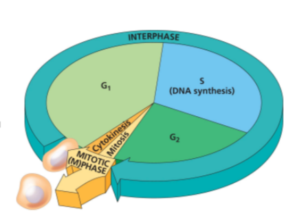
Overall Cell Cycle process
Overall Process
G1 → cell grows
↓
S → DNA is replicated
↓
G2 → cell grows + prepares for division
↓
Mitosis → genetic material divides
↓
Cytokinesis → cytoplasm divides
↓
2 genetically identical cells
Explain the structure of Mitosis & the Mitotic Spindle.
Mitosis divides the nucleus/genetic material so the 2 daughter cells receive identical genetic material.
5 stages: Prophase → Prometaphase → Metaphase → Anaphase → Telophase.
The mitotic spindle begins forming in prophase and consists of microtubules + associated proteins that move/separate chromosomes. In animal cells, spindle microtubules form from 2 centrosomes; each centrosome contains a pair of centrioles, **although centrioles are not essential for mitosis.
Asters are short microtubule arrays around each centrosome.
Mitotic spindle = centrosomes + asters + spindle microtubules.
Don't confuse centrosome (organizes spindle) with centromere (chromosome region).
