Cardiac electrical activity and ECG
 Call Kai
 Learn
Learn Practice Test
 Spaced Repetition
 Match
Match Flashcards
 Knowt Play
1/31

There's no tags or description
Looks like no tags are added yet.
Name | Mastery | Learn | Test | Matching | Spaced | Call with Kai | Chat |
|---|
No analytics yet
Send a link to your students to track their progress
32 Terms
What is cardiac conduction?
The process by which electrical signals are propagated through the heart to coordinate its contractions.
What is the function of autorhythmic regions of the heart?
To initiate and conduct action potentials that regulate the heartbeat.
What are examples of autorhythmic regions of the heart?
- SA node
- AV node
- Bundle of His
- Purkinje fibres
What are pacemaker cells and where are they found?
- Specialised heart cells that initiate and regulate the heart's rhythm.
- Produce pacemaker potential.
- Found in autorhytmic regions e.g., SA node, AV node, Bundle of His and Purkinje fibres
How does the SA node cause contraction of heart?
- The signal travels from the SA node to the AV node, where it is briefly delayed.
- The signal then moves through the Bundle of His and spreads to the Purkinje fibers, stimulating the ventricles to contract.
Why does the AV node delay the signal from the SA node?
The AV node delays the signal for about 100 milliseconds to allow the atria to contract and fill the ventricles with blood.
What is pacemaker potential?
The pacemaker potential is the gradual, spontaneous depolarisation of pacemaker cells in the heart, leading to the initiation of an action potential and the regulation of heart rhythm.

Describe the process of pacemaker potentials.
- Na⁺ enters through funny channels, depolarising the pacemaker cell.
- T-type calcium channels open, allowing Ca²⁺ to enter and continue depolarisation.
- This continues until threshold is reached, causing an action potential.
- L-type calcium channels open, causing rapid Ca²⁺ influx and full depolarisation.
- L-type Ca²⁺ channels close at the peak of depolarisation.
- Potassium channels then open, allowing K⁺ to exit, leading to repolarisation.
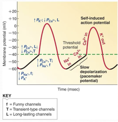
Why is there no resting membrane potential in pacemaker cells?
Pacemaker cells have a continuously drifting membrane potential due to the opening and closing of ion channels, causing self-initiated action potentials.
What is normal pacemaker activity in the SA node?
70-80 action potential per minute
What are cardiac action potentials in cardiomyocytes?
Electrical impulses that pass through cardiomyocytes to neighboring cells, triggering heart muscle contraction.
What causes cardiac action potentials in cardiomyocytes?
Pacemaker potentials
How do pacemaker potentials cause cardiac action potentials?
In pacemaker cells, L-type calcium channels open, causing rapid depolarisation and initiating an action potential in cardiomyocytes.
What are the phases of cardiac action potentials?
- Phase 0 = rapid depolarisation
- Phase 1 = Initial repolarisation
- Phase 2 = Plateau phase
- Phase 3 = Rapid repolarisation
Describe the process of cardiac action potentials.
- Na⁺ influx causes rapid depolarisation (Phase 0), which is partially counteracted by the opening of K⁺ channels (Phase 1), allowing K⁺ to exit the cell.
- Na⁺ channels then close, allowing a plateau phase to occur as L-type Ca²⁺ channels slowly open, allowing Ca²⁺ to enter the cell (Phase 2).
- L-type Ca²⁺ channels close, allowing rapid repolarisation as K⁺ channels open further, allowing K⁺ to exit (Phase 3).
- The cell then returns to its resting potential where K⁺ channels close.

How do cardiac action potentials cause muscle contraction?
- Cardiac action potentials allow Ca²⁺ to enter the cardiomyocyte through L-type calcium channels.
- This calcium binds to ryanodine receptors on the SR, which release more Ca²⁺ into the cell.
- The increased Ca²⁺ binds to troponin, enabling cross-bridge formation between actin and myosin, triggering muscle contraction.

How are action potentials terminated in cardiomyoyctes?
The action potential is terminated by active transport of Ca²⁺ back into the sarcoplasmic reticulum (SR) via Ca²⁺-ATPase pumps (e.g., SERCA2a).

What can affect the action potential in the cardiomyocytes and pacemaker cells?
- Abnormal K+ levels (hypokalaemia, hyperkalaemia)
- Change in extracellular Ca2+
How can hypokalemia affect action potentials?
Low K⁺ makes the resting membrane potential more negative, hindering depolarisation and potentially slowing the heart or causing arrhythmias.
How can hyperkalaemia affect action potentials?
High K⁺ reduces the membrane potential (more positive), making it easier to depolarise, which can lead to abnormal heart rhythms.
How can hypo and hypercalcaemia affect action potentials?
- Hypocalcemia weakens contraction and slows conduction by reducing calcium influx during the plateau phase.
- Hypercalcemia increases calcium influx, shortening action potential duration and potentially raising heart rate.
How does digoxin affect Ca2+ levels and how does this affect cardiac action potentials?
Increases cytosolic Ca2+ and contractility.
What is the contractile response?
The process by which calcium triggers the interaction between actin and myosin filaments in cardiomyocytes, leading to muscle contraction.

Describe the changes in the contractile response during an action potential.
- During the plateau phase, the contractile response peaks as Ca²⁺ enters the cell and triggers contraction.
- As the membrane begins to repolarise, Ca²⁺ decreases as it is pumped back into the SR or out of the cell, causing the contractile response to decrease.

What is the refractory period?
The time when a cardiomyocyte cannot initiate another action potential, preventing a second contraction before the first has finished.
What is an ECG?
- A recording of the electrical activity of the heart
- Made up of a PQRST complex

Describe the P wave of an ECG.
Depolarisation of atria in response to SA node triggering.

Describe PR interval of an ECG.
Delay of AV node to allow filling

Describe the QRS complex of an ECG.
Depolarisation of ventricles triggers main pumping contractions
Describe the the ST segment of an ECG.
Beginning of ventricle repolarisation, should be flat.
Describe the QT interval in an ECG.
The time from the beginning of ventricular depolarization (QRS complex) to the end of ventricular repolarization (T wave), reflecting the duration of a single ventricular action potential.
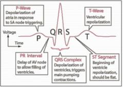
Describe the T-wave of an ECG.
Ventricular repolarisation
