15. Synaptic Integration Part 2 - The Role of Neurotransmitters

0.0(0)
studied byStudied by 0 people
0.0(0)
full-widthCall Kai
learnLearn
examPractice Test
spaced repetitionSpaced Repetition
heart puzzleMatch
flashcardsFlashcards
GameKnowt Play
Card Sorting

1/14

encourage image

There's no tags or description

Looks like no tags are added yet.

Study Analytics
Name
Mastery
Learn
Test
Matching
Spaced

No study sessions yet.

15 Terms

1
New cards

What happens at excitatory and inhibitory synapses during the patellar reflex?

  • Patellar (knee-jerk) reflex involves both excitation and inhibition.

  • Excitation: Ia sensory neuron activates α-motor neuron → quadriceps contract.

  • Inhibition: same Ia sensory neuron activates inhibitory interneuron → inhibits α-motor neuron to hamstrings, preventing antagonist contraction.

  • Ensures coordinated movement (one muscle contracts while the opposite relaxes).

<ul><li><p><strong>Patellar (knee-jerk) reflex</strong> involves both <strong>excitation</strong> and <strong>inhibition</strong>.</p></li><li><p><strong>Excitation:</strong> Ia sensory neuron activates <strong>α-motor neuron → quadriceps contract</strong>.</p></li><li><p><strong>Inhibition:</strong> same Ia sensory neuron activates <strong>inhibitory interneuron → inhibits α-motor neuron to hamstrings</strong>, preventing antagonist contraction.</p></li><li><p>Ensures <strong>coordinated movement</strong> (one muscle contracts while the opposite relaxes).</p></li></ul><p></p>
2
New cards

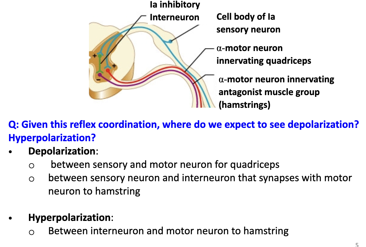
Where do depolarization and hyperpolarization occur in the patellar reflex circuit?

  • Depolarization:

    • Between Ia sensory neuron and α-motor neuron for quadriceps.

    • Between sensory neuron and inhibitory interneuron that affects hamstring motor neuron.

  • Hyperpolarization:

    • Between inhibitory interneuron and α-motor neuron to hamstrings.

  • Allows coordinated contraction and relaxation of opposing muscles.

<ul><li><p><strong>Depolarization:</strong></p><ul><li><p>Between <strong>Ia sensory neuron</strong> and <strong>α-motor neuron</strong> for <strong>quadriceps</strong>.</p></li><li><p>Between <strong>sensory neuron</strong> and <strong>inhibitory interneuron</strong> that affects hamstring motor neuron.</p></li></ul></li><li><p><strong>Hyperpolarization:</strong></p><ul><li><p>Between <strong>inhibitory interneuron</strong> and <strong>α-motor neuron</strong> to <strong>hamstrings</strong>.</p></li></ul></li><li><p>Allows <strong>coordinated contraction and relaxation</strong> of opposing muscles.</p></li></ul><p></p>
3
New cards

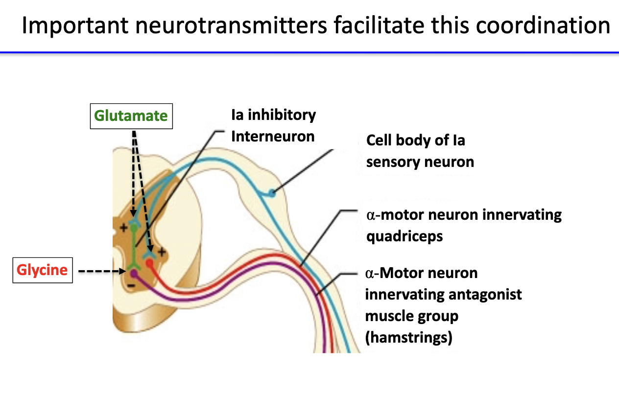
Which neurotransmitters are involved in coordinating excitation and inhibition in the patellar reflex?

  • Glutamate:

    • Released by Ia sensory neurons at excitatory synapses.

    • Activates α-motor neurons to quadriceps and interneurons.

  • Glycine:

    • Released by inhibitory interneurons in the spinal cord.

    • Inhibits α-motor neurons to hamstrings (antagonist).

<ul><li><p><strong>Glutamate:</strong></p><ul><li><p>Released by <strong>Ia sensory neurons</strong> at <strong>excitatory synapses</strong>.</p></li><li><p>Activates <strong>α-motor neurons</strong> to <strong>quadriceps</strong> and <strong>interneurons</strong>.</p></li></ul></li><li><p><strong>Glycine:</strong></p><ul><li><p>Released by <strong>inhibitory interneurons</strong> in the spinal cord.</p></li><li><p>Inhibits <strong>α-motor neurons to hamstrings</strong> (antagonist).</p></li></ul></li></ul><p></p>
4
New cards

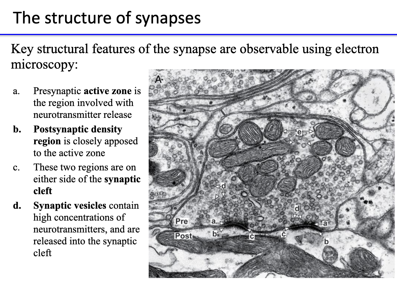
What are the key structural features of a synapse seen under electron microscopy?

  • Presynaptic active zone: site of neurotransmitter release.

  • Postsynaptic density: region containing receptors, directly across from active zone.

  • Synaptic cleft: space between pre- and postsynaptic membranes.

  • Synaptic vesicles: store high concentrations of neurotransmitters, released into cleft upon activation.

<ul><li><p><strong>Presynaptic active zone:</strong> site of <strong>neurotransmitter release</strong>.</p></li><li><p><strong>Postsynaptic density:</strong> region containing <strong>receptors</strong>, directly across from active zone.</p></li><li><p><strong>Synaptic cleft:</strong> space between pre- and postsynaptic membranes.</p></li><li><p><strong>Synaptic vesicles:</strong> store <strong>high concentrations of neurotransmitters</strong>, released into cleft upon activation.</p></li></ul><p></p>
5
New cards

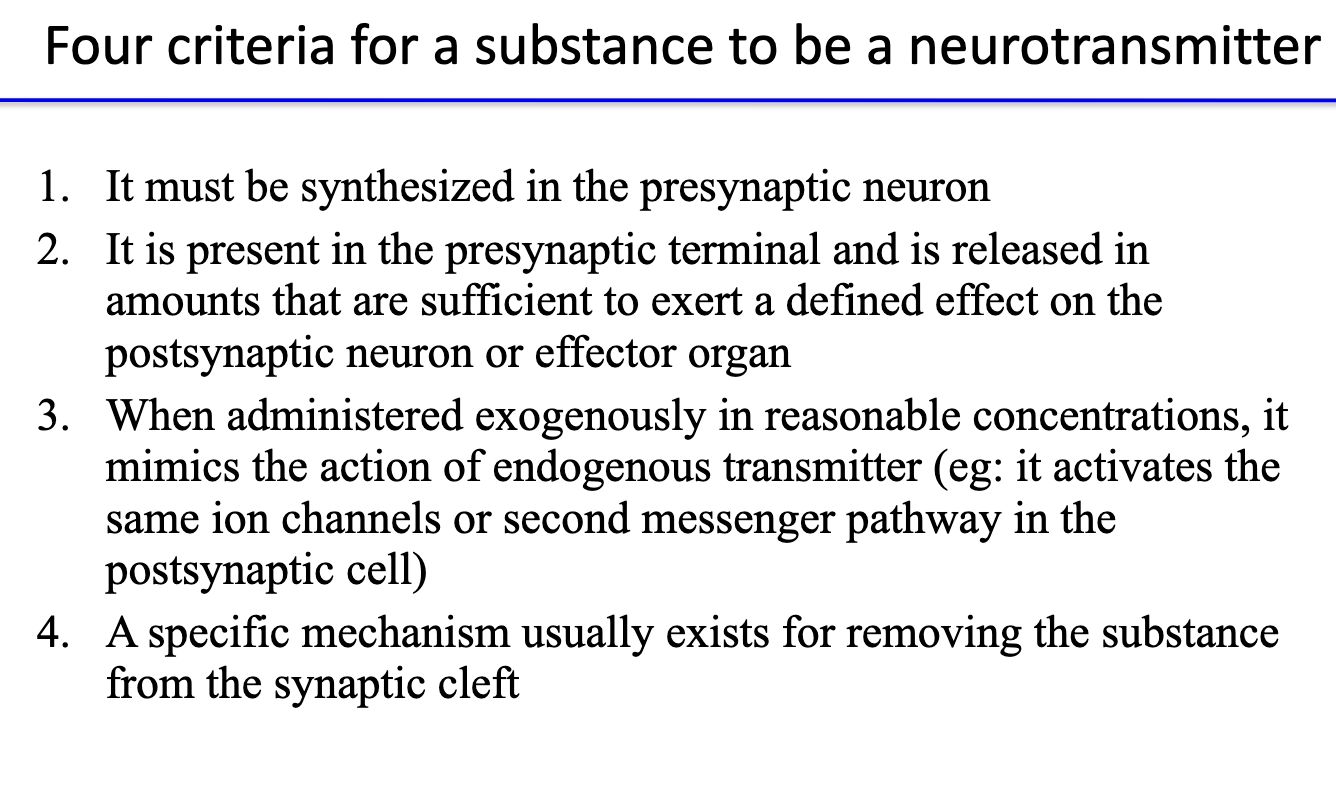
What are the four main criteria for classifying a neurotransmitter?

  1. Synthesized in the presynaptic neuron.

  2. Present in terminal and released in sufficient amounts to affect postsynaptic cell.

  3. When applied externally, mimics the natural transmitter’s action (same receptors/pathways).

  4. Has a specific removal mechanism from synaptic cleft (e.g., reuptake or degradation).

<ol><li><p><strong>Synthesized</strong> in the <strong>presynaptic neuron</strong>.</p></li><li><p><strong>Present in terminal</strong> and <strong>released</strong> in sufficient amounts to affect postsynaptic cell.</p></li><li><p>When <strong>applied externally</strong>, mimics the natural transmitter’s action (same receptors/pathways).</p></li><li><p>Has a <strong>specific removal mechanism</strong> from synaptic cleft (e.g., reuptake or degradation).</p></li></ol><p></p>
6
New cards

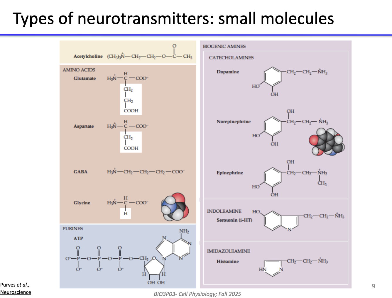
What are small-molecule neurotransmitters?

  • Include acetylcholine, glutamate, GABA, glycine, dopamine, serotonin, norepinephrine.

  • Typically rapid-acting.

  • Synthesized locally in axon terminals.

  • Mediate fast synaptic transmission.

<ul><li><p>Include <strong>acetylcholine, glutamate, GABA, glycine, dopamine, serotonin, norepinephrine</strong>.</p></li><li><p>Typically <strong>rapid-acting</strong>.</p></li><li><p>Synthesized <strong>locally in axon terminals</strong>.</p></li><li><p>Mediate <strong>fast synaptic transmission</strong>.</p></li></ul><p></p>
7
New cards

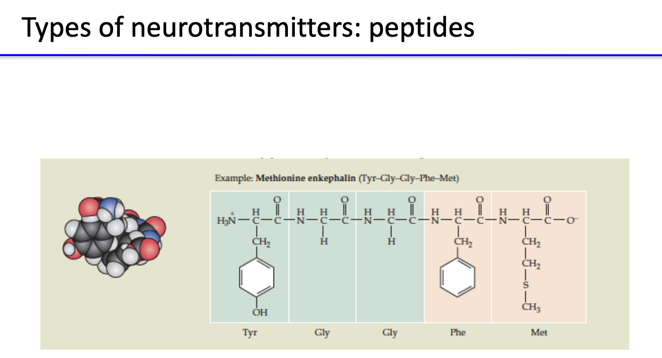
What are peptide neurotransmitters and their general features?

  • Short chains of amino acids (e.g., substance P, endorphins, oxytocin, vasopressin).

  • Synthesized in cell body, packaged into vesicles, and transported to terminals.

  • Usually modulate slower, longer-lasting synaptic effects.

<ul><li><p>Short chains of amino acids (e.g., <strong>substance P, endorphins, oxytocin, vasopressin</strong>).</p></li><li><p>Synthesized in <strong>cell body</strong>, packaged into vesicles, and <strong>transported to terminals</strong>.</p></li><li><p>Usually <strong>modulate slower, longer-lasting synaptic effects</strong>.</p></li></ul><p></p>
8
New cards

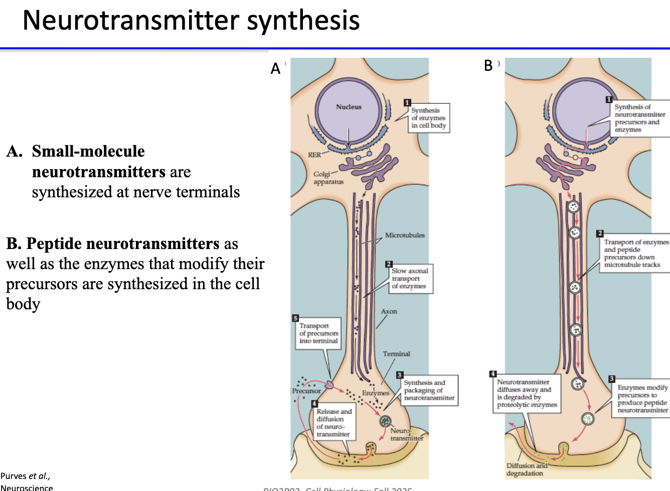
Where are small-molecule and peptide neurotransmitters synthesized?

  • Small-molecule neurotransmitters: synthesized at nerve terminals by local enzymes.

  • Peptide neurotransmitters: synthesized in cell body along with modifying enzymes → transported down axon.

<ul><li><p><strong>Small-molecule neurotransmitters:</strong> synthesized at <strong>nerve terminals</strong> by local enzymes.</p></li><li><p><strong>Peptide neurotransmitters:</strong> synthesized in <strong>cell body</strong> along with modifying enzymes → transported down axon.</p></li></ul><p></p>
9
New cards

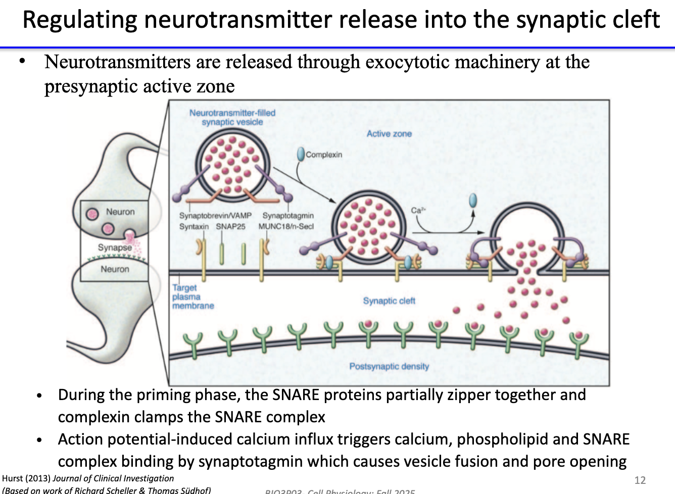
How is neurotransmitter release controlled at the presynaptic active zone?

  • Release via exocytosis controlled by SNARE complex.

  • Priming phase: SNAREs partially zippered; complexin clamps them.

  • Action potential → Ca²⁺ influxsynaptotagmin binds Ca²⁺ + phospholipids + SNAREs → triggers vesicle fusion & pore opening → neurotransmitter released.

<ul><li><p>Release via <strong>exocytosis</strong> controlled by <strong>SNARE complex</strong>.</p></li><li><p><strong>Priming phase:</strong> SNAREs partially zippered; <strong>complexin</strong> clamps them.</p></li><li><p><strong>Action potential → Ca²⁺ influx</strong> → <strong>synaptotagmin</strong> binds Ca²⁺ + phospholipids + SNAREs → triggers <strong>vesicle fusion &amp; pore opening</strong> → neurotransmitter released.</p></li></ul><p></p>
10
New cards

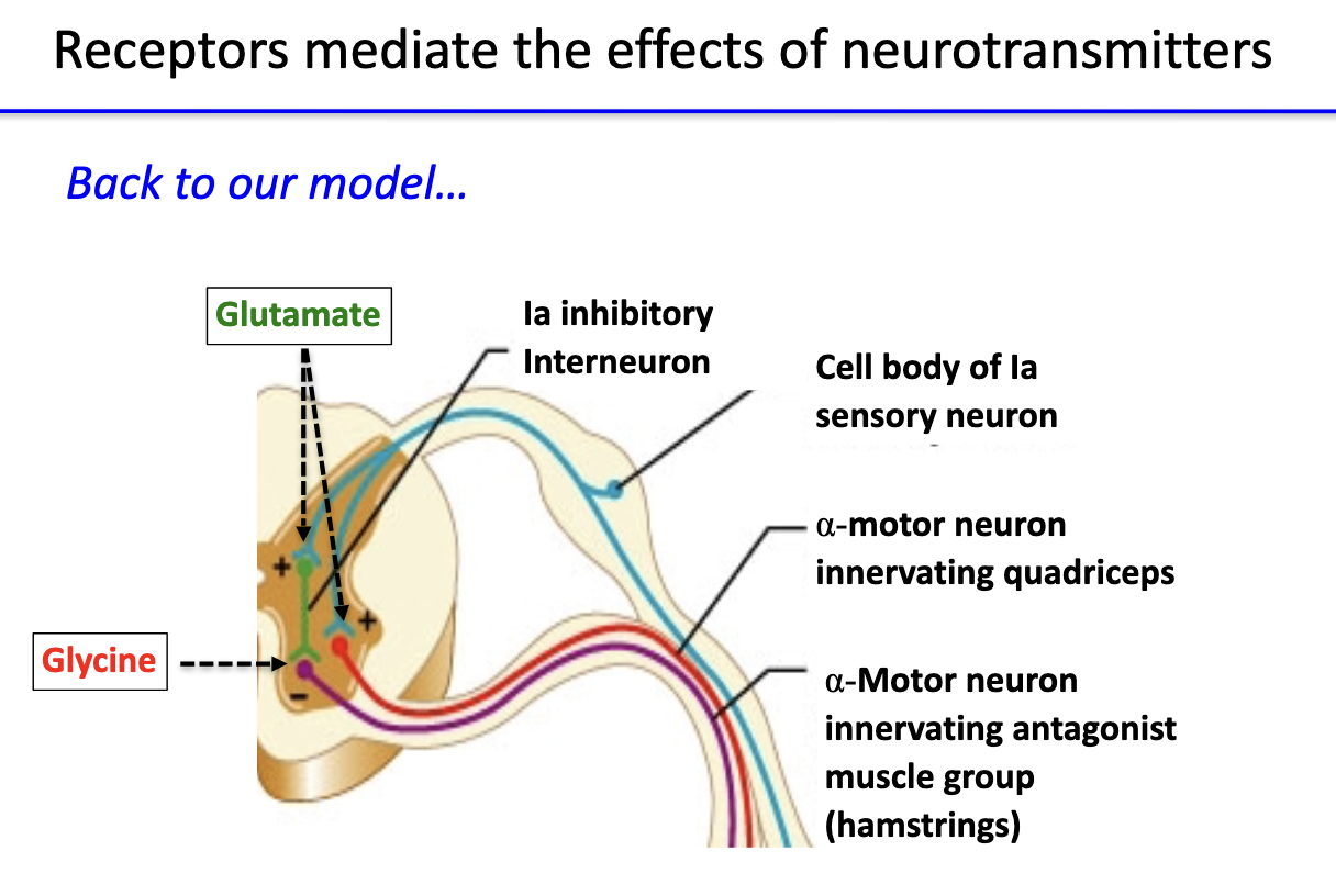
How do receptors mediate the effects of neurotransmitters in spinal circuits?

  • Glutamate receptors: on α-motor neurons to quadriceps → excitatory depolarization.

  • Glycine receptors: on α-motor neurons to hamstrings → inhibitory hyperpolarization.

  • Together, they coordinate excitation and inhibition for proper reflex response.

<ul><li><p><strong>Glutamate receptors:</strong> on <strong>α-motor neurons</strong> to <strong>quadriceps</strong> → excitatory depolarization.</p></li><li><p><strong>Glycine receptors:</strong> on <strong>α-motor neurons</strong> to <strong>hamstrings</strong> → inhibitory hyperpolarization.</p></li><li><p>Together, they coordinate <strong>excitation and inhibition</strong> for proper reflex response.</p></li></ul><p></p>
11
New cards

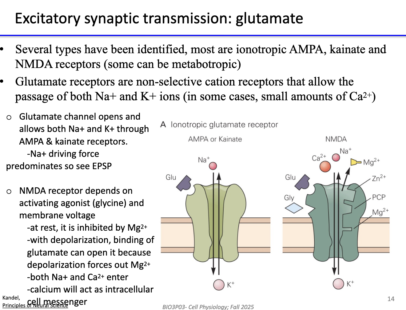
How does glutamate produce excitatory synaptic transmission?

  • Acts on ionotropic receptors: AMPA, kainate, NMDA (and some metabotropic).

  • AMPA & kainate: allow Na⁺ in, K⁺ outEPSP (Na⁺ dominates).

  • NMDA: requires glutamate + glycine + depolarization to remove Mg²⁺ block → allows Na⁺ & Ca²⁺ influx.

  • Ca²⁺ acts as an intracellular messenger, influencing plasticity and signaling.

<ul><li><p>Acts on <strong>ionotropic receptors</strong>: <strong>AMPA, kainate, NMDA</strong> (and some <strong>metabotropic</strong>).</p></li><li><p><strong>AMPA &amp; kainate:</strong> allow <strong>Na⁺ in</strong>, <strong>K⁺ out</strong> → <strong>EPSP</strong> (Na⁺ dominates).</p></li><li><p><strong>NMDA:</strong> requires <strong>glutamate + glycine + depolarization</strong> to remove <strong>Mg²⁺ block</strong> → allows <strong>Na⁺ &amp; Ca²⁺</strong> influx.</p></li><li><p><strong>Ca²⁺</strong> acts as an <strong>intracellular messenger</strong>, influencing plasticity and signaling.</p></li></ul><p></p>
12
New cards

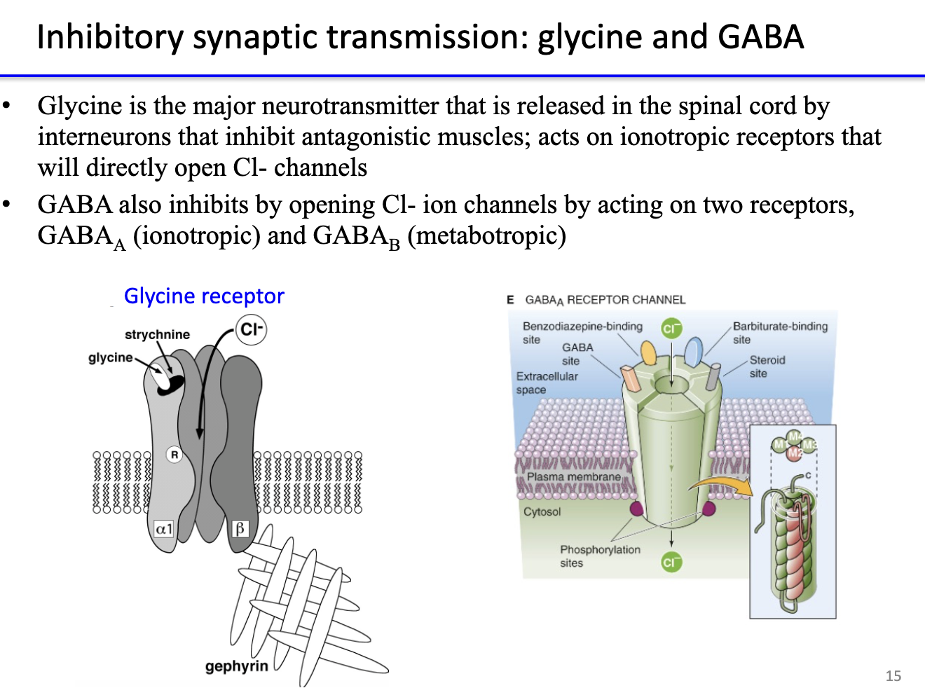
How do glycine and GABA mediate inhibitory synaptic transmission?

  • Glycine: major inhibitory transmitter in spinal cord; opens Cl⁻ channels via ionotropic receptors → hyperpolarization.

  • GABA: major inhibitory transmitter in brain; acts on

    • GABAᴀ: ionotropic → opens Cl⁻ channels.

    • GABAʙ: metabotropic → activates K⁺ channels or inhibits Ca²⁺.

  • Both decrease neuronal excitability.

<ul><li><p><strong>Glycine:</strong> major inhibitory transmitter in <strong>spinal cord</strong>; opens <strong>Cl⁻ channels</strong> via <strong>ionotropic receptors</strong> → hyperpolarization.</p></li><li><p><strong>GABA:</strong> major inhibitory transmitter in <strong>brain</strong>; acts on</p><ul><li><p><strong>GABAᴀ:</strong> ionotropic → opens <strong>Cl⁻ channels</strong>.</p></li><li><p><strong>GABAʙ:</strong> metabotropic → activates <strong>K⁺ channels</strong> or inhibits Ca²⁺.</p></li></ul></li><li><p>Both decrease <strong>neuronal excitability</strong>.</p></li></ul><p></p>
13
New cards

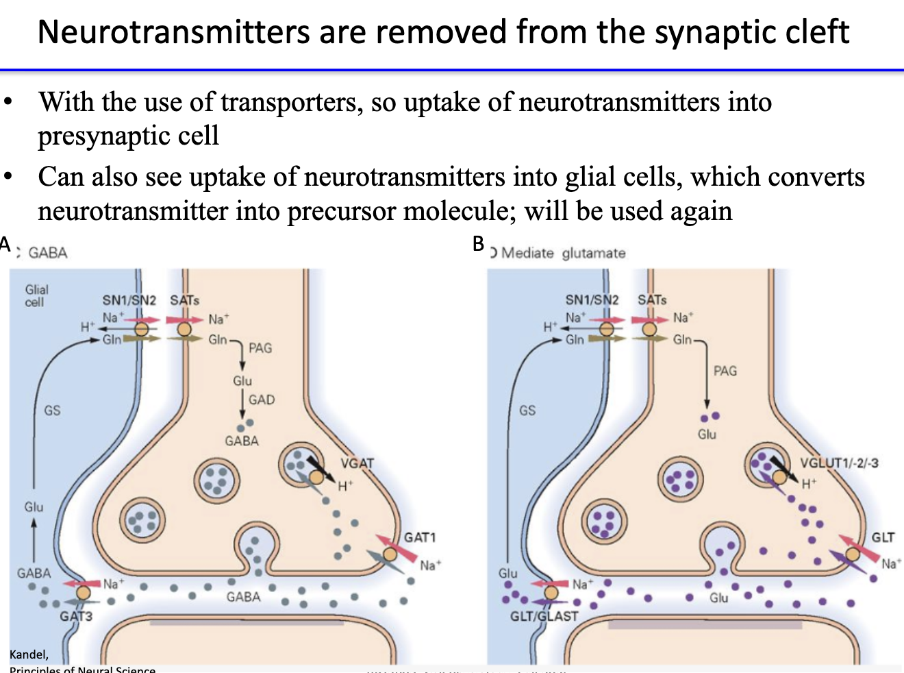
How are neurotransmitters removed from the synaptic cleft?

  • Reuptake: transporters move neurotransmitters back into presynaptic terminal.

  • Glial uptake: glial cells absorb transmitter → convert to precursor form for reuse.

  • Ensures signal termination and recycling of neurotransmitters.

<ul><li><p><strong>Reuptake:</strong> transporters move neurotransmitters back into <strong>presynaptic terminal</strong>.</p></li><li><p><strong>Glial uptake:</strong> glial cells absorb transmitter → convert to <strong>precursor form</strong> for reuse.</p></li><li><p>Ensures <strong>signal termination</strong> and <strong>recycling</strong> of neurotransmitters.</p></li></ul><p></p>
14
New cards

How can drugs alter synaptic transmission?

  1. Alter synthesis, storage, transport, or release of neurotransmitters.

  2. Modify receptor interaction (agonist/antagonist effects).

  3. Influence reuptake or degradation mechanisms.

  4. Replace deficient neurotransmitters with substitutes.

<ol><li><p><strong>Alter synthesis, storage, transport, or release</strong> of neurotransmitters.</p></li><li><p><strong>Modify receptor interaction</strong> (agonist/antagonist effects).</p></li><li><p><strong>Influence reuptake or degradation</strong> mechanisms.</p></li><li><p><strong>Replace deficient neurotransmitters</strong> with substitutes.</p></li></ol><p></p>
15
New cards

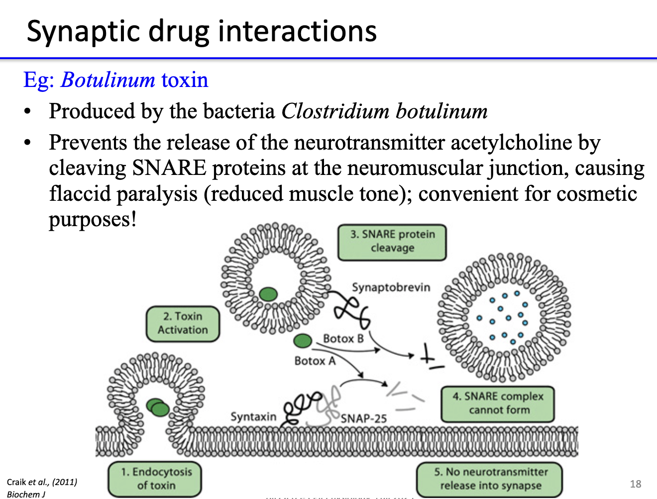
How does botulinum toxin affect neurotransmission?

  • Produced by Clostridium botulinum.

  • Cleaves SNARE proteins → prevents acetylcholine release at neuromuscular junction.

  • Causes flaccid paralysis (muscle cannot contract).

  • Used therapeutically/cosmetically to relax muscles (e.g., Botox).

<ul><li><p>Produced by <strong>Clostridium botulinum</strong>.</p></li><li><p><strong>Cleaves SNARE proteins</strong> → prevents <strong>acetylcholine release</strong> at neuromuscular junction.</p></li><li><p>Causes <strong>flaccid paralysis</strong> (muscle cannot contract).</p></li><li><p>Used <strong>therapeutically/cosmetically</strong> to relax muscles (e.g., Botox).</p></li></ul><p></p>