6.1.14 Assesses signs and symptoms of neurological significance.
 Call Kai
 Learn
 Practice Test
 Spaced Repetition
 Match
 Flashcards
 Knowt Play
1/36

There's no tags or description
Looks like no tags are added yet.
37 Terms
What is a Relative Afferent Pupillary Defect (RAPD), also known as a Marcus Gunn pupil?
Asymmetry in the afferent (sensory) pupillary pathway between the two eyes, detected using the swinging flashlight test.

What are the possible causes and clinical management of RAPD?
Possible causes:
Optic neuritis
Glaucoma (moderate to advanced)
Retinal detachment
Severe macular disease
Optic nerve compression or ischaemia
Clinical management:
- Urgent referral if newly detected or unexplained.
What is Horner's Syndrome and what are its clinical signs?
Disruption of the sympathetic pathway to the eye.
Signs:
Miosis (small pupil)
Ptosis (mild)
Anisocoria greater in dim illumination
Normal direct, consensual and near reflex
Delayed dilation in the dark
What are the possible causes and clinical management of Horner's Syndrome?
Possible causes:
Carotid artery dissection
Brainstem or spinal lesions
Neck or chest trauma
Congenital Horner syndrome
Clinical management:
Urgent referral.
Neuro-imaging often required.
What is Adie's Tonic Pupil and what are its clinical signs?
A benign parasympathetic denervation of the iris sphincter, usually unilateral.
Signs:
Dilated pupil
Poor or absent light reaction
No direct or consensual response
Better (slow) near response

What are the possible causes and clinical management of Adie's Tonic Pupil?
Possible causes:
Idiopathic (most common)
Autonomic dysfunction
Clinical management:
- Usually no treatment required.
What is an Argyll Robertson Pupil?
Clinical signs?
Management?
A classic neurological pupil abnormality associated with neurosyphilis.
Signs:
Small, irregular pupils
No reaction to light
No direct or consensual response from either pupil
Preserved near response
Bilateral
Clinical management:
Urgent referral.
Serological testing and systemic treatment.
What is anisocoria and when is urgent referral required?
Anisocoria is an inequality in pupil size.
Urgent referral if:
Acute onset
Neurological signs present
What must be assessed when investigating anisocoria?
Light and near pupil reactions
Ptosis
Extraocular movements
What are the possible causes of anisocoria?
Physiological anisocoria (most common)
Horner syndrome
Third nerve palsy
Pharmacological agents
Adie's tonic pupil
Trauma
What is Arteritic Anterior Ischaemic Optic Neuropathy (AAION)?
Giant Cell Arteritis:
Results in luminal narrowing or occlusion.
Causes ischaemia of the optic nerve head.
Other associated condition:
- Behçet's disease - a rare, chronic condition causing inflammation in blood vessels (vasculitis) throughout the body.

What are the clinical signs and symptoms of AAION?
Signs:
Pale swollen optic disc
Chalky white disc
RAPD
Cotton wool spots
Altitudinal defect, particularly lower half
Thick and pulsating temporal artery
Symptoms:
Sudden severe vision loss
Headache
Jaw claudication
Scalp tenderness
What is the clinical management of AAION?
Medical emergency!
Stress importance of fast treatment.
Immediate same-day referral to hospital A&E.
What is Non-Arteritic Anterior Ischaemic Optic Neuropathy (NAION)?
Non-inflammatory condition where poor blood flow damages the optic nerve.

What are the possible causes of NAION?
Giant Cell Arteritis - Results in luminal narrowing or occlusion → ischaemia of the ONH Atherosclerosis
What are the clinical signs and symptoms of NAION?
Signs:
Optic disc swelling (often segmental)
Small cup-to-disc ratio
Flame haemorrhages
Altitudinal defect, particularly lower half
Symptoms:
Sudden painless monocular vision loss
Often upon waking
What is the clinical management of NAION?
Medical emergency!
Stress importance of fast treatment.
Immediate same-day referral to hospital A&E.
What is papilloedema and what are its possible causes?
Swollen optic disc due to raised intracranial pressure, due to a tumour in the brain or something else growing in the brain.
Other causes:
Meningitis
Hydrocephalus
What is the clinical management of papilloedema?
Emergency referral.
Urgent referral even if asymptomatic, depending on the degree of swelling.
What are the symptoms of papilloedema?
Severe headache (worse in the morning or when bending over)
Nausea and vomiting
Vision loss / sudden blanking of the vision lasting seconds
What are the mechanical signs of papilloedema?
Elevation of the ONH (3D = 1 mm of elevation)
Blurring of the disc margins
Filling in of the physiological cup
Oedema of the peripapillary nerve fibre layer (NFL)
Retinal and/or choroidal folds
Enlarged blind spot on visual field analysis
What are the vascular signs of papilloedema?
Hyperaemia of the disc
Loss of the spontaneous venous pulsation (SVP) and venous congestion
Papillary/peripapillary haemorrhages
Hard exudates
Cotton wool spots
What are the features of Grade 1 papilloedema?
C-shaped halo surrounding disc
Sparing of temporal disc margin
Radial nerve fibre striation disruption

What are the features of Grade 2 papilloedema?
Halo becomes circumferential
Nasal border elevated
No major vessel obscuration

What are the features of Grade 3 papilloedema?
Obscuration of at least one vessel leaving the disc
Elevation of all borders
Circumferential halo
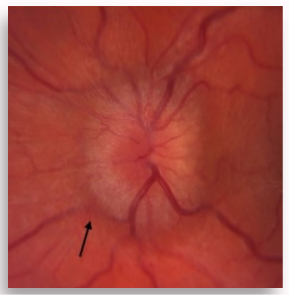
What are the features of Grade 4 papilloedema?
Obscuration of a major vessel on the disc
Complete elevation including the cup
Circumferential halo

What are the features of Grade 5 papilloedema?
Obscuration of a major vessel on the disc and leaving disc
Complete elevation including the cup
Circumferential halo

What are the signs of optic nerve dysfunction?
Reduced VA (both distance and near)
Severe impaired colour vision
RAPD (unilateral/asymmetric bilateral conditions)
VF defect (depending on the condition)
Impaired contrast sensitivity
How do optic nerve disease and macular disease differ clinically?
Optic nerve disease | Macular disease |
|---|---|
RAPD | No RAPD |
Severe dichromatopsia | None-mild dichromatopsia |
Reduced CS | Augmented brightness sensitivity |
What is optic neuritis and what are its possible causes?
An inflammatory or demyelinating disorder of the optic nerve (ON), most commonly the manifestation of multiple sclerosis.
Other causes:
Viral infections
TB
Syphilis
What are the symptoms of optic neuritis?
Rapid loss of vision in one eye
Retro-orbital or ocular pain on movement of the eye
The visual loss is exacerbated by heat or exercise
What are the clinical signs of optic neuritis?
RAPD
VA loss mild to severe
Central scotoma
Severely impaired colour vision (even if VA normal)
Normal fundus
Blurred disc margins
Swollen veins
What is the clinical management of optic neuritis?
Emergency referral.
Treatment: Steroids.
What are the possible causes of monocular transient visual loss?
Aetiology: Anterior to the chiasm.
Gaze-evoked: Lesions of the orbit, cavernous haemangioma, meningioma of the ON sheath.
Lasting seconds-minutes: Amaurosis fugax, ION, vasospasm.
Lasting hours: Thrombi (stops blood flow through vessels), carotid stenosis, migraine.
What are the possible causes of binocular transient visual loss?
Location: Posterior to the chiasm.
Migraine
Arteriovenous (AV) malformation
Emboli
What is the difference between neuro-ophthalmological and non-neuro-ophthalmological causes of transient visual loss?
Neuro-ophthalmological | Non-neuro-ophthalmological |
|---|---|
Problem is behind the eye | Problem is inside the eye |
Pre-chiasmal cause, mainly optic nerve neuropathies | Various ocular causes |
What are the possible non-neuro-ophthalmological causes of transient visual loss?
CRVO
CRAO
AION
RD
Macular disease
Corneal disease
AACG
Uveitis
Other ocular causes